
Abstract
Sheep, particularly lambs, with high circulating levels of Clostridium perfringens type D epsilon toxin develop severe neurologic signs and often die suddenly. On microscopic examination, in the brain, there is microvascular endothelial injury and diffuse vasogenic edema. The aquaporin (AQP) family of membrane water-channel proteins, especially AQP-4, is important in the regulation of water balance in the brain and facilitates reabsorption of excess fluid. In rats given epsilon toxin, generalized cerebral edema was demonstrated by marked albumin extravasation and was correlated with widespread upregulation of AQP-4 in astrocytes. These results suggest that AQP-4 has a role in the clearance of edema fluid from brains damaged by this clostridial toxin.
Keywords
Clostridium perfringens type D enterotoxemia is an important disease of sheep worldwide; the principal clinicopathologic manifestations are neurologic. Absorption into the circulation of large quantities of epsilon toxin produced by these proliferating intestinal bacteria after starch overload causes microvascular endothelial damage in the brain and a severe, generalized, vasogenic edema. After an acute clinical course, death is the usual outcome. 2, 3, 8 Because cerebral edema is a critical component of acute epsilon intoxication, we wanted to examine the role of the water channel protein, aquaporin-4 (AQP4), in this process.
Aquaporins are a family of membrane channel proteins that serve as selective pores through which water traverses the plasma membrane of different cell types. There are 9 different aquaporins, of which 4 (AQP 1, 3, 4, and 9) are found in the brain. AQP-4 is the most abundant water-channel protein in the central nervous system and is believed to play an important role in water homeostasis and the development of cerebral edema. 6, 9 In this study, AQP-4 in the brain was demonstrated by immunohistochemistry. The rat was used because it develops brain lesions that resemble those found in sheep after exposure to epsilon toxin, 2, 4 and we had an antibody known to react with rat AQP-4.
The epsilon toxin used was a partially purified prototoxin prepared from filtrates of broth cultures of Clostridium perfringens type D (Commonwealth Serum Laboratories, Melbourne, Australia). Trypsin-activated toxin was injected intraperitoneally at a dose rate of 0.5 ml after a 1 : 300 dilution as a lethal dose. 1 Four, 10-week-old Sprague-Dawley rats were injected with toxin, and 2 control rats were given a similar volume of physiologic saline solution. At 2 hours after an injection, the brains were fixed by transcardiac perfusion with 4% paraformaldehyde that contained 0.02% heparin. After remaining in situ for 2 hours, the brains were removed and immersion-fixed in 10% buffered formalin for 4 days. Brains were then processed to paraffin wax, and 6-μm sections were cut and stained with hematoxylin and eosin. Duplicate sections were cut for immunohistochemistry.
By using ventral landmarks, 3 coronal levels of brain similar to those used by the National Toxicology Program in the United States for routine toxicologic screening in mice 7 were selected: at the optic chiasm (level 1), mammillary bodies (level 2), and the widest part of the cerebellum (level 3). At level 1, the cingulate and parietal cortices, caudate-putamen, and corpus callosum were examined; at level 2, the occipital and temporal cortices, thalamus and internal capsule; and, at level 3, the cerebellum and pons. Brain sections were assessed independently by 2 pathologists blinded to whether the slides were from the treated or control group.
To evaluate the severity and distribution of cerebral edema, extravasation of endogenous albumin was detected immunohistochemically by using a goat anti-rat albumin (Cat. no. 0113–0341; Cappel, West Chester, PA) at a dilution of 1 : 20,000. This was followed by a biotinylated rabbit anti-goat immunoglobulin secondary (Vector, Burlingame, CA), at a 1 : 250 dilution for 30 minutes then washed in phosphate buffered saline. To detect AQP-4, a rabbit polyclonal anti-rat antibody (Cat. no. AB3068; Chemicon, Temecula, CA) was used at a dilution of 1 : 4000, followed by a biotinylated goat anti-rabbit immunoglobulin secondary (Vector), at a 1 : 250 dilution for 30 minutes, then washed in PBS. Both the albumin and the AQP-4 were incubated with a streptavidin conjugated peroxidase tertiary, washed, and visualized with 3,3′-diaminobenzidine tetrahydrochloride. Sections were counterstained with hematoxylin, dehydrated, cleared, and mounted. No antigen retrieval was necessary for the albumin antibody, but ethylenediaminetetraacetic acid antigen retrieval solution was used for the AQP-4. Positive and negative controls were included in these protocols.
In control brains, albumin extravasation was negligible (Fig. 1) and confined to non-blood-brain–barrier (BBB) regions but, in all toxin-treated brains, albumin leakage was widespread (Fig. 2) in gray and white matter of the cerebrum, cerebellum, and brainstem. In control brains, AQP-4 immunopositivity was expressed as widespread fine granularity in astrocytic processes (Fig. 3) of both gray and white matter, especially the former. Immunoreactivity was more abundant where astrocytic end-feet were in contact with capillaries and postcapillary venules (Fig. 3) and the outer (pia mater) and inner (ependymal) surfaces of the brain (glia limitans). The ependyma was strongly immunopositive. In toxin-treated brains, the distribution of AQP-4 immunoreactivity was similar, but granules were more coarse and numerous (Fig. 4), particularly in perivascular (Fig. 4) and subpial sites. In all brains exposed to epsilon toxin, and in all regions examined, AQP-4 expression in the neuropil was more robust than in control brains and correlated with marked albumin leakage from blood vessels.
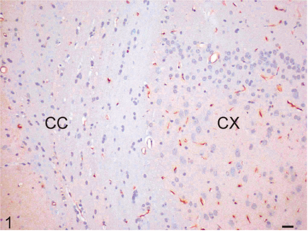
Control brain; rat. Minimal albumin extravasation in the cerebral cortex (CX) and corpus callosum (CC). Immunohistochemistry for endogenous albumin, hematoxylin counterstain. Bar = 10 μm.
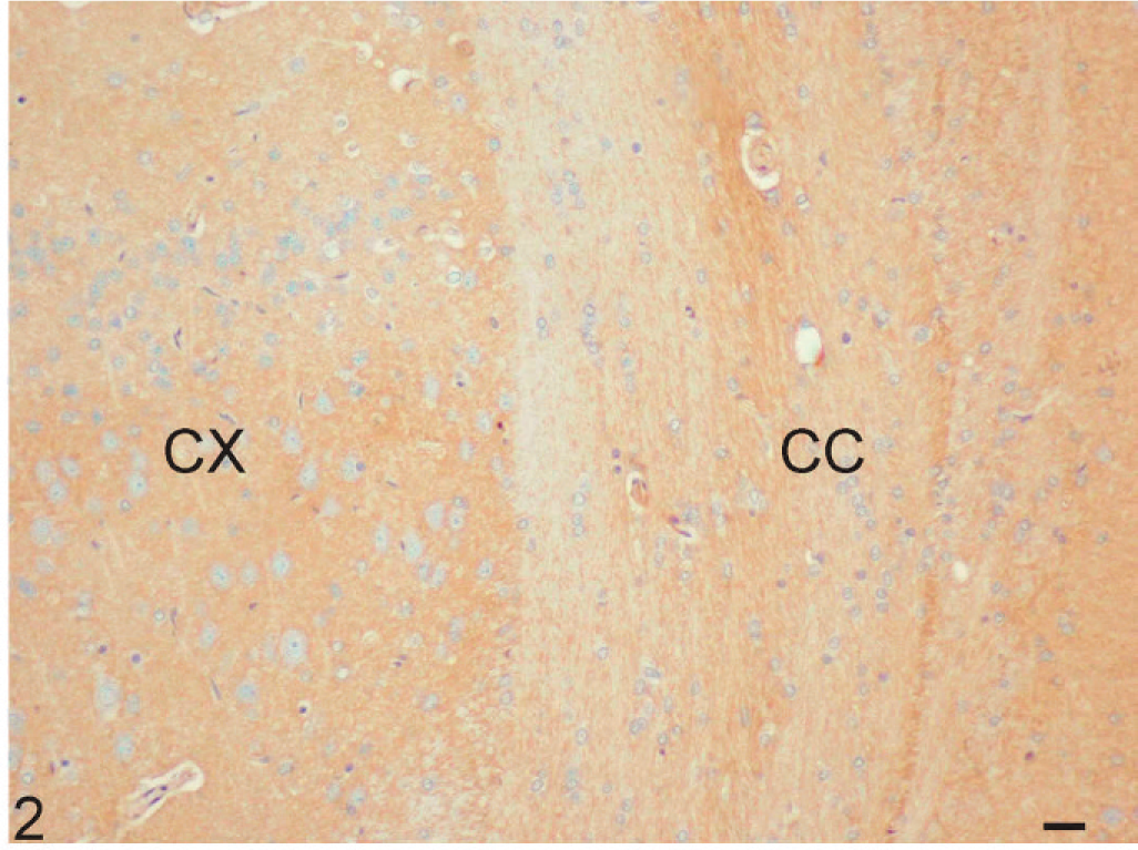
Toxin-treated brain; rat. Marked albumin leakage into the cerebral cortex (CX) and corpus callosum (CC). Immunohistochemistry for endogenous albumin, hematoxylin counterstain. Bar = 10 μm.
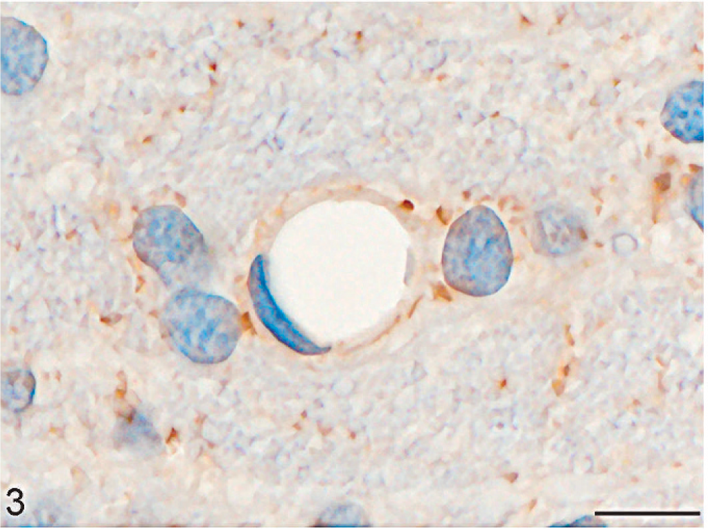
Control brain; rat. Finely granular AQP-4 immunopositivity in astrocytic processes and perivascular end-feet of a postcapillary venule. Immunohistochemistry for AQP-4, hematoxylin counterstain (oil immersion). Bar = 10 μm.
Toxin-treated brain; rat. Abundant, coarsely granular, astrocytic AQP-4 immunoreactivity, with marked upregulation in a postcapillary venule end-feet. Immunohistochemistry for AQP-4, hematoxylin counterstain (oil immersion). Bar = 10 μm.
In rats exposed to epsilon toxin, cerebral edema was severe and widely distributed, as shown by abundant albumin extravasation. Astrocytes are particularly prone to swelling in edematous states. 5 The high concentration of AQP-4 in perivascular end-feet and the dense network of astrocytic processes at the brain surface beneath the pia mater is consistent with regulation of water permeability at the BBB and cerebrospinal fluid-brain barrier, respectively. 6, 9 Although AQP-4 has an important role in water homeostasis in the brain, control mechanisms are poorly understood. 9 However, this water-channel protein probably moves water from the extracellular space into astrocytes, thus reducing osmotic stress on surrounding neurons. 6, 9
In summary, AQP-4 was widely upregulated in edematous epsilon toxin-treated brains compared with controls, which suggested that, after microvascular endothelial damage, the water-channel protein AQP-4 plays a role in attempting to resolve the edema by astrocytic uptake. A better understanding of aquaporin water channels may lead to new targeted therapies for ameliorating the devastating effects of brain edema.