
Abstract
An outbreak of acute, fatal, hemorrhagic pneumonia was observed in more than 1,000 mixed breed dogs in a single animal shelter. The Department of Anatomic Pathology at the University of California at Davis School of Veterinary Medicine performed necropsies on dogs that were found moribund in acute respiratory distress or found dead with evidence of nasal bleeding. All dogs had hemothorax and an acute, fibrinosuppurative pneumonia. Large numbers of gram-positive cocci were observed within the lungs of all dogs and within septic thromboemboli of remote organs in about 50% of cases. Bacterial cultures from the dogs and their environment revealed widespread beta-hemolytic Streptococus equi subspecies zooepidemicus (Lancefield Group C). Extensive diagnostic testing failed to reveal the consistent presence of copathogens in individual cases. The clinical, epidemiologic, molecular biologic, and pathologic data indicate that a single clone of S. zooepidemicus was the cause of an acutely fatal respiratory infection in these dogs.
Canine infectious respiratory disease (CIRD, “kennel cough”) is a clinical syndrome nearly ubiquitous in animal shelters and other densely housed canine populations. Clinical signs of disease last days to weeks, and, although death is a rare sequela to disease, intractable and even mild respiratory disease can be a criterion for euthanasia in some shelters. The etiology of CIRD is multifactorial, and host and environmental factors such as stress and crowded conditions are likely to contribute to morbidity. In the past, the most commonly associated bacterial agent was Bordetella bronchiseptica. 4 However, as a result of some combination of enhanced detection, sampling site selection, vaccination, and/or natural evolution, other bacterial agents have been recently or increasingly implicated. 2,3,5 In one study, isolation of S. equi subsp. zooepidemicus from the respiratory tract of both healthy and diseased dogs increased dramatically with increasing clinical respiratory disease. 2
In a single shelter with an intake of ∼50,000 animals/year, CIRD was an ongoing problem of high morbidity but low mortality. In the summer of 2006, veterinarians at the shelter noticed an increasing number of dogs found acutely moribund and in respiratory distress or dead with bleeding from the nose and/or mouth. In February 2007, more than a thousand dogs were estimated to have suffered or died from severe “hemorrhagic pneumonia.” Dogs with low-grade respiratory disease were not included in the estimate. The Humane Society of the United States and members of the Koret Shelter Medicine Program at the University of California at Davis inspected the facility in February of 2007. During the 2-week inspection period, the necropsy service at the School of Veterinary Medicine at UC Davis performed necropsies on 8 dogs found dead or acutely moribund and euthanized.
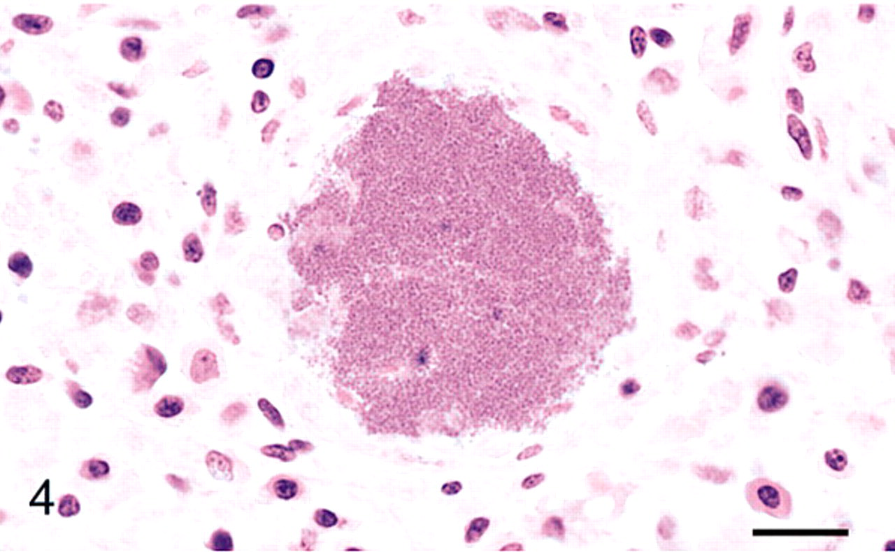
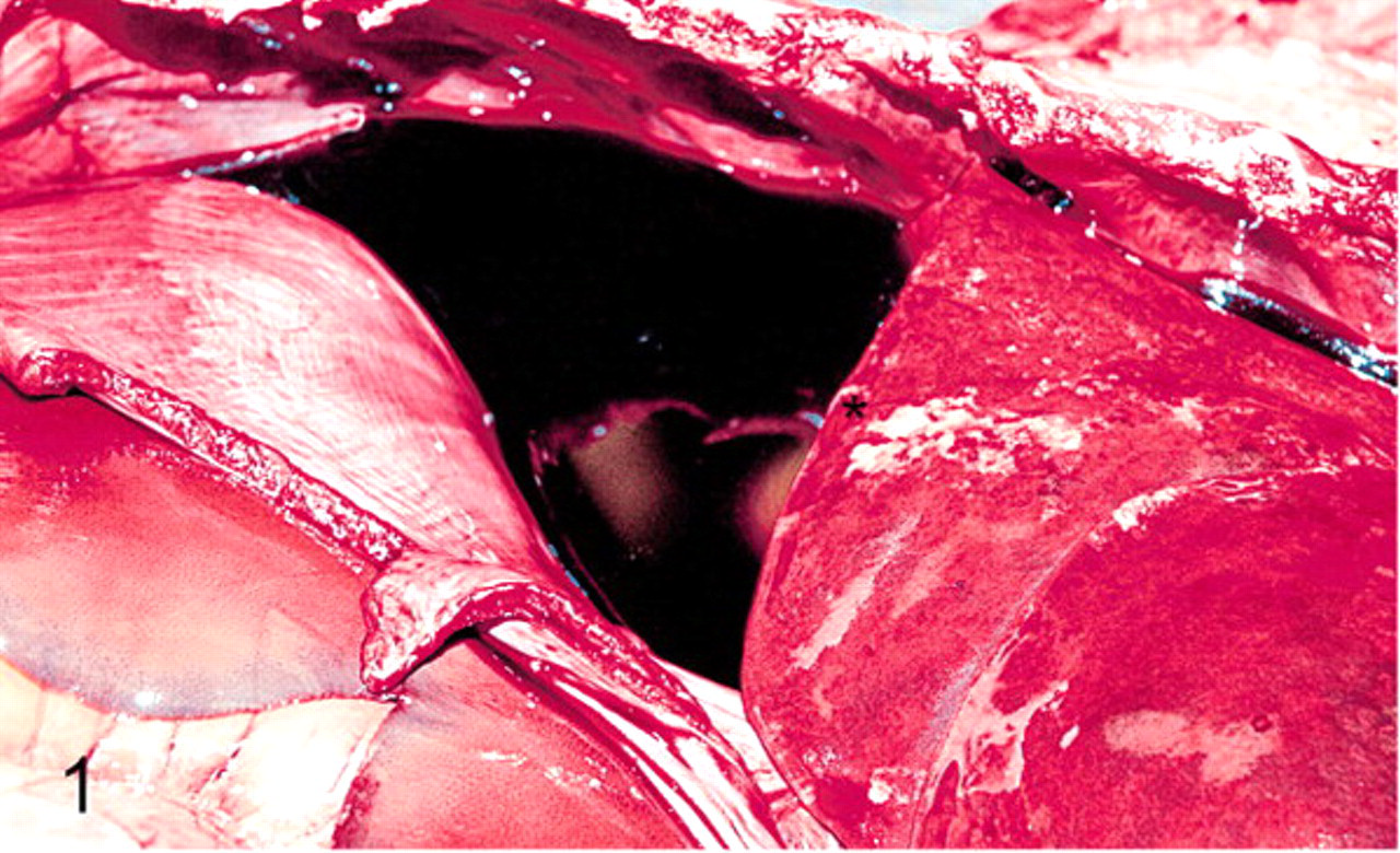
On gross examination all dogs had hemothorax (200–900 milliliters) and rubbery, mottled dark to bright red lungs (Fig. 1). Variably sized and poorly demarcated areas of collapse were present in all lung lobes. Three of the animals had a concurrent acute necrotizing rhinitis and sinusitis. Histologically, there was acute to peracute fibrinosuppurative pneumonia with extensive to lobular obliteration of alveolar spaces with neutrophils (Fig. 2). Large and medium-sized airways were unaffected, but inflammation was intense around most terminal airways. Occasional small vessels were thrombosed with fibrin-enmeshed erythrocytes and leukocytes. Variable to large numbers of intralesional gram positive cocci were present within the cytoplasm of macrophages and free within the extracellular space in pairs, strings, and large colonies (Fig. 3 and inset). Septic thromboemboli were less consistently observed (3/8) in the spleen and renal glomeruli (Fig. 4). No evidence was detected of prior (chronic) lung inflammation or bronchiolar injury on histologic examination of these cases.
Spleen; Case 3. A large cluster of coccoid bacteria distends a sinusoid within the red pulp. HE. Bar = 50 μm.
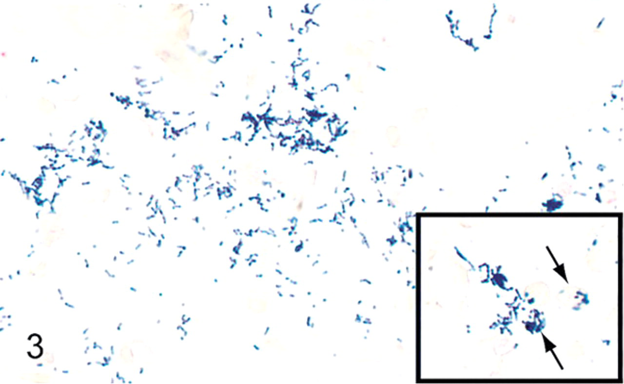
Lung; Case 1. Numerous chains and clusters of gram-positive cocci are free within alveolar spaces and engulfed by alveolar macrophages (inset, arrows). Brown and Brenn Gram stain.
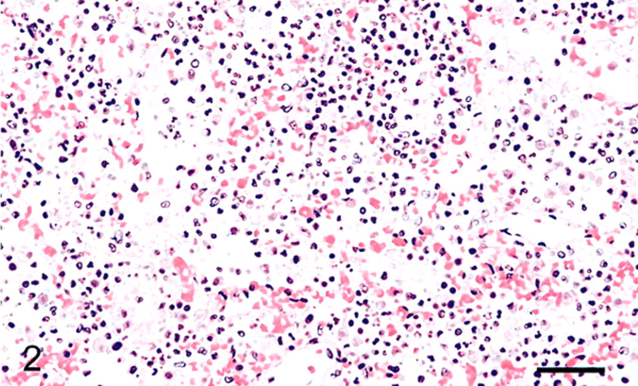
Lung; canine; Case 1. Alveolar septae are obscured, and alveolar spaces are filled with neutrophils and fibrin. HE. Bar = 100 μm.
Thoracic cavity, canine; Case 1. The space between the lung (asterix) and the diaphragm in the caudal and dorsal thoracic cavity is filled with unclotted, hemmorrhagic fluid.
Polymerase chain reaction (PCR)-based amplification of common canine respiratory pathogens was performed on the lung tissue of 5 of 8 necropsied dogs (Table 1). Canine adenovirus, canine herpesvirus, and distempervirus were inconsistently detected. Canine influenza virus, canine parainfluenza virus, and B. brochiseptica were undetectable by this method. Under the aegis of the Koret Shelter Medicine Program, similar panels were performed on oropharyngeal samples from dogs with respiratory disease at this shelter in 2003 and 2005. At those times, hemorrhagic pneumonia was not present, death was uncommon, and B. bronchiseptica but not S. zooepidemicus was commonly detected in dogs with respiratory disease.
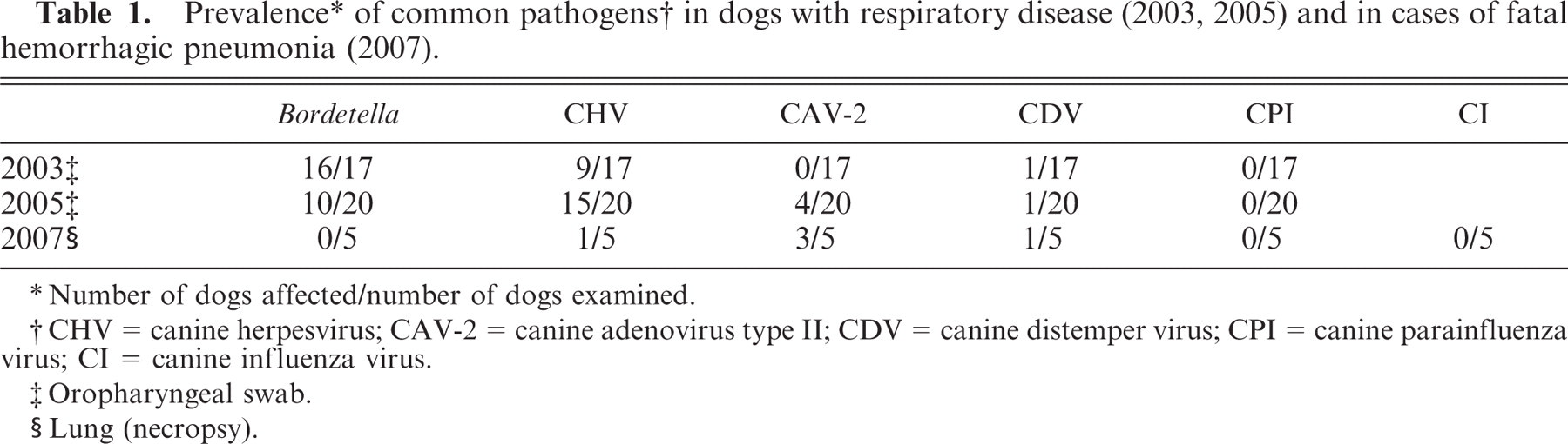
∗ Number of dogs affected/number of dogs examined.
† CHV = canine herpesvirus; CAV-2 = canine adenovirus type II; CDV = canine distemper virus; CPI = canine parainfluenza virus; CI = canine influenza virus.
‡ Oropharyngeal swab.
§ Lung (necropsy).
Lung tissue from 6 of 8 necropsied animals was submitted for culture to the Microbiology Service, VMTH, UC Davis. Two samples from the environment (sink, cage floor), 2 samples from the upper respiratory tract of neighboring dogs (oropharynx), and 1 sample from a foot abscess of an affected dog were also submitted. S. equi subsp. zooepidemicus was cultured from all samples. In 3 of 6 samples cultured from lung and from the foot abscess, S. zooepidemicus was the sole bacterial isolate. Bacterial identification was based on observation of gram-positive, catalase-negative, chain-forming cocci, which fermented lactose and sorbitol but not trehalose. The bacteria were not susceptible to doxycycline, the antibiotic most commonly used in the affected shelter. Each bacterial isolate was also screened by PCR for Se18.9, 7 including its flanking sequences, and by immunoblot of mutanolysin-extracted proteins for reactivity with rabbit antiserum to recSzPW60, 6,8 the Moore and Bryans typing antigen of S. zooepidemicus. All isolates from dogs at the time of the outbreak (lanes 3–11, Fig. 5) yielded a similar-sized amplicon by PCR (Fig. 5). This result and the observed similarity in size of the SzP proteins (data not shown) indicated that the shelter outbreak of hemorrhagic streptococcal pneumonia was caused by a single clone of S. zooepidemicus and that the environment was contaminated with the same clone. S. zooepidemicus was also cultured from the spleen of a cat that died during the canine epizootic and from the lung of a dog that died 2 months after the outbreak had ended. However, these isolates yielded 2 different amplicons distinct from both the outbreak isolate and each other (lanes 1–2, Fig. 5).
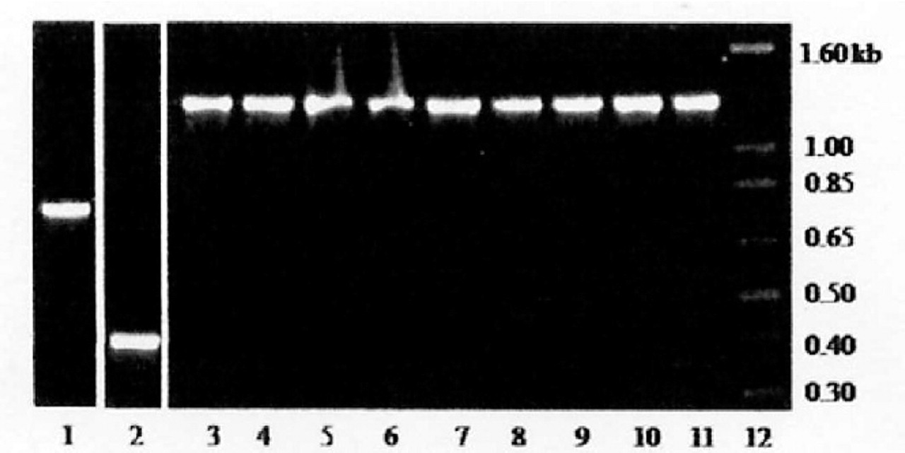
Polymerase chain reaction amplification of DNA fragments from S. zooepidemicus isolated from dogs and the shelter environment using primers FUS and FDS located upstream and downstream, respectively, of se18.9. 7 Lanes 2–12 are fragments amplified from colonies isolated during the time of the outbreak. Lanes 6, 8, and 11, lung (cases 1–3); lane 10, foot abscess (case 4); lanes 5 and 7, oral swabs of neighboring dogs; lane 4, 9 environmental isolates (sink, cage floor); lane 3, spleen from a cat that died acutely with respiratory failure during the canine respiratory outbreak (lane 2 is from an isolate from the nasal cavity of this cat). Lane 1 shows a fragment amplified from an isolate of a dog that died 2 months after the outbreak had ended.
In horses, S. zooepidemicus is a commonly isolated mucosal commensal that opportunistically invades as a sequela to virus infection, high ambient temperature, or prolonged transportation. Infections are endogenous in that a preexisting clone in tonsillar tissue is selected, is amplified, and eventually dominates in the lower respiratory tract. 1,6 Unlike the shelter epizootic described here, the clone found in pneumonic lungs varies from horse to horse. Dominance of a single clone of an organism only infrequently detected in healthy household dogs indicates that environmental conditions in combination with stress and possibly other as yet unknown microbial agents provided a setting for rapid invasion into densely populated and highly susceptible hosts. Many of the cases described in the shelter were in dogs admitted to the environment as early as 2 days prior to their death. The short interval between entry to the kennel and onset of fatal pneumonia suggests either a challenge that was overwhelming or selection of a clone with enhanced virulence. In this outbreak, the most common canine respiratory copathogens either were absent or were inconsistently present. In addition, there was no histologic evidence of predisposing viral damage, which was consistent with the death of several dogs within 2 days of admission to the shelter. In a previous report, an outbreak of hemorrhagic pneumonia in kenneled research dogs was attributed to S. zooepidemicus. 3 Although the outbreak described in this paper was more protracted and involved more animals, the pathologic and microbiologic features were otherwise similar.
The outbreak ended subsequent to imposed husbandry changes, including a dramatic depopulation, facility cleaning, and staff education. Attempts to identify potential virulence factors in the clone from this shelter, including antiphagocytic proteins, proteases, and pyrogenic exotoxins, are ongoing.