
Abstract
A spontaneous lung tumor in a 5–year-old goat of the Murciano-Granadina breed is described in this paper. Clinical signs of cachexia and tachypnoea were evident, and a considerable amount of white mucous foamy fluid was discharged from the nostrils when the animal's head was lowered. A lung tumor with the characteristics of bronchioloalveolar carcinoma was detected during histopathologic examination. The tumor cells were positive for surfactant proteins C and B, confirming that alveolar type II cells were the origin of the neoplasia. Tumor samples were tested by polymerase chain reaction, immunoblotting, and immunohistochemistry for the presence of Jaagsiekte sheep retrovirus (JSRV) and enzootic nasal tumor virus (ENTV), another retrovirus very closely related to JSRV, but all tests were negative. Therefore, this is the first reported case of spontaneous bronchioloalveolar carcinoma not related to JSRV or ENTV infection in a goat.
A 5-year-old goat of the Spanish Murciano-Granadina breed with a history of progressive respiratory illness and deterioration was submitted to the diagnostic service of the Veterinary Faculty of Zaragoza (Spain). The goat presented with severe cachexia, tachypnoea, and accumulation of fluid in the respiratory airways as detected by auscultation. A considerable amount of white, mucous, and foamy fluid was discharged from the nostrils when the animal's head was lowered. Comprehensive clinical examination revealed no other abnormalities in the animal. The goat was humanely euthanatized based on the level of deterioration.
Postmortem examination confirmed that the animal was in poor body condition and revealed lesions confined to the lungs. A large quantity of white, frothy fluid was present in the trachea, and the cranioventral regions of the lobes of both lungs were consolidated. They were reddish or gray in color, with clear delineation between the affected and nonaffected areas. Lung sections showed small coalescent gray nodules with foamy fluid pouring from sections of the respiratory airways when under slight pressure (Fig. 1). No other notable changes were observed in the remainder of the respiratory system or in any other organ system.
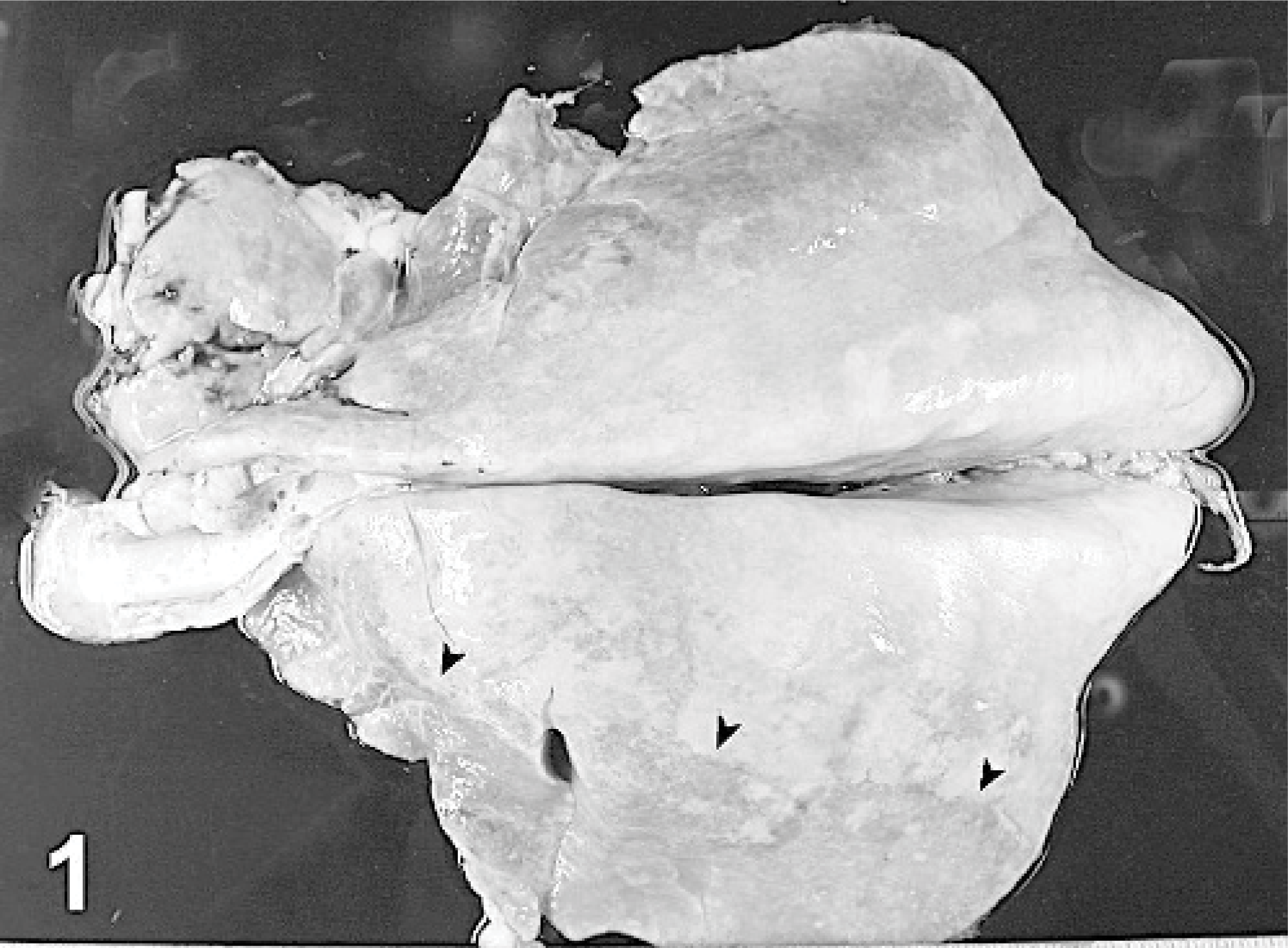
Lungs; goat. Cranioventral portions of both lungs (arrowheads) are shown to be consolidated, and a whitish fluid is pouring from the trachea.
Samples of the lung lesions were taken at necropsy, snap frozen in liquid nitrogen, and stored at −70°C. Representative lung sections were fixed in 10% neutral-buffered formalin and embedded in paraffin wax, using routine procedures, and 4- to 6-μm sections were stained by the hematoxylin and eosin method. Microscopic observation revealed a proliferation of cells growing on the line of normal alveolar structures. These cells were cuboidal or columnar, aligned in small groups or found single and binucleated. Some of them were dome shaped, containing tiny cytoplasmic vesicles. Round or oval prominent nuclei with a slight degree of anisocariosis were characteristic, found singly or in groups, and mitotic figures were occasionally found. The architecture of the lung was generally preserved, and the limits between the proliferation areas and normal alveoli were not clearly delineated. Proliferating tissue coexisted with an intense inflammatory exudate rich in neutrophils (Fig. 2). The histopathology of the lung was consistent with a bronchioloalveolar carcinoma. 6
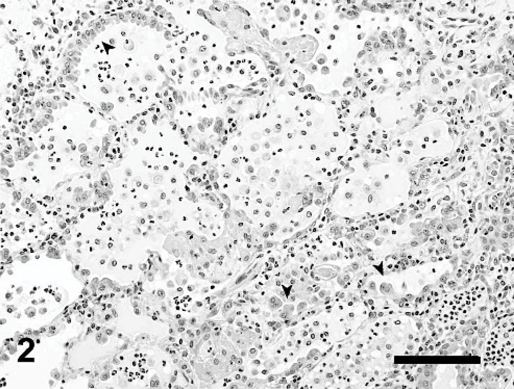
Lung; goat. Histopathology of lung lesions showing the features of a bronchioloalveolar carcinoma. Cuboidal and columnar cells, aligned in small groups, single or even binucleated, were proliferating in the alveolar walls (arrowheads). The architecture of the lung was generally preserved, and an inflammatory reaction rich in neutrophils was also found. Bar = 100 μm.
To identify the type of cells proliferating, an immunohistochemical (IHC) study of the sections was performed using antisera against human surfactant proteins (SPs) B and C (Chemicon International) and human Clara cell 10-kd protein (CC-10, kindly provided by Gurmukh Singh, VA Stars and Stripes Healthcare Network, Department of Veterans Affairs, Pittsburgh, Penn.), following routine IHC methods. Briefly, 4- to 6-mm lung sections were cut onto positively charged slides (SuperFrost Plus; Menzel-Gläser, Braunschweig, Germany), which were dewaxed and rehydrated by routine procedures. Antigen retrieval was performed by microwaving the sections at 800 W for a total of 10 minutes (5 minutes, ×2) in 10 mM citrate buffer adjusted to pH 6.0. After cooling, endogenous peroxidase was quenched with 0.3% hydrogen peroxide in methanol. Positively labeled cells were detected using the EnVision System (Dako, Denmark) according to the manufacturer's instructions. Diaminobenzidine was the substrate used to identify positively labeled cells, and the sections were counterstained with Carrazzi's haematoxylin. Tissue sections of sheep lung naturally affecetd by ovine pulmonary adenocarcinoma (OPA) and unaffected sheep lung were used as positive and negative controls, respectively. Substitution of the primary antibody by TRIS buffer at pH 7.5 was an additional control for residual endogenous peroxidase activity.
Most of the tumor cells were strongly positive to SP-C (Fig. 3) and negative to CC-10 (data not shown) labeling. SP-B was only detected in a minor proportion of tumor, cells and the intensity of the labeling was very light (data not shown). These IHC results revealed that type II pneumocytes were the proliferating cells. The identification of surfactant proteins in the tumor cells meets the criteria defining bronchioloalveolar carcinoma. 6
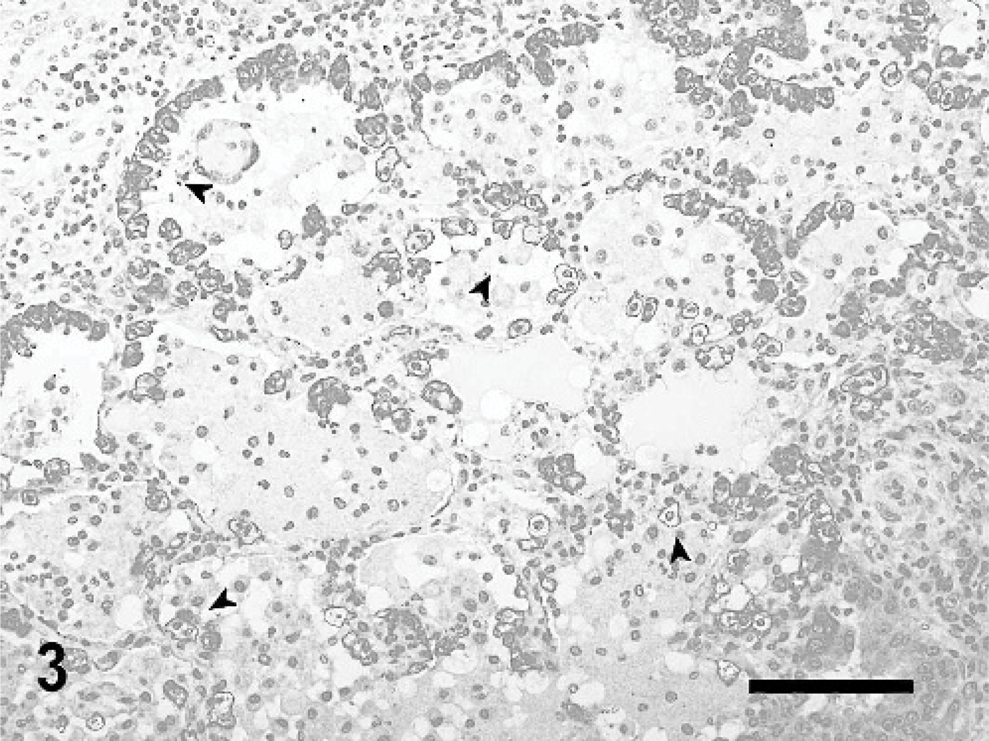
Lung; goat. Immunohistochemical evaluation of the tumor using rabbit anti-human SP-C. Most of the tumor cells are strongly positive to SP-C labeling (arrowheads). Bar = 100 μm.
Bronchioloalveolar carcinomas are infrequent lung neoplasias in any of the domestic species except sheep, which are affected by OPA (previously termed sheep pulmonary adenomatosis, ovine pulmonary carcinoma, or Jaagsiekte), a contagious lung neoplasm caused by the Jaagsiekte sheep retrovirus (JSRV). 9 Just a few cases of OPA-like changes have also been described in spontaneous tumors in goats in Peru, 4 India, 11 or Saudi Arabia. 1
In accordance with the traditional farming methods in this region of Spain, the goat in our study was from a flock cohabiting with sheep. OPA is a common disorder in sheep in this area, but a case of OPA has never been recorded in a goat, although JSRV infection is relatively common in domestic goats (authors' unpublished results). In order to determine whether JSRV was implicated in the lung neoplasia of the goat in this study, genomic DNA was extracted from fresh tumor tissue collected at necropsy and examined for the presence of proviral JSRV by hemi-nested polymerase chain reaction (PCR). 8 The presence of ENTV-2 (enzootic nasal tumor virus-2), another retrovirus very closely related to JSRV and associated with nasal adenocarcinoma in goats, was also investigated by specific hemi-nested PCR. 7 Several pulmonary tumor samples were tested with both of the PCR primer sets, and all of them were negative but were positive for amplification of the GAP-DH gene, 8 demonstrating the integrity of the extracted DNA. Total protein from fresh tumor tissue was also obtained, and capsid protein from JSRV was checked by sodium dodecyl sulfate–polyacrylamide gel electrophoresis/Western blotting, 9 with negative results. As a corroborative test, pulmonary tumor sections were tested by IHC methods as previously described, 3 using rabbit antisera against different JSRV proteins: capsid protein, matrix, and surface protein. No positive labeling to any of the sera was detected in the goat tumor, whereas positivity was detected in all OPA controls (results not shown).
As far as we know, this is the first case of bronchioloalveolar carcinoma reported in a goat that is not related to JSRV infection. The descriptions in Peru, India, and Saudi Arabia were based on histopathologic examination only, and the existence of JSRV retrovirus was not demonstrated in these cases. Despite this, the histologic architecture of the tumors described and the pictures shown in these reports are consistent with OPA histopathology. 5 Meanwhile, the histologic characteristics of the tumor described in this paper showed neither bronchiolar involvement nor the acinar or papilary patterns of growth characteristic of OPA or caprine lung tumors described in the other countries. Furthermore, OPA has been experimentally reproduced in goats using lung fluid rich in JSRV, confirming that this species can develop the disease. However OPA was reproduced less efficiently and after a longer incubation period than in sheep, indicating a lower susceptibility of goats to the disease. 12 The low susceptibility of goats to develop OPA experimentally may explain why the incidence of naturally occurring OPA is low in the goat.
Other caprine pulmonary tumors are also very infrequent, and only a few cases of lung primary tumors have been described, defined as squamous cell tumors 10 or pulmonary mucoepidermoid tumors. 2 Thus, the lung tumor described in this paper can be added to the short list of primary lung tumors reported in goats and, as far as we know, it is the first reported case of bronchioloalveolar carcinoma unrelated to JSRV or ENTV infection.
Footnotes
Acknowledgements
This work was supported by grants from the European Union (QLK 2-CT-2001-02380) and the Spanish Comisión Interministerial de Ciencia y Tecnología (AGL 2001-1812 GAN). Thanks are due to Dr. Christina Summers for critical discussion of the manuscript, to Rosario Puyo for technical assistance, and to the veterinary surgeons of the Gabinete Técnico Veterinario of Zaragoza for their collaboration.