
Abstract
Malignant mixed Müllerian tumor (MMMT) is a rare neoplasm of the female genital system. A case of MMMT in the uterus of an 8-year-old female dwarf rabbit, which died with clinical signs associated with severe acute dyspnea and anorexia, is described. At necropsy, an oval, firm tumor was found in each of the 2 uterine horns. Numerous metastases were scattered throughout the mediastinum, thoracic diaphragm, and all pulmonary lobes. Microscopically, the tumors consisted of a poorly demarcated, unencapsulated neoplasm, composed of closely associated carcinomatous and sarcomatous components and areas of osteosarcomatous differentiation. Metastases were composed entirely of the sarcomatous component with osteosarcomatous differentiation. Immunohistochemically, the neoplastic epithelial component was positive for cytokeratin and negative for α-smooth muscle actin (α-SMA), vimentin, and desmin. The sarcomatous component was diffusely and strongly positive for vimentin, focally positive for α-SMA (<20% of cells positive), and negative for desmin. The neoplasm was diagnosed as a heterologous MMMT with metastases to the lung, mediastinum, and thoracic diaphragm.
In domestic animals, primary uterine neoplasms are classified as epithelial or mesenchymal tumors. 8 In women, in addition to these 2 types, a very rare neoplasm with epithelial and mesenchymal components has been described and classified as a mixed Müllerian tumor. A mixed neoplasm consisting of 2 components that are both histologically malignant is termed a malignant mixed Müllerian tumor (MMMT), sarcomatoid carcinoma, carcinosarcoma, or malignant mixed mesodermal tumor. 9 Such neoplasms are generally highly aggressive and mainly occur in postmenopausal women. 7
Histologically, MMMTs are composed of closely associated malignant epithelial and sarcomatous components. Depending on the sarcomatous component, MMMTs are classified as either homologous or heterologous types. In homologous MMMTs, the sarcomatous element consists predominantly of spindle, round, or giant cells, whereas in heterologous tumors, foci of rhabdomyosarcoma, chondrosarcoma, osteosarcoma, or liposarcoma are included.7,9
Uterine MMMTs have been described in cats,3,10,11 in a sow, 1 and in rodents.2,6,16 In the veterinary literature, a uterine MMMT has been described once in a rabbit, 4 but the report had no data concerning the history of the disease, the clinical treatment, or eventual metastases. In the current paper, the clinical, gross, microscopic, and immunohistochemical findings of a primary uterine MMMT in a rabbit with metastasis to the lungs, mediastinum, and thoracic diaphragm are described.
An 8-year-old, 2-kg, female dwarf rabbit (Oryctolagus cuniculus) was presented to the Department for Birds, Small Mammals and Reptiles at the Institute for Poultry Health, Veterinary Faculty of Ljubljana (Ljubljana, Slovenia), with a 4-day history of malodorous and occasionally red urine. On clinical examination, a large abdominal mass was palpable in the caudal abdomen, and malodorous urine was detected. Urine examination with urine dipstick analysisa detected the presence of leukocytes, nitrites, and erythrocytes. Bacterial cystitis was suspected and treated with 20 mg/kg of trimethoprim–sulfamethoxazole b twice daily orally. Because of the palpable abdominal mass, radiographic examination was also advised but was refused by the owner. In the following days, the health of the rabbit improved, and the animal was dismissed after 5 days of hospitalization. Two months later, the rabbit was readmitted with severe acute dyspnea and anorexia. Due to the poor clinical status of the patient, only a laterolateral radiograph was performed. On the radiograph, a large soft tissue mass with calcification occupied the caudal ventral abdomen (Fig. 1A). Moreover, the heart silhouette was not clearly visible, and there was soft tissue opacity of the lungs. The rabbit died shortly after initial treatment, and a necropsy was performed.
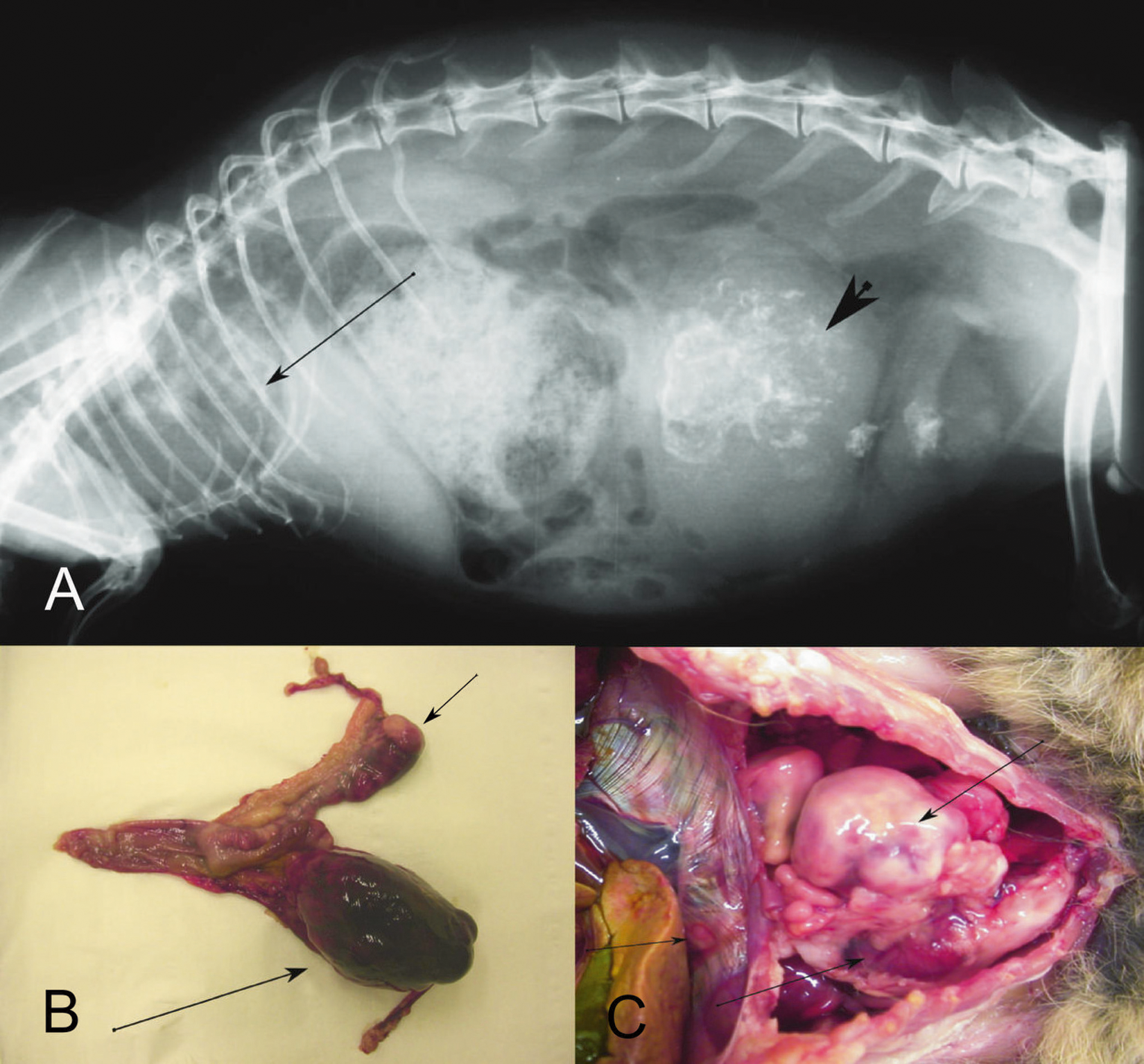
Radiographic and macroscopic features of a uterine neoplasm from a dwarf rabbit (Oryctolagus cuniculus).
At necropsy, a bulky, ovoid, firm, smooth, red–black, 7.5 cm × 4.5 cm × 3 cm mass was present in the right uterine horn. The cut surface was smooth, red–black, and slightly convex. An oval, irregularly lobulated, firm, smooth, white, 3.5 cm × 2 cm × 1.5 cm mass with a white, smooth, glistening, and homogenous cut surface was noticed at the end of the left uterine horn (Fig. 1B). The uterus corpus, vagina, and both ovaries were normal in size and shape. Numerous firm, smooth, white, glistening, poorly demarcated nodules with a white, smooth, and flat cut surface, from 1 mm to 2.5 cm in diameter, were scattered throughout the mediastinum, thoracic diaphragm, and all lung lobes (Fig. 1C). There were no additional masses in other organs.
Samples of masses from the uterus, lung, and thoracic diaphragm were promptly fixed in 10% neutral buffered formalin and routinely embedded in paraffin. Approximately 4-µm-thick sections were stained with hematoxylin and eosin and examined microscopically. Immunohistochemistry was performed with antibodies raised against cytokeratin c (diluted 1:100), α-smooth muscle actin c (α-SMA; diluted 1:25), desmin c (diluted 1:20), and vimentin c (diluted 1:300). Human uterine tissue was used as a positive control. Sections treated without primary antibodies served as negative controls.
Microscopically, large, poorly demarcated, unencapsulated neoplasms, consisting of closely associated epithelial and sarcomatous components, characterized the uterine tumors detected grossly (Fig. 2A, 2B). The neoplasm extended into the adjacent myometrium and protruded into the uterus lumen. A large area of the intraluminal mass was necrotic, with multifocal dystrophic calcification.
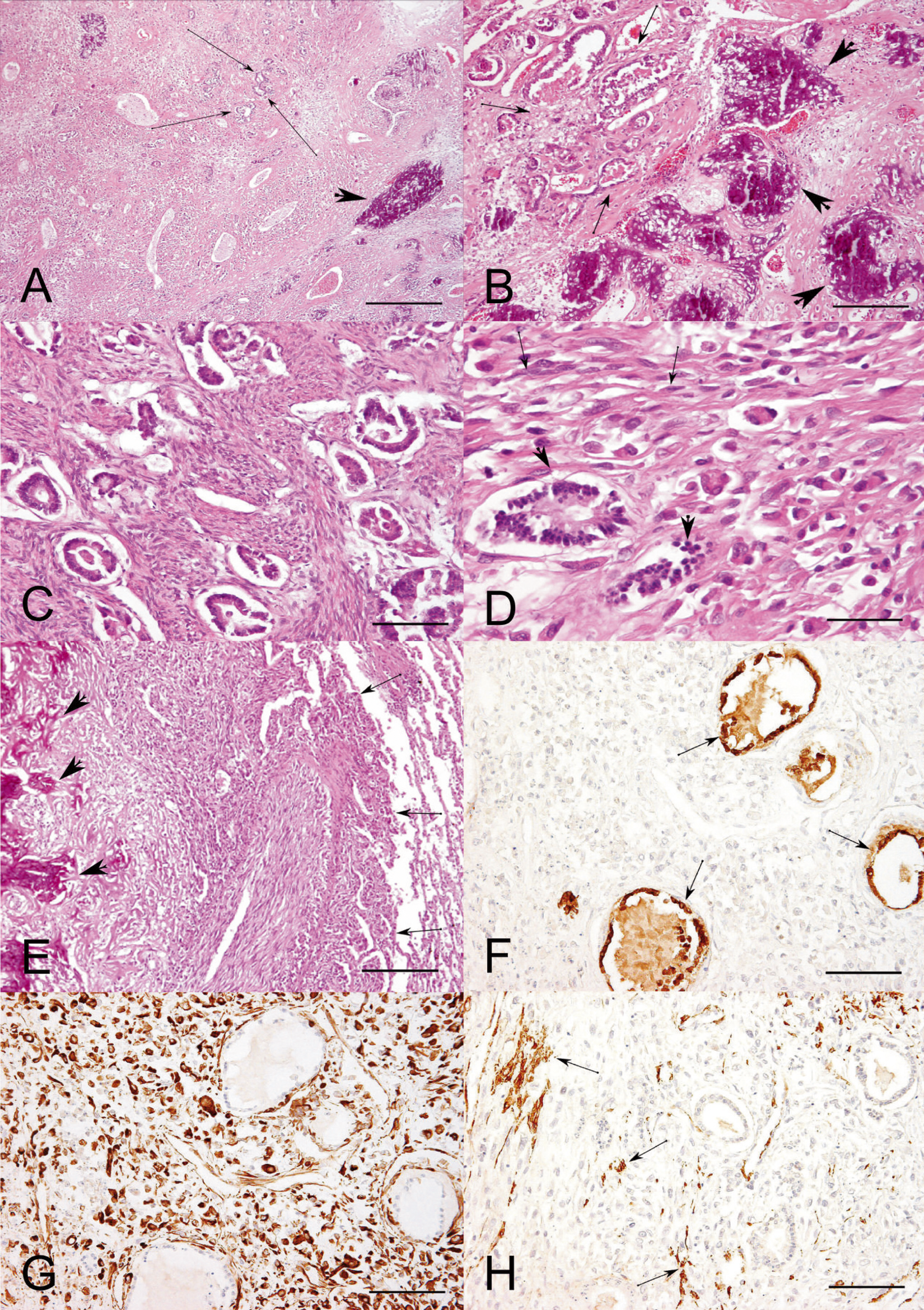
Histopathologic and immunohistochemical features of a uterine neoplasm from a dwarf rabbit (Oryctolagus cuniculus).
The epithelial component formed small groups of widely spaced tubules with narrow lumina, in which neoplastic epithelial cells were arranged in 1 or several rows, supported by, or intermixed with, neoplastic sarcomatous mesenchymal stroma. Neoplastic epithelial cells consisted of highly pleomorphic, polygonal cells, with moderately abundant eosinophilic cytoplasm and distinct cell borders (Fig. 2C, 2D). The hyperchromatic nuclei varied from oval to round, with moderate anisokaryosis and distinct nucleoli. Mitoses were rare (5 per 10 high power fields). The sarcomatous mesenchymal component of the neoplasm was composed predominantly of undifferentiated spindle cells, with foci of heterologous osteosarcomatous differentiation that contained osteoid and calcified bone matrix (Fig. 2A, 2B). Neoplastic spindle cells were moderately pleomorphic, with slightly or deep eosinophilic fibrillar cytoplasm and indistinct cell borders. The hyperchromatic nuclei varied from oval to round with moderate anisokaryosis and inconspicuous nucleoli (Fig. 2D). Five mitotic figures were found in 10 random fields at 400×. Discrete foci of necrosis and several collections of lymphocytes were scattered in the sarcomatous component of the tumor. A large group of neoplastic cells was found in a myometrial lymphatic vessel. Metastatic foci were composed entirely of sarcomatous mesenchymal component (Fig. 2E), where neoplastic cells were more pleomorphic and larger than the cells in the primary tumor.
Immunohistochemically, neoplastic epithelia were cytokeratin positive (Fig. 2F) and negative for α-SMA, vimentin, and desmin. Neoplastic mesenchymal cells were diffusely and strongly positive for vimentin (Fig. 2G), focally positive for α-SMA (<20% of cells positive; Fig. 2H), and negative for desmin. Based on gross, microscopic, and immunohistochemical staining features, the uterine neoplasm was diagnosed as a heterologous MMMT, with metastases to the lung, mediastinum, and thoracic diaphragm.
The incidence of uterine tumors is very high in rabbits; adenocarcinoma, adenoma, leiomyoma, and leiomyosarcoma have been described. Among these tumors, uterine adenocarcinomas prevail, and their incidence increases considerably with age.14,17 To the authors’ knowledge, MMMT has only once been described in a rabbit. 4 MMMTs are rare neoplasms in women and even rarer in domestic animals.1,3,4,10,11,16 Because some MMMTs have been diagnosed on samples submitted after hysterectomy, information regarding the history, clinical data, and metastases in animals are very limited.
Malodorous and occasionally red urine, with leukocytes, nitrites, and erythrocytes, was the first clinical sign in the rabbit case described herein. After 2 months, the rabbit’s health worsened dramatically, and it became anorexic and severely dyspneic. In the veterinary literature, hematuria, serosanguineous vaginal discharge, mammary cysts and neoplasms, skin masses, abdominal pain, anorexia, dehydration, and/or poor general health are described as common clinical features in rabbits with uterine tumors.14,17 When lung metastases occur, anorexia and difficult breathing are commonly observed. 13 In women, the most common symptoms of MMMT are postmenopausal bleeding and enlargement of the uterus.5,7
In the current report, the rabbit was 8 years old. To date, MMMT has been diagnosed in an 8-year-old cat, 10 in a 10-year-old sow, 1 and in an 11-year-old cat. 11 In women, MMMTs most commonly occur during the postmenopausal period. 9
At necropsy, an oval tumor was found in each of the uterine horns of the rabbit. Similarly, neoplastic proliferations into the uterine lumen, causing dilatation of the affected uterine horn, have been described in a sow, cats, and rats.1,6,10,11,16 In women, MMMTs are usually fleshy, necrotic masses that often fill the uterine cavity with myometrial invasion, similar to the case described herein. Numerous metastases were found in the lung, mediastinum, and thoracic diaphragm in the rabbit in the current study. In women, metastases are more common in the peritoneum, omentum,9,15 and pelvic organs. 5 Metastases to the mesosalpinx, ovary, 1 and peritoneum, omentum, and middle ligament of the bladder, 10 and mesentery of the small and large intestines, 6 have been reported in a sow, a cat, and a rat. Distant metastases have occurred to the liver, bone marrow, brain, 15 lung,5,12 lymph nodes, and spine 5 in women and to the lungs, diaphragm, 10 pleura, liver, 1 lymph nodes, pancreas, and spleen 6 in animals.
The histopathological features of the uterine neoplasm described herein had both carcinomatous and sarcomatous components and was consistent with MMMT. 9 Because the sarcomatous mesenchymal component had osteosarcomatous differentiation, the MMMT was considered to be of heterologous type. In women, heterologous MMMTs predominate 15 ; in animals, heterologous components have only been reported in rats and in a sow.1,6,16 In the current case, metastases were only of the sarcomatous mesenchymal component. This is in contrast to metastases of MMMT in women, where metastases consist only of epithelial cells.15 In rats, however, metastases have consisted of both components. 6 The results of immunohistochemical examination were in accordance with the results of previous studies and confirmed the diagnosis of MMMT.1,10,16
In summary, a uterine MMMT neoplasm, with metastases to the mediastinum, thoracic diaphragm, and lungs was described in a rabbit. The diagnosis was based on histological examination and supported by immunohistochemistry. Although such neoplasms are very rare in domestic animals, they should be taken into consideration when a large mass is palpable in the caudal abdomen of older female rabbits and perhaps also in other domestic animals.
Footnotes
Acknowledgements
Immunohistochemical examinations were performed at the Institute of Pathology, Medical Faculty, University of Ljubljana, by Dr. Jože Pižem, as well as by Mr. Dane Velkavrh, whose contribution is greatly appreciated.
a.
Multistix® GP, Bayer AG, Leverkusen, Germany.
b.
Primotren®, Lek d.d., Ljubljana, Slovenia.
c.
Cytokeratin (clone MNF116), α-smooth muscle actin (α-sm-1), desmin (clone D33), vimentin (clone V9); Dako Denmark A/S, Glostrup, Denmark.
The author(s) declared no potential conflicts of interest with respect to the research, authorship, and/or publication of this article.
The author(s) received no financial support for the research, authorship, and/or publication of this article.