
Abstract
Cementoma is a very rare odontogenic neoplasm of mesenchymal origin. Clinically, in 3 horses, multiple bony enlargements of the upper and lower jaw extending into the oral cavity were observed. Radiographically, multiple, well-circumscribed, radiopaque masses surrounding the roots of the upper and lower incisors or an upper premolar tooth were present. Due to malocclusion and local pain, single teeth were extracted in each case. Grossly, a hard grayish-white mass surrounding the root of the incisors and the premolars was identified. Histopathologically, the tumors consisted of excessive deposition of cementum-like tissue. Cells, resembling cementoblasts, lined irregularly shaped lacunae, which were present in the tumor tissue, and showed minimal cellular pleomorphism. Mitotic figures were not present. Macroscopically and histologically, all 3 equine cases displayed great similarities to cementomas in other species. However, due to their high degree of differentiation, hypercementosis must be considered as a differential diagnosis.
Cementoma is a very rare odontogenic neoplasm of mesenchymal origin. 2, 8 It is a slowly growing tumor arising from cementoblasts, first described by Norberg in 1930 in humans. 7 Until now fewer than 100 human cases have been reported. 1, 2, 9 The neoplasm is characterized by deposition of differentiated cemental matrix around the tooth root, proliferation of cementoblasts, and destruction of the lamina dura, which represents the alveolar cortical bone consisting of dense osteoid material. 3, 5, 9 Although cementomas have been described as benign solitary neoplasms with low growth activity, there are reports of more rapidly growing and aggressive variants. 1 Cementomas in humans are proliferations of cementoblasts with cement deposition and are classified according to their macroscopic and histologic features in 3 categories: periapical cemental dysplasia, benign cementoblastoma (true cementoma), and gigantiform cementoma. 8, 9 In humans the neoplasms embed a single root or multiple roots of an erupted permanent tooth in a mass of cementum-like matrix. 9 In horses, cementoma-like lesions have been reported only as incidental radiologic and macroscopic findings and have not been characterized in detail until now (http://www.poolhousevets.co.uk/equine_dentistry_casestudies4.html). Three independent cases of cementomas in horses are described in this report.
An 18.7-year-old neutered standard bred male horse (case No. 1; Fig. 1), a 17-year-old male standard bred horse (case No. 2), and a 2-year-old female Holstein horse (case No. 3) displayed upon clinical and radiographic examination multiple, well-circumscribed, round-shaped opaque masses surrounding the roots of several incisors (case No. 1 and 2) or premolars (case No. 3). In all 3 cases deformation of the surrounding bone and lack of a detectable lamina dura were observed. Moderate malocclusion and pain were present during clinical examination. In each case 1 affected tooth (case No.1, first left upper incisor, 201; case No. 2, first left lower incisor, 301; case No. 3, second left upper premolar, 206) was extracted surgically using extraction forceps and submitted for pathologic examination.
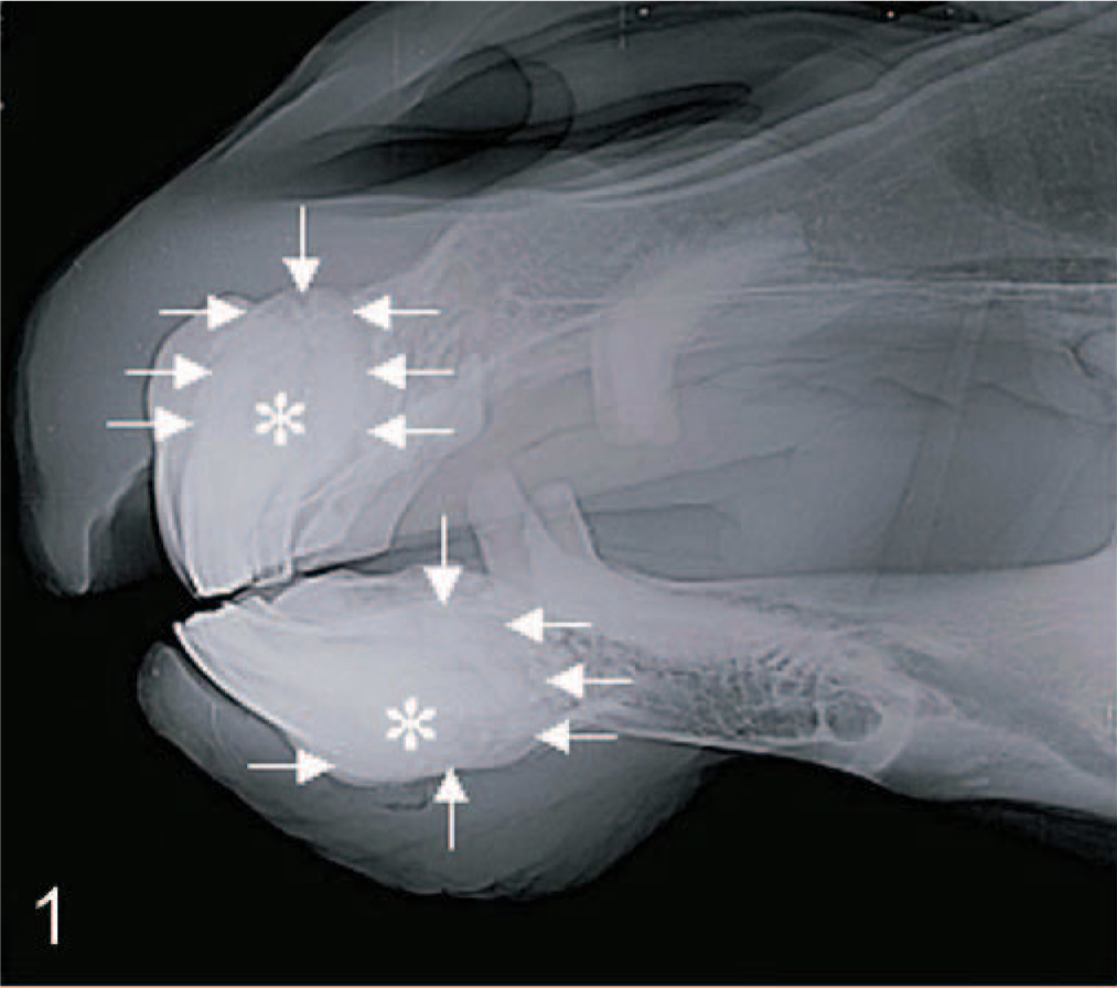
Upper and lower incisors; 18.7-year-old castrated standard bred male horse, case No. 1. A panoramic radiograph reveals well-circumscribed radiopaque masses (arrows) embedding the roots of affected teeth (asterisks).
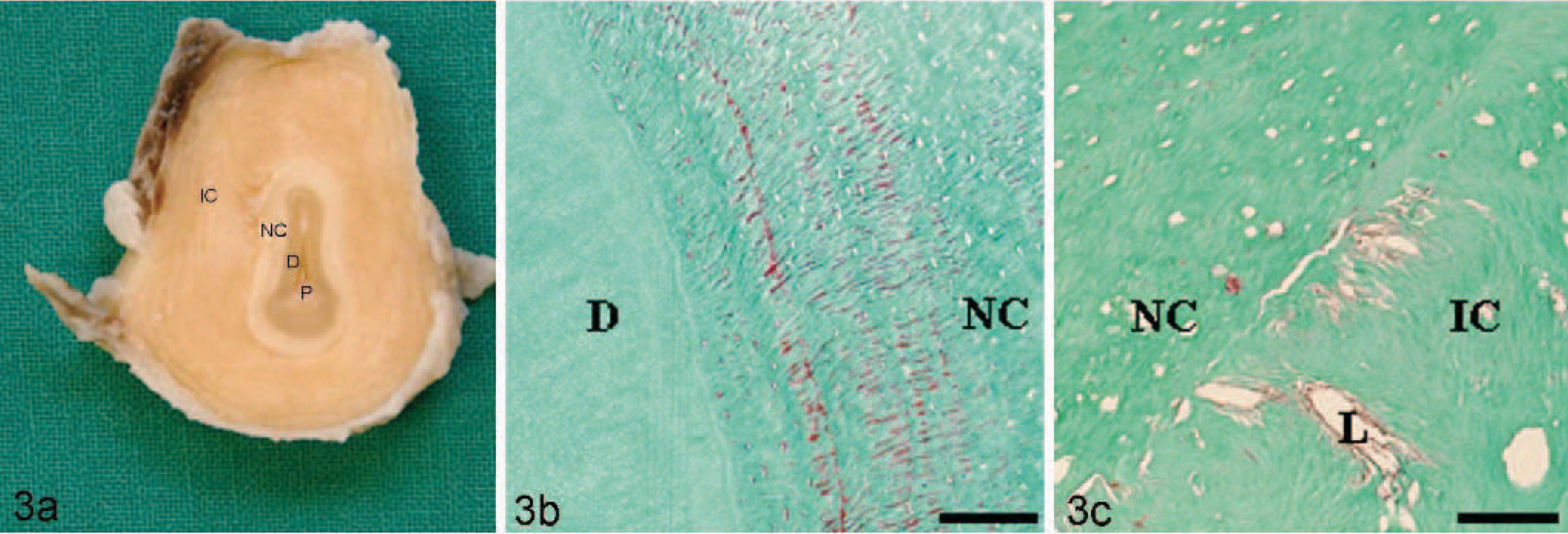
Grossly, a normal tooth root was surrounded by a round well-circumscribed, grayish-white, bony mass of 6.5 × 3 × 1.5 cm (case No. 1; Fig. 2), 3 × 2 × 2 cm (case No. 2), and 4 × 2 × 2 cm (case No. 3). On transversal section a demarcation between normal cementum and cementum-like tissue was visible (Fig. 3). The teeth with neoplastic masses were fixed in neutral buffered formalin, and representative slices of tissue samples were decalcified in 5% nitric acid at 37°C (cases No. 1 and 3) for 7 hours or in 25% EDTA-disodium salt solution for 4 weeks at room temperature (case No. 2). After dehydration through graded alcohols and embedding in paraffin wax, 4-μm thick sections were prepared and stained with HE and Masson's trichrome stain.
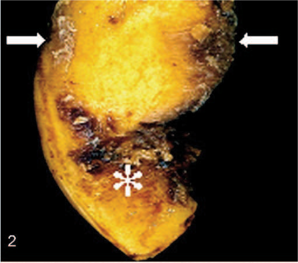
First left upper incisor (201); 18.7-year-old neutered standard bred male horse, case No. 1. A bony, hard, well-circumscribed 6 × 2 cm sized cementoma (arrows) embedding the dental root (star) was observed.
First left lower incisor (301); 17-year-old male standard bred horse, case No. 2.
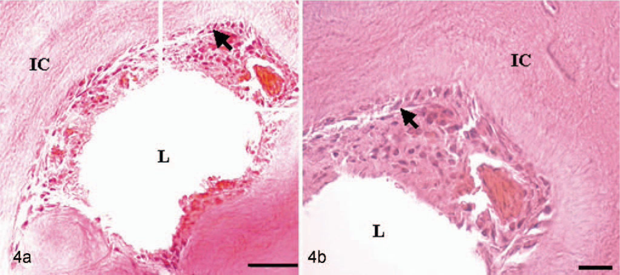
Histologically, all 3 neoplasms displayed similar histologic features. The masses consisted of a prominent, well-differentiated and demarcated proliferation of cementum-like material with abundant collagen fibers and abnormal lacunae formation surrounding normal dental structures, including dental pulp, dentin, and cementum (Fig. 3). Well-differentiated cementoblast-like cells lined the abnormal lacunae within the tumor masses in all 3 cases. The cementoblast-like cells were characterized by medium-sized oval nuclei and a moderate eosinophilic cytoplasm and displayed minimal cellular pleomorphism. No mitotic figures were detectable (Fig. 4). Very mild reversal line formation was also present in all examined cases. In case No. 1, severe inflammation with neutrophils, plasma cells, and multinucleate giant cells at the periphery of the tumor were observed. Whether the multinucleate giant cells are inflammatory cells or cementoclasts remains unclear. Radiographic, clinical, macroscopic, and histologic findings supported the diagnosis of a benign cementoma.
Second left upper premolar (206); 2-year-old female Holstein horse, case No. 3.
All 3 cases of an equine cementoma reported here were characterized by abundant amounts of cementum-like tissue surrounding the dental root. The tumors consisted of mineralized trabeculae of cementum-like tissue fused with the root of the tooth. Histologic findings resembled benign cementoblastomas (true cementomas) of humans. 9 Benign cementoblastomas (true cementomas) in humans may develop due to a reactive and/or hyperplastic process after periodontal inflammation, or they might have a genetic background. 4, 10 The pattern of occurrence of the trait in humans is consistent with an autosomal dominant mode of inheritance with variable expressivity of the phenotype, whereas the etiology and pathogenesis in domestic animals are still unknown. 4, 10 Human benign cementoblastomas (true cementomas) and bovine cemental abnormalities are predominantly located in premolar and molar teeth. 3, 5, 6 The equine cementomas described in the present report were located at the incisors and premolars.
Cementoblastomas (true cementomas) have been described in most human cases as benign, solitary, slow-growing neoplasms. 7, 9 In contrast to the findings in other species, in which predominantly single molars were affected, several incisors and premolars were simultaneously affected in the examined horses. 1, 2, 3, 5 Similar to other species, the prognosis of the present cases, based on histology and clinical recovery after surgery, is favorable. However, the 2-year-old female Holstein horse (case No. 3) was euthanized after surgery due to therapy-resistant wound healing complications. The present report showed that cementomas may occur in both young (2-year-old) and old (17-year-old and 18.7-year-old) horses. In humans 75% of benign cementoblastomas (true cementomas) were observed in young adults and children and only 25% in adults older than 30 years. 1, 4
Hypercementosis must be considered as a differential diagnosis. 6 Although cementoma and hypercementosis usually have characteristic findings, atypical cases are diagnostically challenging. Hypercementosis, a non-neoplastic condition, is characterized by accumulation of excessive cementum in continuation with the normal radicular cementum. 5, 6 In addition, there is no demarcation between normal and newly formed cementum. Radiographically, hypercementosis is an incidental finding because of its relatively small size and absence of clinical symptoms.11 Radiographically, a radiolucent shadow of the periodontal membrane and the radiopaque lamina dura at the outer border of hypercementosis are consistent findings. 6 In contrast, the human benign cementoblastoma (true cementoma) exhibits a destruction of the lamina dura due to the expansile growth of the tumor. 6 In some cases of atypical hypercementosis, the cementum deposition can show cementoma-like architecture. 6 In these cases the histopathologic examination reveals an increased production of an acellular regularly mineralized cementum. The excessive cementum is attached to the dental root and associated with a thin connective tissue without cementoblasts. In contrast to hypercementosis, benign cementoblastoma (true cementoma) consists of irregularly mineralized cementum fused to the root of the tooth. 6
In the presented horses the masses consisted of abnormal cementum-like tissue surrounding a well-demarcated normal tooth. Radiographically, an intact lamina dura could not be observed. Histologically, the neoplasm consisted of irregularly mineralized cementum-like tissue fused to the root of the affected teeth and neoplastic cells resembling normal cementoblasts lined the lacunae. Moreover, lesions were associated with malocclusion and local pain. 6 Based on clinical, radiographic, and pathologic findings, the final diagnosis of a benign cementoma was made in each case. Hypercementosis should be considered as a differential diagnosis of oral cavity tumors in horses. The pathogenesis underlying the mentioned neoplastic changes needs to be addressed in further studies.