
Abstract
A spontaneous case of unilateral true hermaphroditism was observed during the routine necropsy of a 9–week-old presumed female Sprague-Dawley rat on a repeat-dose toxicity study. There were no drug-related effects observed. True hermaphroditism is rare in rats, and despite the large numbers of rats examined annually, few cases are reported in the literature.
Despite the large numbers of rats examined annually during safety assessment studies, unilateral true hermaphroditism is rarely observed. In this report, the condition was observed during the routine necropsy in a 9-week-old, intermediate-dose, presumed female Sprague-Dawley rat [Crl:CD(SD)BR] in a repeat-dose toxicity study and is intended to bring awareness of this rare developmental condition.
True hermaphroditism is an intersex condition in which the affected individual has elements of both ovarian and testicular tissue that may combine to form an ovotestis. The condition is classified as bilateral, testes and ovaries or ovotestis on both sides; unilateral, ovotestis on 1 side and ovary or testis on the other side; or lateral, testis on 1 side and ovary on the other side. Affected individuals have the phenotypic appearance of a male or female based on the external genitalia, which is often accompanied by abnormalities in the reproductive system reflective of feminized males (mammary development) or masculinized females (clitoromegaly). This condition is very rare in rats.1–9 As in this report, most rats with true hermaphroditism are phenotypically female.
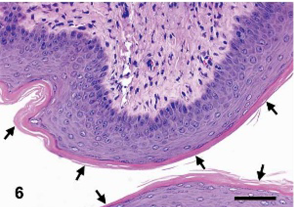
In this report, the testicular tissue was unilateral (right) and was characterized by light microscopy as well-formed seminiferous tubules lined by Sertoli cells with occasional spermatogonia (Fig. 1). Interstitial (Leydig) cells were prominent between the seminiferous tubules. Sperm formation was not evident (Fig. 2). Ovarian tissue occurred bilaterally and was characterized by numerous well-developed follicles with oocytes in some follicles, but no corpora lutea were observed (Fig. 3). The left oviduct histologically resembled the tortuous ducts of an epididymis (Fig. 4). The uterus and uterine glands were lined predominantly by stratified squamous epithelium intermixed with areas of columnar epithelium. Cornification was evident in a few areas (Fig. 5). The vagina was lined by stratified squamous keratinized epithelium, typical of estrogenic stimulation (Fig. 6). In the mammary gland, ductal and alveolar proliferation was abundant. Persistent estrus and excessive mammary gland stimulation have been reported previously.3,8 The presence of stratified squamous epithelium in the uterus and keratinized lining of the vagina in this animal is consistent with hyperestrinism (estrus). An enlarged clitoris with an osseous core was observed upon further examination of the formalin-fixed external genitalia (Fig. 7). Castration cells characterized microscopically as vacuolated pituitary gonadotrophs were a conspicuous feature in the pituitary gland and suggest exposure to increased levels of luteinizing hormone releasing factor (Fig. 8). Long bone growth was altered with notable retention of cartilaginous cores within most trabeculae of the metaphysis (Fig. 9). The bone changes were considered to be consistent with antiresorptive effects of excessive estrogen production, resulting in a disruption of normal bone turnover.
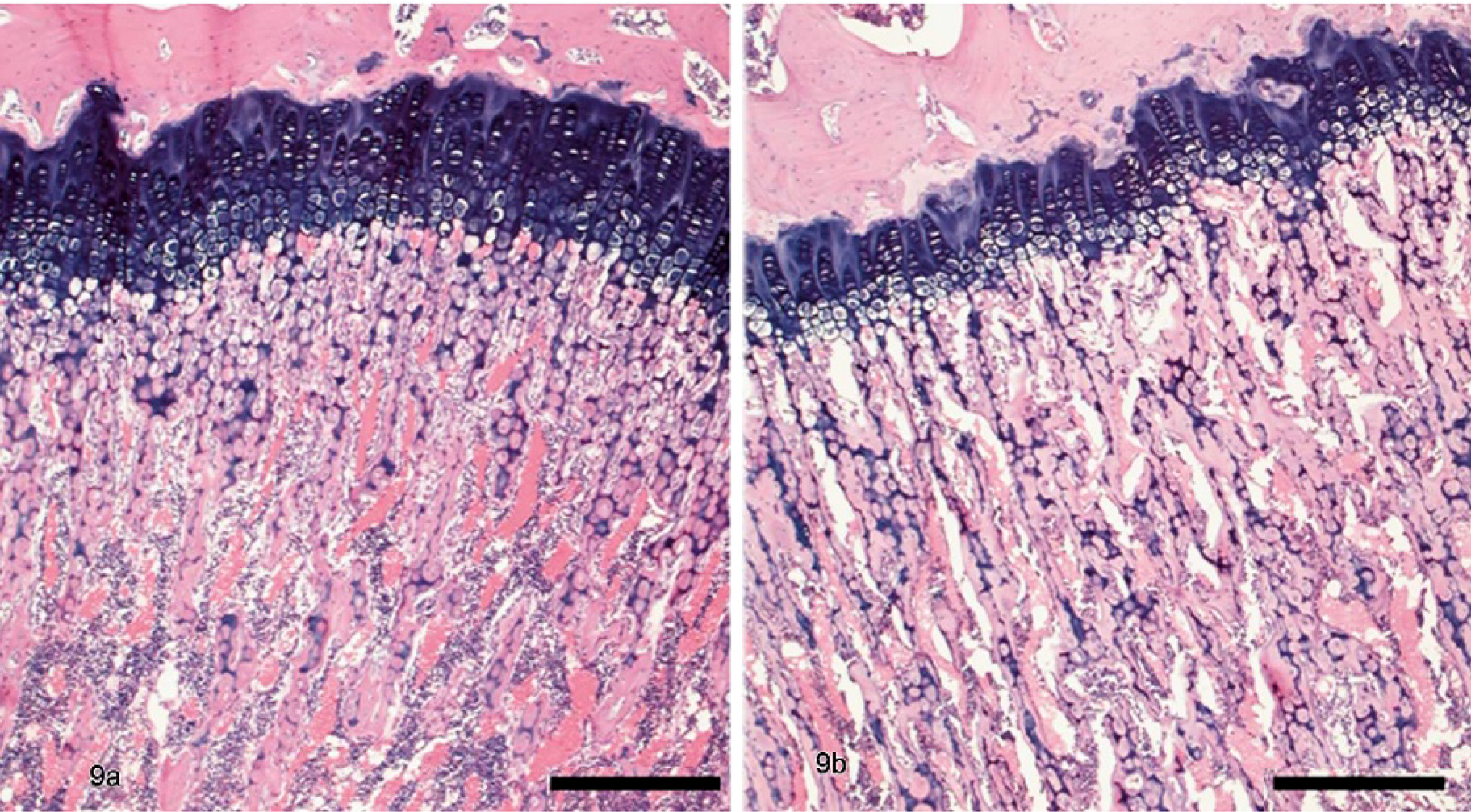
Metaphysis, long bone; rat. Note retention of cartilage cores in the hermaphroditic rat (9b) versus a control rat (9a). HE. Bar = 400 μm.
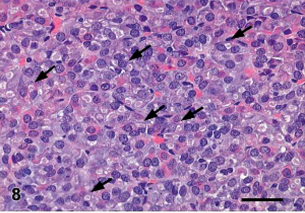
Pituitary; rat. Prominent castration cells are seen (arrows). HE. Bar = 50 μm.
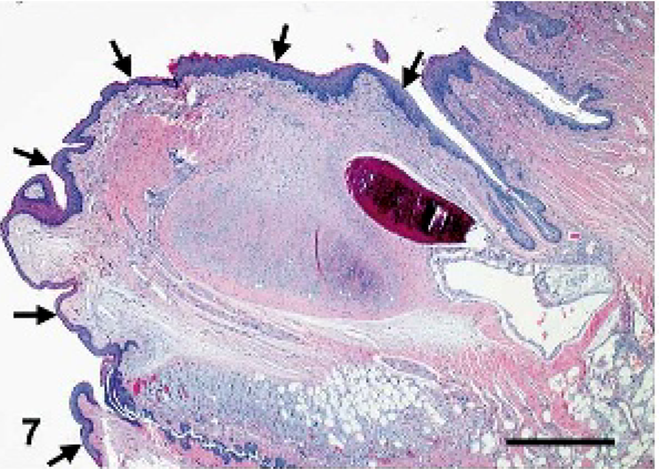
Clitoris; rat. Clitoromegaly is noted (arrows). HE. Bar = 1,000 μm.
Vagina; rat. Cornified epithelial lining is seen (arrows). HE. Bar = 100 μm.
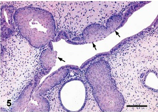
Uterus; rat. Multiple areas of the uterus are lined by stratified squamous epithelium (arrows). HE. Bar = 200 μm.
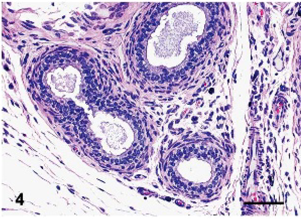
Epididymis; rat. Well-formed ducts have no sperm. HE. Bar = 200 μm.
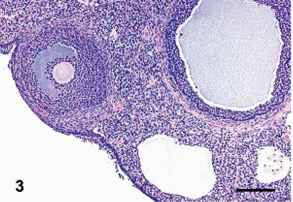
Ovary; rat. Mature follicles are present, but corpora lutea are absent. HE. Bar = 200 μm.
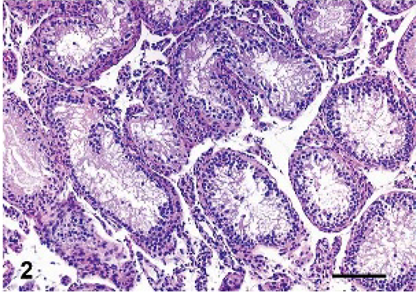
Testis; rat. Spermatogenesis and prominent interstitial cells are absent. HE. Bar = 200 μm.
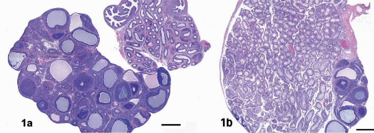
Ovary, right side (1a); ovotestis, left side (1b); rat. Spermatogenesis and corpora lutea are absent. Epididymis is adjacent to the ovary (1a). HE. Bar = 500 μm.
Development of this malformation is poorly understood. Embryonal development of the genital ridge into either an ovary or a testis depends on the absence or presence of an intact Y-chromosome. For testicular differentiation, the SRY gene (sex determining region on the Y-chromosome) is essential and is responsible for the induction of testicular development and is necessary for Sertoli cells to produce Müllerian-inhibiting substance, which results in Müllerian duct degeneration. Zygogenetic investigations have shown that in humans, 80% of the hermaphrodites have a female karyotype 46-XX, while a smaller number of patients have 46-XX/46-XY mosaicism and a very small number have 46-XY.10