
Abstract
Subendothelial heart valve angiectasis has been reported in cows, dogs, pigs, rats, mice, and in human fetuses and newborns. We observed a high incidence (62 in 208 animals examined) of spontaneous angiectasis on the atrioventricular (AV) valves in 10– to 40–week-old Sprague-Dawley rats. The angiectasis was observed predominately on the septal cusp of the right AV valve and located near the AV ostium in 57 of 62 animals. Of the remaining 5 valvular angiectases, 2 were present on the parietal cusp of the right AV valve and 3 were on the left AV valve. The angiectases were single or multiple, ranging from 40 to 300 um in diameter and were characterized by light microscopy as blood-filled dilatations lined by endothelium. Spontaneously occurring abnormalities in normal laboratory animals, such as the spontaneous valvular angiectasis reported here, need to be differentiated from drug-related lesions.
Spontaneous valvular angiectases have been described in aborted human fetuses,2,12 calves and cattle,7–9 pigs,5 dogs,3,11 and bioprosthetic valves.1,4,6 Spontaneous valvular cysts have also been reported in rats and mice.10 We observed a relatively high incidence of spontaneous focal-to-multifocal blood-filled cysts on the atrioventricular (AV) valves in otherwise normal laboratory Sprague-Dawley (SD) rats. These valvular changes occurred at a higher frequency on the right AV valves. Some of these cysts had apparent connections to smaller vessels. Similar to those in other reports,7,11 these blood-filled valvular cysts were endothelial-lined, as confirmed by positive immunohistochemical staining for von Willebrand's factor (vWF)–related antigen, indicating that they may represent dilated blood vessels. Similar findings to those described here have been given various descriptive names, including blood-filled cysts, angiectases, hematocysts, hemocysts, and hematomas. However, the evidence of a continuous endothelial lining in the examples described in this report suggests the term “angiectasis” as the most appropriate for lesions of this morphology and location.
The initial findings of valvular angiectasis were discovered in the control group of naïve rats from an investigative toxicology study. To further investigate the findings, we evaluated stock male and female SD rats, 10–40 weeks old, obtained from commercial sources (Crl:CD[SD], rats from Charles River Laboratories, Raleigh, NC, and [Hsd:SD], rats from Harlan Sprague Dawley, Inc., Indianapolis, IN and Frederick, MD) (Table 1). Animals were housed and euthanatized in accordance with the standards of the American Association for Accreditation of Laboratory Animal Care and the institutional Animal Care and Use Committee. At necropsy, the heart was immersion-fixed in paraformaldehyde and examined under a stereomicroscope after the atria were removed. Valvular angiectasis could occasionally be identified without the aid of magnification. The vast majority (57 out of 62) of angiectases were present on the right AV valve cusp that attaches to the interventricular septum (septal) and were located proximally near the site of valvular attachment to the fibrous ring of the AV ostium (Figs. 1, 2). In some instances, angiectasis was not evident by gross examination, whether it was located on the valve leaflet or on the valve fibrous ring. Angiectases were present on the right AV valve cusp that attaches to the ventricular free wall (parietal) in 2 rats, and in 3 other rats, the angiectasis was present on the left AV valve. Typically, the cysts were dark red, irregularly round to oval, elevated, 40–300 μm in diameter, and single or multifocal (Figs. 1, 2).
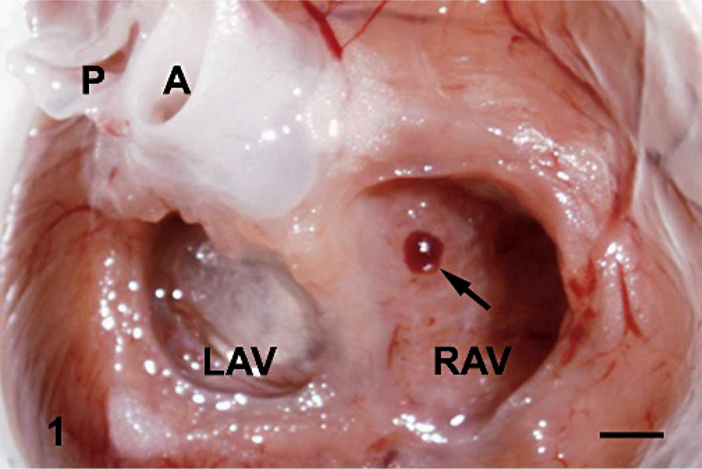
Heart; rat. Stereomicroscopic examination of focal angiectasis (arrow) on the right atrioventricular (AV) valve is shown. P = pulmonary artery; A = aorta; LAV = left AV valve; R = right AV valve. Bar = 200 μm.
Summary of the spontaneous angiectasis on atrioventricular valves in Sprague-Dawley rats (lesion/total number of rats).
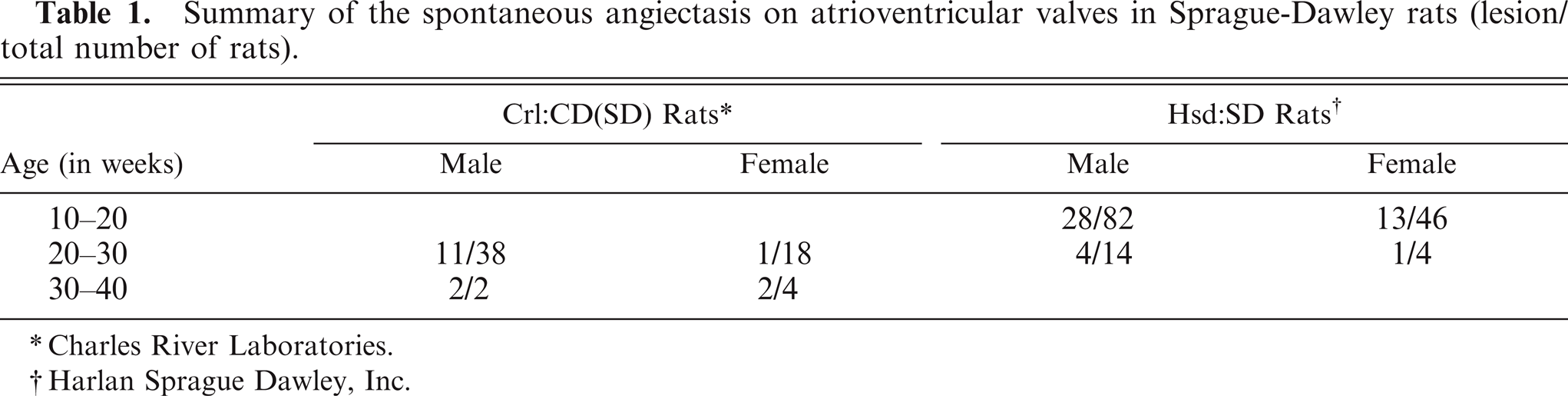
Charles River Laboratories.
Harlan Sprague Dawley, Inc.
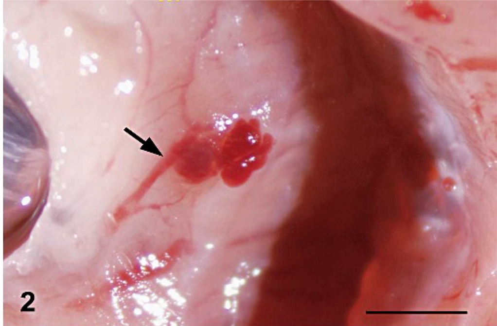
Heart; rat. Focal angiectasis on the septal leaflet of the right atrioventricular valve is shown. The arrow indicates the connecting vessel to the angiectasis. HE. Bar = 300 μm.
Representative samples of AV valves with macroscopic findings were transferred to 10% neutral-buffered formalin for additional overnight fixation, then embedded in paraffin, sectioned at 4 μm longitudinally along the valve leaflet, and stained with HE. Valvular cysts were lined by a continuous single layer of endothelium and filled with blood (Fig. 3a, b). There was no identifiable smooth muscle surrounding the structure or any inflammatory cell infiltration. Thrombosis was not observed in any of the angiectases. Serial sections of 1 left AV valvular angiectasis, which included multiple blood-filled cysts, demonstrated that the cysts were interconnected. Immunohistochemical staining with an endothelial cell marker, vWF–related antigen (Factor VIII, Chemicon, Temecula, CA), using an indirect chromogenic immunoperoxidase detection assay (Dako, Carpinteria, CA) demonstrated that these angiectases were lined by a continuous layer of endothelium (Fig. 3c).
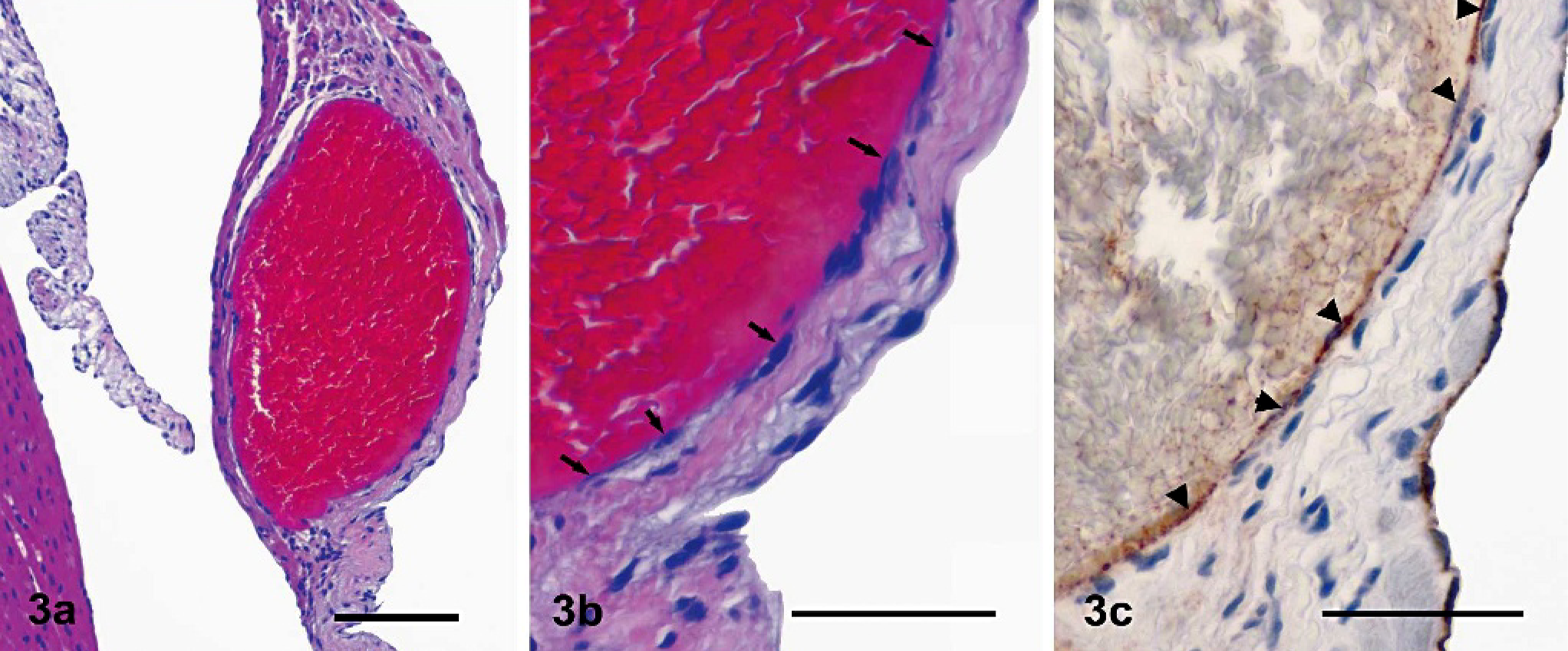
Heart atrioventricular (AV) valve; rat.
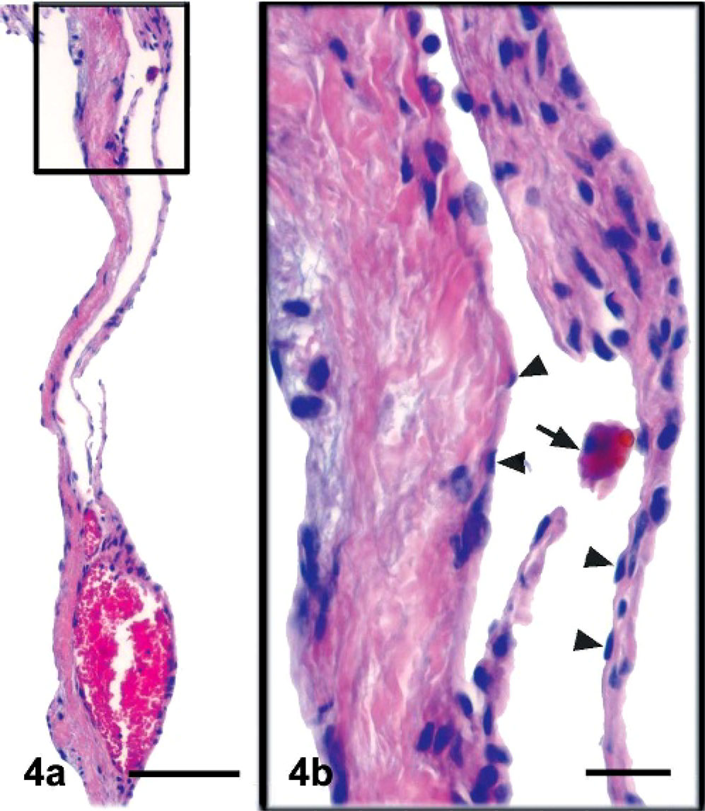
In the dog, partially arrested blood flow in the cystic lesion followed by degeneration and necrosis has been reported in valvular hemocysts/angiectases.11 In our evaluation of angiectases in SD rats, we did not observe degeneration of any blood cell components or of the vessel itself (Fig. 3). Erythrophagocytosis was occasionally observed in the lumen of a blood vessel connected to an area of angiectatic change (Fig. 4). There was no evidence of calcification in these angiectases.
Atrioventricular (AV) valve; rat.
It is unclear why the vast majority of angiectases occurred in the right AV valve as compared with the left AV valve. A similar occurrence involved the interventricular septum (septal) cusps as compared with the ventricular free wall (parietal) cusps.
A variety of confusing and often inaccurate terms have been used to describe valvular cysts in animals. For example, a “hematoma” is an abnormal localized collection of blood, in which the blood is clotted or partially clotted, is usually within an organ or a soft-tissue space, and is generally the result of a break in the wall of a blood vessel. A “hematocyst” is described as an effusion of blood into a cyst that develops abnormally, while “angiectasis” implies dilatation of an intact blood vessel. The valvular hematomas/hematocysts described previously2,3,5,7–9,11–12 and the valvular blood-filled cysts in this report have a complete endothelial lining; therefore, these lesions are most accurately termed “angiectases.”
Serial sectioning of an angiectasis on the left AV valve enabled us to identify a vessel connecting multiple cysts (Fig. 4); however, we could not establish the existence of definitive channels between the angiectases and the ventricular lumen. Potential connections, which were most likely dilated capillaries or small veins, between the cysts and small blood vessels were noticed (Figs. 2, 4). It has been reported that valvular hematocysts/angiectases in calves9 and human fetuses12 have vascular channels linking them to the ventricular lumen and that they eventually heal by resorption and organization. On the contrary, Marcato et al.7 hypothesized that AV valvular cysts in cattle were derived from the dilation of blood and lymphatic vessels, did not regress with age, and were the result of mechanical effects. In dogs, studies of serial sections revealed that valvular cysts communicated with blood vessels but showed no connection with the valvular surface.11 Additionally, the presence of an extensive, dense arrangement of blood vessels in the septal cusp of the tricuspid valve was usually the site of development of blood cysts.11
Most reports suggest that valvular angiectasis occurs at higher incidences in young animals and humans (e.g., 47–56% in autopsied human fetuses and newborns2,12) as compared with adults and is thus defined as congenital.8 However, Marcato et al.7 reported that older cows had a higher incidence (16.2%) than younger animals (11.5% in calves, 7.9% in steers, and 6.4% in heifers) and suggested that valvular angiectasis in this species may be acquired. In this study, the angiectasis was not strictly age-related since this change was observed in both young (10 weeks) and middle-aged (40 weeks) SD rats (Table 1) and there was no increased incidence in older animals.
The exact etiopathogenesis of spontaneous valvular angiectasis in the rats of this report is unknown; anoxia and the establishment of a pressure gradient between the atrium and ventricle at the valve cusp base during valve closure are possibilities.2,12 There is no evidence that development of valvular angiectasis was induced by trauma or associated with the necropsy procedures routinely utilized by our laboratory.
Heart valves are critical structures regulating blood flow in the circulatory system. Any valvular finding can be of concern for scientists using animals in drug-safety studies. It is critical to differentiate spontaneously occurring abnormalities in normal laboratory animals from drug-related lesions. These vascular angiectases may not represent a true pathologic condition in humans or animal species, but rather may represent inconsequential incidental findings of an indeterminate cause.
Footnotes
Acknowledgements
We thank William Janusz, Susan Wells, Diane Pecht, Martin Slade, Virginia Eppolito, and Kathy Petersen for their excellent technical contributions.