
Abstract
Amputation is commonly performed in an attempt to both treat and diagnose conditions affecting the digits of cats. The records of multiple veterinary diagnostic laboratories were searched to identify submissions of amputated digits from cats. Eighty-five separate submissions were reviewed for diagnosis, age, sex, limb of origin, and digits affected; and the original submitting clinics were surveyed to determine clinical outcome. The Kaplan-Meier product-limit method was used to determine the disease-free interval and survival time. Neoplastic disease was identified in 63 of 85 submissions, with exclusively inflammatory lesions composing the other 22 cases. In 60 (95.2%) of the neoplastic cases, a malignant tumor was identified. Squamous cell carcinoma was the most commonly identified malignant tumor (n = 15; 23.8%) and was associated with a median survival time of 73 days. Other diagnoses included fibrosarcoma (n = 14; 22.2%); adenocarcinoma, likely metastases of a primary pulmonary neoplasm (n = 13; 20.6%); osteosarcoma (n = 5; 7.9%); mast cell tumor (n = 4; 6.3%); hemangiosarcoma (n = 5; 7.9%); malignant fibrous histiocytoma (n = 2; 3.2%); giant cell tumor of bone (n = 2; 3.2%); and hemangioma (n = 2; 3.2%). Giant cell tumor of bone has not been previously described in the digits of cats. Various neoplasms can occur in the digits of cats, and submission of the amputated digit for histopathologic diagnosis is essential to determine the histogenesis and predict the clinical outcome.
Amputation is commonly used as a method of both diagnosis and treatment for diseases in feline digits. Due to the restricted anatomic construction of the digit, neoplastic and inflammatory diseases may have similar clinical appearances and require histopathologic evaluation to determine the nature of the underlying condition. Thus, these amputated digits, or portions there of, should always be sent to a veterinary diagnostic laboratory to obtain a specific diagnosis and information about the expected clinical course of the disease. Few studies on the nature of masses in feline digits have been reported. Those reports that do exist consist primarily of case reports or retrospective studies on the occurrence of epithelial tumors, primarily carcinomas of pulmonary origin.8,25 To our knowledge, no published data on the prevalence of neoplasms in feline digits currently exist. The purpose of this study was to determine the types and prevalence of neoplastic disease occurring in amputated feline digits submitted to a veterinary diagnostic laboratory for diagnosis and to determine the clinical outcome associated with these diseases.
Materials and Methods
With the exception of amputated feline digits being examined rather than canine digits, materials and methods used in this study were identical to those previously described in Wobeser et al.27
Results
Amputated digits from 85 cats were evaluated. Multiple digits were affected in 14 cats (16.5%), and 1 cat had 2 different neoplastic processes in the same digit. A total of 12 different diagnoses were present (Table 1). Malignant neoplastic processes were present in 60 (70.5%) submissions, benign neoplasms in 3 (3.5%), and inflammation only in 22 (26%).
Summary data of diagnoses in amputated digits from 85 cats.
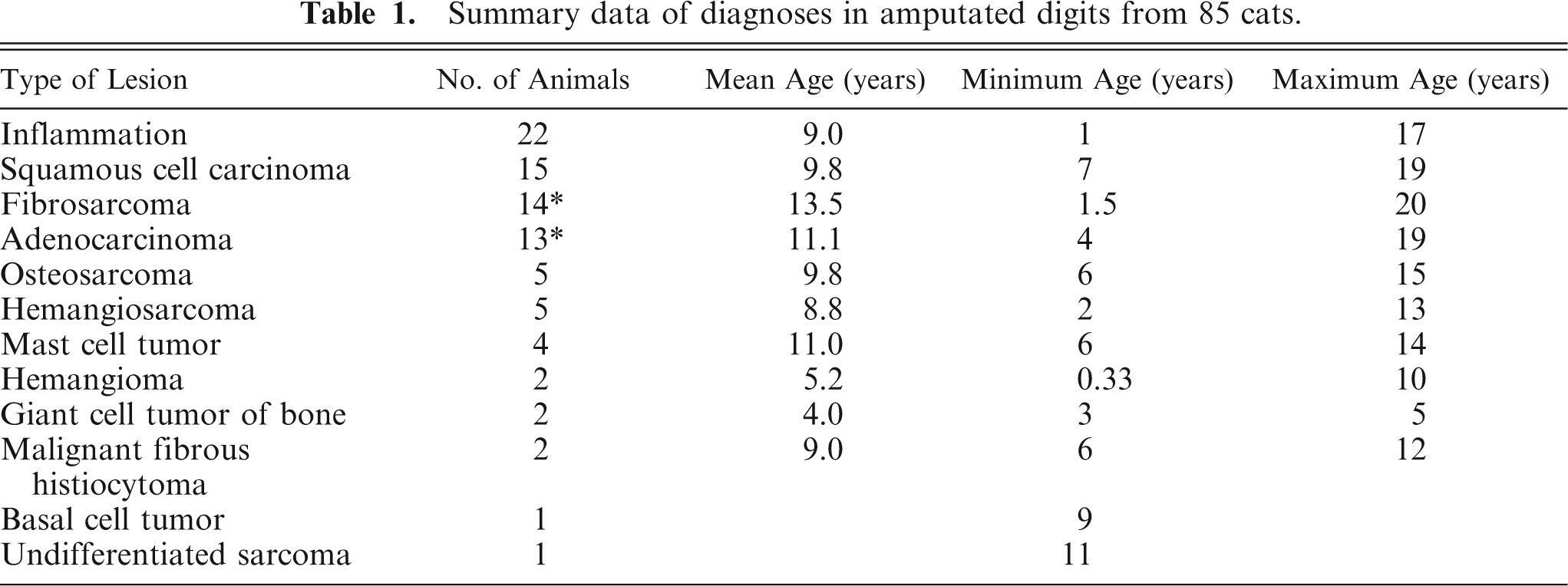
The overall study population consisted of 7 breeds of cats: 50 domestic short-haired cats, 12 domestic long-haired cats, 9 Siamese, 6 Persians, 4 domestic medium-haired, 1 Himalayan, 1 Turkish angora, and 2 cats whose breed was not identified. Forty-five cats were females, of which 39 were spayed; and 40 were males, 35 of which were castrated.
Fifty-nine submissions specified the leg of origin in the clinical history. The front leg was significantly more often affected (n = 39; 66.1%) than the hind leg (n = 20; 39.9%) (P = .01). This pattern was present in both neoplastic and inflammatory lesions. Lesions were evenly distributed among digits.
Surveys were sent to the original submitting veterinarian for each of these 85 submissions. Thirty-eight of these were completed and returned for a response rate of 44.7%. Adjunct therapy beyond further surgical excision consisted of an unidentified type of chemotherapy and was reported to be performed in only 2 cats.
Squamous cell carcinoma (SCC) (n = 15; 23.8%) was the most commonly diagnosed neoplasm. Ten female cats and 5 male cats were affected. Multiple digits on the front paws or both the front and hind paws were affected in 4 cats. Surveys were returned on 7 of these cats. Two cats had local recurrence, and 3 cats had metastasis of the tumor with a median disease-free interval of 45 days, a median survival time of 73 days, and a 3-month survival rate of 33% (Fig. 1).
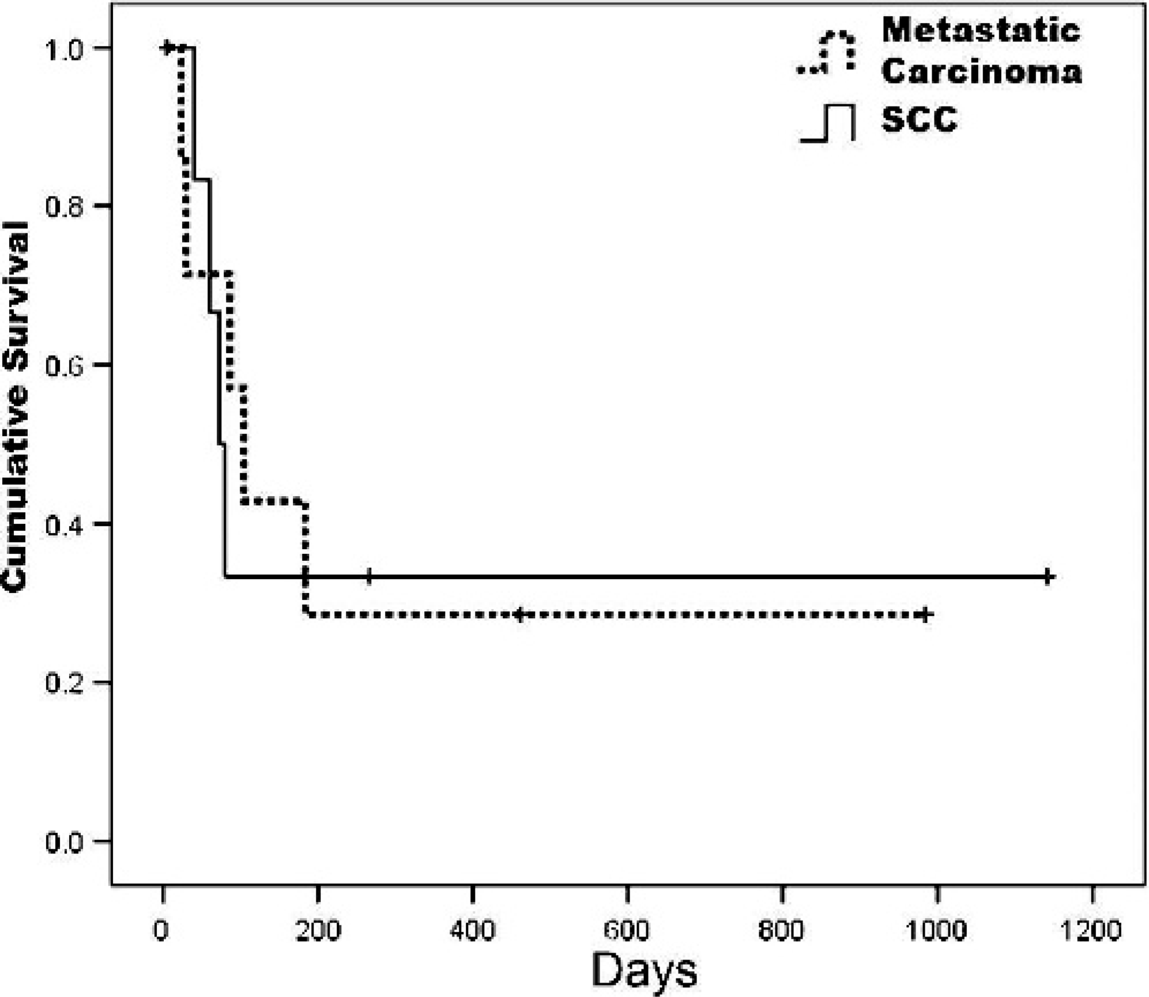
Kaplan-Meier survival curve for cats with primary squamous cell carcinoma of the digit (SCC, n = 7) and metastatic carcinomas to the digit (n = 7).
Fibrosarcoma (n = 22.2%) was the second most commonly diagnosed neoplasm. Seven female and 7 male cats were affected. Multiple digits on the front paws were affected in 2 cats at the time of surgical submission. Three surveys were returned on these animals, with a single animal having metastasis and subsequent euthanasia at 900 days. The other 2 cats were alive at 14 and 29 days, respectively, postamputation.
Adenocarcinoma was identified in 13 cats (20.6%). Females were significantly more likely to be affected (P = .05), with 10 female and 3 male cats diagnosed. Multiple digits of both the front and hind paws were affected in 4 cats at the time of surgical submission. Seven surveys were returned, with 5 cats reported to have metastatic disease, and 3 of these cats had radiographic evidence of pulmonary masses at the time of amputation. The median disease-free interval was 24 days, and median survival time was 104 days (Fig. 1).
Osteosarcoma and hemangiosarcoma were each diagnosed in 5 cats. Mast cell tumor was diagnosed in 4 cats. Giant cell tumor of bone (GCTB), hemangioma, and malignant fibrous histiocytoma were each diagnosed twice. One basal cell tumor and 1 sarcoma of undetermined cellular origin were also diagnosed.
Discussion
Information on the prevalence and clinical outcome of lesions of the feline digits is scarce. Previous studies on neoplastic disease in the digits of cats have focused primarily on pulmonary adenocarcinomas metastatic to the digits.8,21,25 In this study, 63 of 85 submitted amputated digits from cats contained 1 or more neoplasms, and the other 22 contained inflammation, primarily pyogranulomatous in nature, with no evidence of neoplasm. This varies dramatically from a study of diseases of the claw and claw bed in cats where only 3 of 65 cases were neoplastic.20 The vast majority (60/63, 95.2%) of neoplasms in this study were malignant. While SCC and adenocarcinoma were 2 of the most common neoplasms identified, overall most of the neoplasms, 35/63 (55.6%) in this study, were mesenchymal in origin, which is in contrast to the preponderance of literature on feline digital neoplasms. Affected digits in this study were primarily from the front limbs. A similar pattern is described in articles on neoplasia in the digits of dogs,11,14 which speculate that possible reasons for this may be increased weight bearing on the front-versus-hind limbs and increased exposure to carcinogens during digging behavior.11 No sex predilection for the development of neoplasia was present apart from adenocarcinoma, where females were overrepresented.
SCC was the most common neoplasm and accounted for 15 of the 63 neoplasms found. Reports of the frequency of SCC in the digits of cats are contradictory, with some suggesting the digit is the second most common site of SCC involving bone,14 while others suggest that SCC in the digits is rare.8,18 Differentiation of primary digital SCC from metastatic carcinoma can be difficult, but in the largest study of carcinomas in the digits of cats, it was found that histopathologic evaluation of digits with metastatic pulmonary carcinomas showed ciliated cells, goblet cells, and secretory material,25 which were lacking in the SCC diagnosed in this study, and thus the SCCs in this study were determined to be primary digital tumors. The cats with SCC in this study were generally older, with the youngest being 7 years of age at the time of diagnosis. The median disease-free interval and the median survival time were both very short, 45 days and 75 days, respectively. Although the number of neoplasms in this sample is small, these data compare well with literature that suggests that digital SCC is aggressive and metastasizes rapidly18 or affects multiple digits at the time of diagnosis.5
Fibrosarcoma was the next most commonly diagnosed neoplasm, affecting 14 animals. Although this is a common neoplasm in cats and has been reported to occur on the limbs, no case reports describing its occurrence on the digits could be found. Two animals had multiple digits affected at the time of diagnosis, and 1 other cat had local recurrence and was subsequently euthanized due to this recurrence 900 days after diagnosis. The average age of cats diagnosed with fibrosarcoma was 13.5 years, but the age range was very broad, with the youngest cat being 1.5 years of age at the time of diagnosis.
Adenocarcinoma was the third most commonly diagnosed tumor, with 13 animals diagnosed. Although it is reported to be a rare event, there are a considerable number of case reports describing metastatic pulmonary adenocarcinoma to the digits—so-called “lung-digit syndrome.”6,8,12,16,21,25 In many of the digits diagnosed with adenocarcinoma in this study, a ciliated columnar epithelium with goblet cells reminiscent of pulmonary bronchial epithelium was present. As well, 3 cats had confirmed pulmonary masses on radiographs. Cats with metastatic pulmonary carcinoma may not exhibit any clinical respiratory signs, and the lesions developing in the digits may be the first indication of disease.8,12,16,18 The reason for the digit as a site of metastasis is unclear, but in humans, it is suggested that trauma, temperature, hormone influences, and plentiful vasculature may play a role.2,17
The average age of cats with digital adenocarcinoma was 11.1 years in this study, and female cats were over-represented. Other studies contained larger numbers of animals, and the sex bias in this study may be an artifact of the lower number of cases. The median survival time was 104 days in cats in this study. As such, the prognosis for adenocarcinoma in the digits of cats must be considered to be poor.
In this study, 4 of 13 cats had multiple digits involved at the time of diagnosis, which is similar to 1 study where 6 of 19 cats had multiple digit involvement.8
Osteosarcoma was a relatively common diagnosis in this study, representing 5 of 63 (7.9%) neoplastic masses diagnosed. Osteosarcoma in cats is not as well described as that in dogs, with fewer studies being reported.4,13,22 The largest study of feline osteosarcoma found that 9 of 145 cases of osteosarcoma arose in the digits.10 The distribution of lesions in the current study was roughly evenly split with 3 in the front legs and 2 in the hind legs. In this study, 3 of 5 cats affected were male. Surveys were returned on only 2 cats, with 1 being lost to follow-up at 758 days, while the second had metastatic disease and was euthanized at 152 days.
Hemangiosarcoma was also diagnosed in 5 cats. Typically hemangiosarcomas are reported to occur more frequently in older male cats. In this study, 3 tumors were in 13-year-old male cats, but 2 were in 2-year-old female cats. The pathogenesis of vascular tumors is unclear, but trauma has been suggested as a possible predisposing factor.15 This theory would fit well as this is likely to be a common occurrence in the digits.
Other tumors less frequently diagnosed composed the remaining lesions in submitted digits. Mast cell tumors were present in 4 cases; malignant fibrous histiocytoma, hemangioma, and GCTB were each diagnosed twice; and a single basal cell tumor and 1 sarcoma of unidentified cellular origin were present. One of the hemangiomas was diagnosed in multiple digits in a 4-month-old cat and may represent vascular malformation rather than a neoplasm.
Of note, were the 2 GCTBs diagnosed in this study. This is a rarely reported neoplasm in domestic animals, and few reports exist in cats.3,7,19,23,24,26 The relative frequency of its diagnosis in the digits in this study was unexpected, as GCTB has never previously been reported in the digits of cats and is uncommon in humans. Only 13 cases of GCTB were seen in the hand in a retrospective study of 50 years of submissions at the Mayo Clinic.1 Typically, this tumor is reported to occur primarily in the epiphysis of tubular long bones in humans and animals,22 and reported cases in cats originated in the femur,24 ulna,3,19 rib,23 and tibia.7 Both tumors in this study occurred in young adult cats (3 and 5 years of age); the age range in reported literature was from 1 to 12 years of age.3,19 The histogenesis of GCTB is unclear, but the current hypothesis is that the fibroblast-like stromal cells in the tumor are the proliferating component and the giant cells that give the tumor its name are non-neoplastic reactive cells with immunohistochemical staining properties consistent with osteoclasts.7
In summary, the amputated digits from cats reviewed in this study contained primarily neoplastic disease, most of which was malignant. SCC was the most common diagnosis with fibrosarcoma and adenocarcinoma, likely of pulmonary origin and metastatic to the digits, the next most common. Both SCC and adenocarcinomas of the digit have poor prognoses. The scope of neoplastic disease in the digit is much greater than previously reported, and more than just metastatic pulmonary tumors should be considered when one is confronted with masses on the digits requiring surgical amputation. Due to the similar gross appearance of lesions of the feline digit and the variable prognoses, submission of amputated digits for histopathologic examination is required to obtain an accurate diagnosis and prognosis.