
Abstract
A 5-month-old mixed-breed filly presented with diarrhea due to Salmonella typhimurium infection and subsequently developed pneumonia in addition to ischemic necrosis of distal limbs. Pulmonary lesions were characterized by numerous discrete, disseminated pyogranulomas with intralesional fungal hyphae. The morphologic characteristics of fungal hyphae were consistent with Aspergillus spp., and large numbers of A fumigatus were isolated from lung tissue via fungal culture. Lesions in all 4 limbs were similar in distribution, duration, and severity and were characterized by coagulation necrosis accompanied by occasional thrombi in small vessels without evidence of thrombosis of larger limb arteries. Thus, limb lesions are consistent with symmetrical peripheral gangrene, a potential complication of sepsis.
Keywords
Pulmonary aspergillosis, although relatively uncommon in horses, has been associated with enterocolitis, often due to Salmonella infection13 or less often other enteric infections.4 Factors that contribute to the development of Aspergillus pneumonia in horses with acute enterocolitis include neutropenia and immunosuppression, as well as enteric mucosal compromise. The latter allows invasion of fungal elements from the intestinal ingesta into the bloodstream.13 In a retrospective study of pulmonary aspergillosis, 86% of the horses with mycotic pneumonia also had disease compatible with loss of gastrointestinal mucosa integrity.15 Another reported complication of enterocolitis in horses is acute thrombosis of digital arteries with subsequent ischemic distal limb necrosis.1,3 Gram-negative sepsis and endotoxemia, possibly accompanied by enteric loss of antithrombin III, results in hypercoagulability that is presumed to contribute to arterial thrombosis.1 In contrast to distal limb necrosis associated with digital artery thrombosis, symmetrical peripheral gangrene6 is a disease entity in which gangrenous necrosis of distal extremities occurs without evidence of large vessel occlusion or vasculitis.8 Symmetrical peripheral gangrene, often associated with disseminated intravascular coagulation, is a well-documented but not well-understood condition.8 It is a potential but rare complication of sepsis and has been reported in human infants with Salmonella spp. septicemia.11 Gangrene of distal limbs has also been reported in calves with salmonellosis10 and in a foal with dehydration and hypoxemia.14 Here we describe macroscopic and histopathologic findings of invasive pulmonary aspergillosis and ischemic distal limb necrosis, consistent with symmetrical peripheral gangrene, in a 5-month-old foal that presented with Salmonella typhimurium enterocolitis.
A 5-month-old mixed breed filly was presented to the Boren Veterinary Medical Teaching Hospital at Oklahoma State University for acute onset of profuse watery diarrhea. On presentation, the filly was dehydrated (capillary refill time 10 sec), febrile (104.2°F), tachycardic (110/min), and mildly tachypneic (20/min). Laboratory analysis revealed hypoalbuminemia (2.1 g/dl; reference range 2.5−3.8 g/dl), hyperfibrinogenemia (1, 200 mg/dl; reference range 100−400 mg/dl), and neutropenia (PMN 1.835/μl; reference range 2,500−8,500/μl), with a degenerative left shift (bands 496/μl; reference range 0−300/μl) and toxic changes in neutrophils. S typhimurium was cultured from the feces during the first few days of hospitalization. After the initiation of therapy, fecal cultures became negative for Salmonella spp. and diarrhea clinically improved but did not completely resolve. After several days of hospitalization with moderate improvement, the filly began to develop edema of the distal limbs, became tachypneic with pulmonary crackles and wheezes, and died. The total time between presentation and death was 10 days.
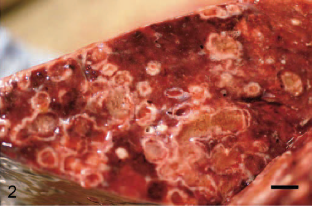
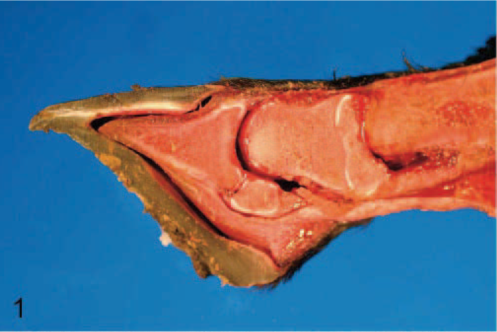
At necropsy, external examination revealed dry alopecic foci over the fetlocks of all 4 limbs. Subcutaneous tissues of all limbs distal to well-defined circumferential lines at the level of the midmetacarpus/metatarsus were dark red to purple and diffusely softened. Sagittal sections of the limbs revealed complete separation of the distal phalanx from the hoof lamina, as well as softening and collapse of articular surfaces of phalanges (Fig. 1). Lesions in all 4 limbs were similar in character and severity. The pleural cavity contained approximately 500 ml of turbid serosanguineous effusion, and the tracheal lumen contained copious stable white foam; 1 segment of the tracheal mucosa was interrupted by 2 well-circumscribed 0.5-cm diameter dull tan and slightly roughened foci. Numerous multifocal to coalescing 0.5 to 1.5 cm, firm, tan, nodular foci, often with slightly caseous centers, were disseminated throughout all lung lobes (Fig. 2). Intervening pulmonary parenchyma was dark red and wet. The kidneys were moderately swollen with pale cortices. The cecum and large colon exhibited diffuse and marked thickening of the submucosa by clear, gelatinous edema. Otherwise, mucosal surfaces were unremarkable, and the contents were watery.
Lung; foal. Multifocal to coalescing, well-demarcated, tan firm foci, many of which have caseous centers, are disseminated throughout the lung. Bar = 1 cm.
Distal limb; foal. Sagittal section illustrating collapse of articular surfaces of proximal interphalangeal joint and separation of distal phalanx from hoof lamina.
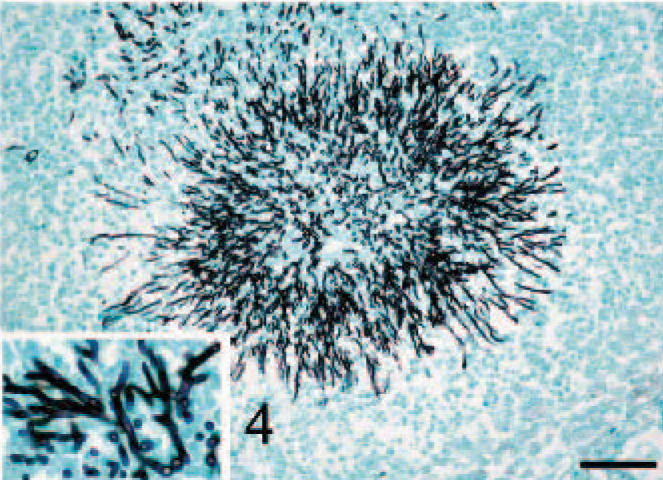
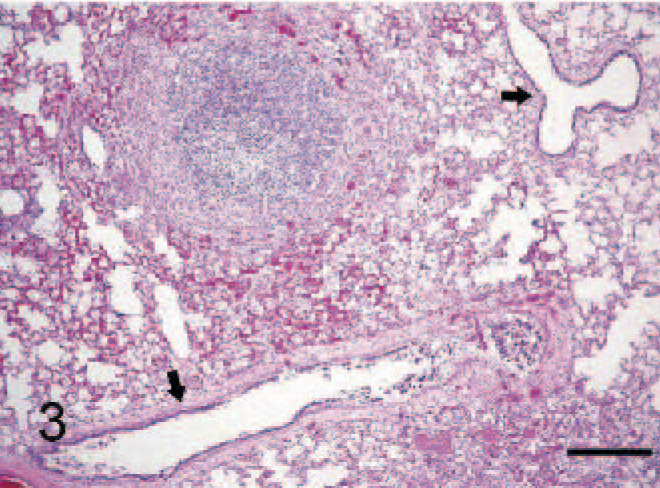
Microscopically, the disseminated foci in the lung obscured or completely effaced the open alveolar architecture but did not involve bronchi or bronchioles (Fig. 3). Lesion centers were composed of necrotic debris admixed with intense infiltrates of neutrophils that often surrounded radiating aggregates of fungal hyphae. Necrotic centers were rimmed by a thin zone of epithelioid macrophages and occasional multinucleate giant cells with a thin peripheral zone of fibrosis. Grocott methenamine-silver nitrate (GMS) stains for fungi revealed sunburst arrangements of 5 to 7 μm wide, dichotomously branching septate hyphae with roughly parallel walls (Fig. 4). Large numbers of Aspergillus fumigatus were isolated via fungal culture of lung tissue.
Lung; foal. Radiating arrangements of numerous Aspergillus fumigatus hyphae are present within the centers of discrete pulmonary pyogranulomas. Bar = 80 μm. Inset illustrates dichotomous, 45° branching of septate hyphae with roughly parallel walls. GMS stain.
Lung; foal. Focally intense pyogranulomatous inflammation effaces the pulmonary parenchyma and often spares adjacent bronchioles (arrows). HE stain. Bar = 325 μm.
Lesions in the tracheal mucosa were histopathologically characterized as discrete foci of necrosis of the lamina propria and submucosa accompanied by suppurative inflammation, intralesional hyphal invasion, and ulceration of the overlying epithelium. Other microscopic findings included full-thickness coagulation necrosis of the skin and subcutis of distal limbs with occasional thrombi in small caliber vessels (Fig. 5). Histologic lesions in the small intestine and colon were limited to mucosal and submucosal edema accompanied by diffuse and mild mixed inflammatory infiltrates. A moderate number of dilated crypts containing necrotic debris were scattered within the small intestinal mucosa. In one focus, the gastric serosa was expanded by an intense accumulation of neutrophils accompanied by macrophages, lymphocytes, and plasma cells in a background of necrosis and granulation tissue. GMS-positive hyphae (similar to those described in lung) were present within the inflammatory focus. In the kidney, proximal convoluted tubule epithelia exhibited necrosis with segmental disruption of basement membranes, demonstrated by periodic acid−Schiff reaction.
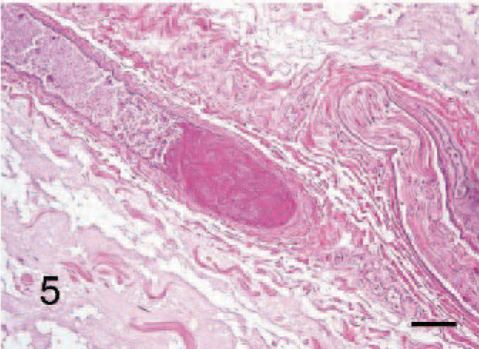
Subcutis of distal limb; foal. A thrombus is present in a small-caliber blood vessel. Note the obscured cellular detail of the vessel wall and surrounding tissues due to coagulation necrosis. HE stain. Bar = 70 μm.
The gross and microscopic distribution of pulmonary pyogranulomas indicates hematogenous dissemination of Aspergillus. The focus of necrosis and inflammation with intralesional fungi in the gastric serosa is further support of hematogenous fungal dissemination in this case. Although inflammatory intestinal lesions had almost completely resolved prior to death, clinical history of severe diarrhea accompanied by multiple Salmonella-positive fecal cultures was consistent with a diagnosis of enterocolitis secondary to Salmonella infection. The pyogranulomatous character of the of the pneumonia with mild fibrosis is consistent with a chronic inflammatory process that correlates with the 10-day or longer duration of clinical disease and indicates that embolic seeding of the lungs likely occurred during the acute stage of enteric disease.
Regionally extensive coagulation necrosis of distal-limb soft tissues accompanied by severe, diffuse necrosis of the hoof laminae and collapse of subchondral bone in phalangeal joints is consistent with avascularization and ischemic necrosis of distal extremities. Frostbite, fescue foot, and ergotism are some potential causes of distal limb necrosis that affect all 4 limbs in ungulates and were ruled out on the basis of history and lack of possible exposure. Thrombosis of digital arteries1,3,16 as well as aorto-iliac thrombosis,2,9 has been reported in foals with gram-negative sepsis, the latter often associated with enterocolitis. Endotoxemia and septic shock may lead to arterial thrombosis, especially of distal limbs, by inducing a hypercoagulable state in addition to poor peripheral perfusion.1 The distribution of ischemic limb necrosis in cases of arterial thrombosis is variable and ranges from involvement of a single limb to involvement of all 4 limbs.1 In this foal, the absence of occlusive thrombi in large arteries of the distal limbs, as well as the symmetrical involvement of all 4 limbs, is consistent with the disease entity referred to as symmetrical peripheral gangrene. Thrombi in small vessels, as seen in this foal, is a reported feature of symmetrical peripheral gangrene12 and is believed to be associated with disseminated intravascular coagulation.8 Other predisposing factors for the development of poor peripheral perfusion and ischemic necrosis of distal extremities include dehydration, hypovolemia, and hypoxemia7,14 as well as vasospasticity or vasoconstriction.5 Hypercoagulability and poor perfusion are evidenced in this foal by coagulation necrosis of distal limbs with thrombosis of small vessels; patchy tubulorrhectic renal necrosis is consistent with poor perfusion and may be due in part to terminal hypoxia.
This report describes the simultaneous occurrence of pulmonary aspergillosis and ischemic distal limb necrosis, both of which are uncommon sequelae of Salmonella enterocolitis in horses. Distal limb necrosis in this foal was not accompanied by thrombosis of major digital arteries and thus is similar to symmetrical peripheral gangrene rather than previously reported digital artery thrombosis in septic foals.1,3,16 Laminitis is another common sequela to enterocolitis in horses and is characterized acutely by ischemic necrosis restricted to soft laminae of the hoof. Thus, typical laminitis differs greatly from regionally extensive necrosis, in this case affecting all soft and bony tissues of the distal limbs. Both embolic Aspergillus pneumonia and symmetrical peripheral gangrene should be considered as potential complications of sepsis in foals.
Footnotes
Acknowledgements
We thank Drs. Roger J. Panciera and Jerry W. Ritchey for critical review of the manuscript.