
Abstract
Hamartomas of the liver and biliary system are extremely rare entities in both animals and humans. Biliary hamartomas in humans are usually multiple and constitute the von Meyenburg complexes. This report describes the presence of a large solitary mass arising from the edge of the right medial liver lobe of a domestic rabbit. Histologically, the mass was composed of an extensive network of large varying sized cystic structures lined by simple cuboidal to columnar epithelium within an abundant fibrous stroma. Within many of the cyst lumina were varying sized, pale white to greenish hard concretions identified as choleliths and were analyzed and found to be composed of calcium carbonate. This is the first known report of biliary hamartoma with cholelithiasis in rabbits.
Hamartomas are defined as focal malformations that are composed of an abnormal mixture of tissue elements or an abnormal proportion of a single element, normally present at that site. 26 Hamartomas have been described in a number of organ systems, including the liver and associated biliary tract of animals and humans. In humans, mesenchymal hamartomas of the liver, composed of well-vascularized mature connective tissue intermixed with elongated branching bile ducts and hepatocytes, have been described. 2 A bile-duct hamartoma, also known as von Meyenburg complex in humans, is composed of a multiple, varying sized, focal disorderly collection of bile ducts and ductules surrounded by abundant fibrous stroma, and scattered throughout the hepatic parenchyma of numerous lobes. 2 In this report, a solitary large biliary hamartoma with cholelithiasis in a domestic rabbit is described.
A recently euthanized adult male intact transgenic LCAT (lecithin°cholesterol acyltransferase) New Zealand white rabbit was presented to the diagnostic pathology services branch of the Division of Veterinary Resources at the National Institutes of Health in Bethesda, Maryland. Before submission, the animal had been demonstrating ataxia and incoordination of 1 month's duration. Because of progression of the clinical signs, the animal was humanely euthanized. On necropsy, the animal was in fair nutritional condition. Upon opening the peritoneal cavity, an approximately 4 × 2 × 1-cm irregularly shaped mass was adhered by fibrous connective tissue to the caudal extent of the right medial liver lobe and partially adhered by fibrous adhesions to the cranial pole of the right kidney. The proximal 25% of the mass closely adhered to the liver was greenish blue, whereas the remaining mass was opaque whitish-tan. The surface of the mass was gyrose (Fig. 1). The mass was teased away from the liver, at which point a vascular or biliary communication between the liver lobe and the mass was not observed. The mass was easily removed from the renal capsule. Upon cutting into the mass, hard gritty concretions were present. Upon further evaluation, it was noted that the gyrose sections were elongate cystic structures in which the concretions resided. The concretions were pigmented and gave the mass its various coloration patterns (Fig. 2). A few of the concretions were submitted for stone analysis, and the remainder of the mass was placed in 10% formalin. The gall bladder appeared within normal limits, with patent communication of the common bile duct to the duodenal papilla.
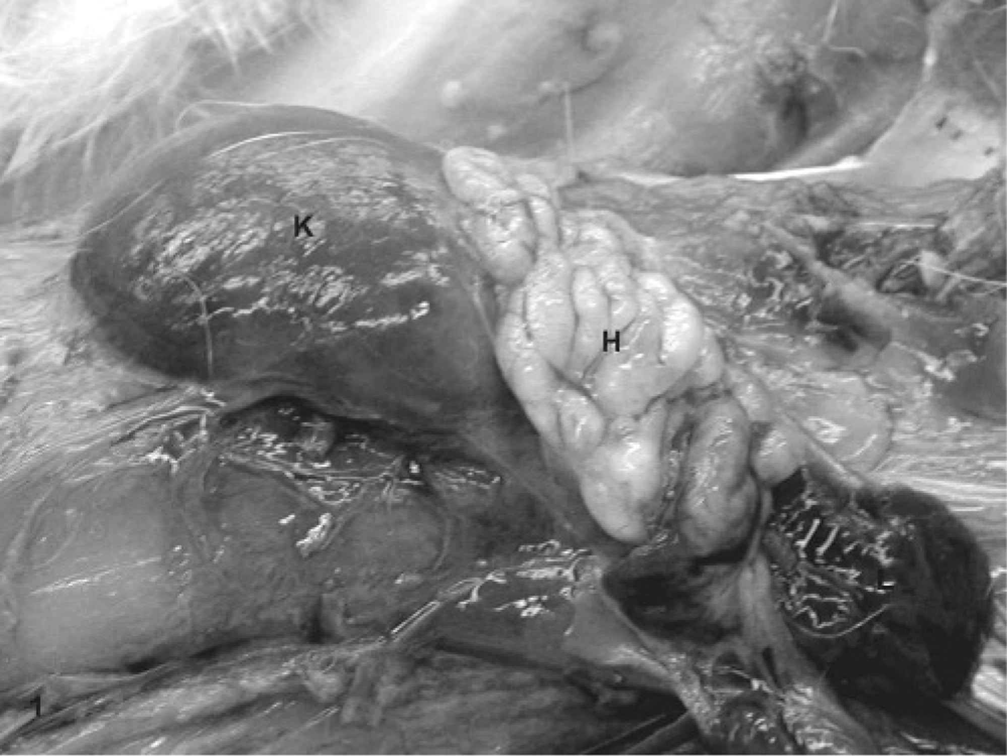
Abdomen; domestic rabbit. Gross photograph of hamartoma in situ. Kidney (K), hamartoma (H), liver (L).
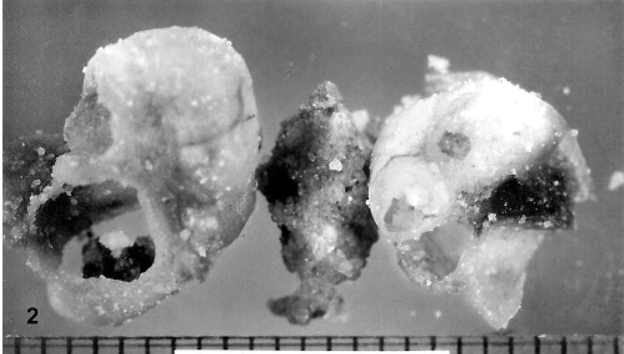
Hamartoma; domestic rabbit. Gross photograph of 1 large cholelith (center), and numerous smaller ones present in cystic spaces of hamartoma.
Normal bile content and volume were observed. The liver was of normal size and lobulation. Further evaluation of the animal, including brain, entire spinal cord and spinal column, and both hind legs, did not reveal a cause for the ataxia noted clinically. Select tissues were collected along with the central nervous system and the peripheral nerves of the hind legs and placed in formalin. After fixation, tissues were sectioned, processed, embedded in paraffin and 5-um hematoxylin and eosin (HE) sections were made. Masson's trichrome, alcian blue at pH 2.5, mucicarmine, periodic acid-Schiff (PAS), Gomori's methylene silver (GMS), acid fast, and Gram stained sections were also made of the mass. Microscopically, the mass was composed of numerous varying sized cystic structures within a loosely arranged collagenous network (Fig. 3). The cysts were lined by a simple cuboidal to columnar epithelium that in areas was partially thrown into folds and had a well-defined brush border. In a few areas, the mucosa had the appearance of columnar pseudostratification. The cells had oval nuclei that were uniform in size and located at the base of the cells; contained condensed finely stippled chromatin; on occasion, 1 small indistinct nucleolus; and moderate amounts of lightly eosinophilic cytoplasm. A few cysts had multiple small areas of the mucosa folded into gland-like structures. At the base of the gland-like structures, the mucosa transitioned from simple cuboidal/columnar to simple plump cuboidal with a mucinous-like cytoplasm (Fig. 4). These cells were positive for mucicarmine, PAS, and alcian blue 2.5. Scattered throughout the mucosa were individual goblet-like cells that also stained positive for mucicarmine, PAS, and alcian blue 2.5. In the proximal aspect of a few of the gland-like structures, as well in a few isolated areas of the mucosa, eosinophilic apical blebs were observed. Present within the lumina of many of the cysts were varying sized, irregularly shaped, amphophilic to basophilic mineralized concretions.
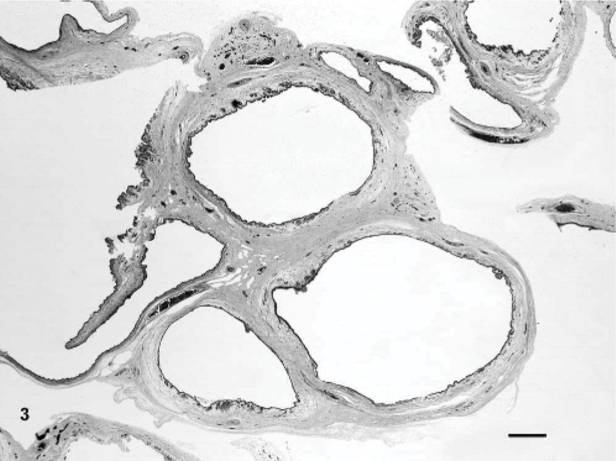
Hamartoma; domestic rabbit. Numerous cystic structures within a fibrous stroma. HE. Bar = 460 um.
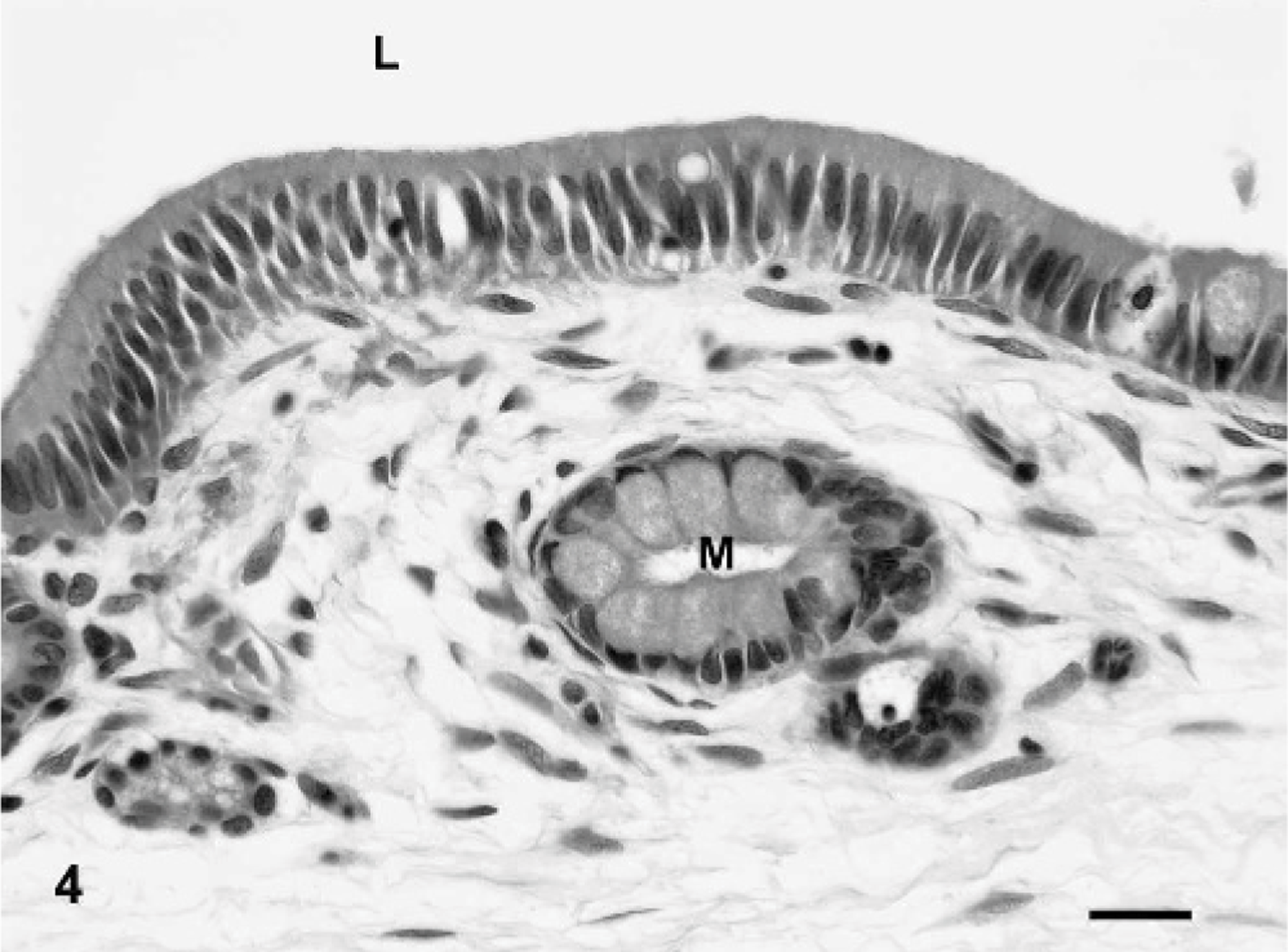
Hamartoma; domestic rabbit. Note the lumen (L) of one cyst lined by simple columnar epithelium. A mucosal crypt with mucus secreting cells (M) is present. HE. Bar = 45 um.
The large concretions removed from the cysts were analyzed by X-ray diffraction analysis by the Urolithiasis Laboratory of Houston, Texas. The concretions were composed of 100% calcium carbonate.
Microscopically, the sections of liver examined were within normal limits. The gall bladder was not examined microscopically. Microscopic evaluation of the spinal cord demonstrated a few dilated myelin sheaths. No other abnormalities were noted to explain the ataxia.
Hamartomas are rare tumors in animals and humans. The majority of reports in animals are centered around vascular hamartomas; 3, 21, 25 however, hamartomas of abomasal 32 and dermal smooth muscle, 12 retinal astrocytes, 16 myocardium, 13 pulmonary alveoli, 27 and ovarian interstitial cells 14 have been reported. Hamartomas of the liver and biliary system in the veterinary literature are extremely rare, with only 1 case of vascular hamartoma in a dog, 15 8 cases of vascular hamartoma in cattle, 9 1 case of mixed hamartoma in an equine fetus, 20 and 1 case of bile-duct hamartoma in a calf. 5 In humans, hamartomas of the liver and biliary system are primarily designated as either biliary-duct hamartomas, also known as von Meyenburg complexes, or mesenchymal hamartomas. Human biliary hamartomas are multifocal discrete masses of biliary ducts within the hepatic parenchyma. Mesenchymal hamartomas are solitary, usually small, yet can be large, 17 intrahepatic masses composed of immature mesenchymal connective tissue interspersed among biliary ducts and tubules, with focal aggregates of hepatocytes. In this rabbit case, the mass was solitary, attached to the hepatic capsule, without involvement of the hepatic parenchyma, and was composed of cystic structures within a mature collagenous stroma. A single case of solitary intrahepatic bile duct hamartoma of the liver has been diagnosed in a human. 8 The mass in this rabbit had similar microscopic appearances to the human case; however, it was extrahepatic, and, in areas, the cysts had gland-like structures similar to mucosal crypts seen in the gall bladder. Also, the mass had areas of mucus-producing cells indicative of the extrahepatic biliary system. Two cases of biliary adenofibroma in man 29, 30 closely resembled the rabbit tumor. Similar features included cystic structures lined by simple cuboidal epithelium, intraluminal concretions, and a fibrous stroma. However, the adenofibromas also had mitotic figures of the fibrous stroma, as well as the glandular structures; the concretions were microscopic; and the masses had dense focal tubuloglandular architecture within the hepatic parenchyma. The rabbit tumor was composed solely of cystic structures, many of which contained grossly visible concretions; mitotic figures were not observed; and the hepatic parenchyma was not involved.
The anatomic outline of the extrahepatic biliary tract in rabbits varies widely, and is considered normal. 10, 18 The gall bladder has been described as being absent, bilobed, and duplicated, as well as variable in size in rabbits. 22 These variations indicate differences among rabbits in the developmental stages of the extrahepatic biliary tract. This may possibly explain the cause of the hamartoma seen in this rabbit and provide stronger evidence for the hamartoma being of extrahepatic biliary origin. The gall bladder has mucus-secreting cells that are not observed in the intrahepatic biliary tree. Similar mucus-secreting cells were observed in the hamartoma. A single human case of mucinous hamartoma of the extrahepatic biliary duct system with nonmucinous intrahepatic biliary hamartomas has been described 4 and demonstrates the difference in cellular phenotypes in the biliary system. Bile-duct hamartomas are hypothesized to arise from ductal-plate malformations of the peripheral interlobular bile ducts in the late phase of bile-duct embryogenesis. 28 This leads to the development of multiple intrahepatic hamartomas, which were not observed in this case. One interesting note is that some biliary hamartomas and bile cysts can develop from hepatic ischemia. 19 This rabbit had the genetic propensity to develop atherosclerosis, which can cause ischemic lesions. However, atherosclerotic lesions were not observed in this animal.
Bile contains 4 calciums anions that are readily precipitated by calcium to form choleliths: bilirubinate, palmitate, carbonate, and phosphate. 6 The concretions within the cysts were identified as choleliths composed solely of calcium carbonate. Cholelithiasis is a rare natural disease in rabbits; however, it can be induced by alterations in diet. 6 The choleliths are thought to have developed because of stasis of mucosal secretions. The cysts were considered to be close-ended sacs, thus prohibiting release of secretions, and they were devoid of bile. Luminal communications between hamartomas and the normal biliary ductal system are controversial. Two different entities are thought to exist; one consisting of hamartomas connected to draining bile ducts and the second having no connections. 31 Extrahepatic biliary mucosal epithelium has the ability to secrete fluid that has an ionic composition similar to that of serum. 7 Normally, when bile enters the gall bladder, the acidity of bile causes an increase in solubility of calcium salts, most namely carbonate, bilirubinate, phospate, and long-chain fatty acids. 23 However, with the depletion of bile, because of the lack of communication between the hamartoma and the hepatic biliary system, the secretions present are less acidic, decreasing the solubility of calcium. This alteration in pH along with secretory stasis could lead to calcium precipitation, which would include the formation of calcium salts, including calcium carbonate. A rare condition known as limy bile exists in humans where a pasty white material composed solely of calcium carbonate accumulates in the gall bladder and common bile ducts. 1 A nidus was not observed in any of the stones analyzed from this rabbit. The lack of a nidus, along with negative stains for acid fast organisms, fungal elements (GMS), and bacteria (Gram's), and no evidence of inflammation, strengthen the likelihood that the concretions formed by stasis of secretions. The gallbladder secretes a mucoprotein that is a major component of human choleliths. 24 This protein has pronucleation activity, 11 potentially producing the core for all stones formed in the hamartoma.
Given the extrahepatic location, the presence of mucinous-secreting cells, the formation of calcium carbonate concretions, and the paucity of bile, the mass observed in this rabbit was identified as a noncommunicating extrahepatic biliary hamartoma that contained intraluminal calcium carbonate choleliths. This is the first known hamartoma identified in the rabbit, and the first biliary hamartoma with choleliths in animals.
Footnotes
Acknowledgements
The author thanks Jorge Chavez and Annie Merriweather for technical assistance, and Dr. Mark Bryant for his critical review of the manuscript. This research was supported in part by the Intramural Research Program of the National Institutes of Health, Office of Resource Services.