
Abstract
Medullary sponge kidney was diagnosed in a 10-year-old male Shih Tzu dog with a long history of hyposthenuria, but with no other findings indicating renal failure or hormonal aberration. At the dog's death from heart failure, an autopsy was performed. On gross morphology, bilateral kidneys were normal size and had many cysts ranging from the corticomedullary junction to renal papillae. Histopathologic findings showed that almost all of the cysts were lined by monolayered or multilayered and columnar or cuboidal epithelium with chilium similar to epididymis. immunohistochemically, all of these cells were strongly positive for AE1/AE3 and negative for vimentin. Many of these cells were positive for cytokeratin 7 (CK7), and only a few cells were positive for desmin. The results of staining are the same as those for epithelium of the collecting duct of normal canine kidney. This is the first report of this pathologic entity in the canine kidney.
Medullary sponge kidney is characterized in humans by dilation of the collecting ducts of the renal pyramids in association with multiple, often communicating papillary cysts. 3, 15 Although occurrence among siblings or within families has been reported, 2, 3, 7, 8, 15 the usual incidence is considered to be sporadic. 3, 15 Many instances of this disease are detected incidentally at necropsy or x-ray examination. The patient is usually asymptomatic but often presents with a decrease in urinary concentrating ability. 3, 15
In dogs, morphologic abnormalities of the kidney were reported in many breeds, such as the Golden Retriever, Labrador Retriever, Boxer, and Finish Harrier, and Shih Tzu. 1, 4– 6, 9– 14 But all of these abnormalities were categorized as congenital renal dysplasia in which the abnormality appeared mainly in the cortex or cortex and medulla. Picut and Lewis recommended the histopathologic criteria for the diagnosis of canine renal dysplasia 6, 13 : asynchronous differentiation of nephrons, persistent mesenchyme, persistent metanephric ducts, atypical tubular epithelium, and dysontogenic metaplasia. A dog presenting with such dysplasia symptoms usually has renal insufficiency from an early age. 1, 4– 6, 9– 14 The present report is an immunohistochemic and histopathologic study of medullary sponge kidney in a dog.
A 10-year-old male Shih Tzu dog had manifested hyposthenuria (urine specific gravity, <1.015) from 1 year of age, but he had no other findings that indicated renal failure or hormonal aberration. The dog died of heart failure at 10 years of age. Necropsy was performed. Tissues were fixed in 10% buffered formalin and embedded in paraffin by the routine procedure. Paraffin sections were cut 4-μm thick and stained with hematoxylin and eosin (HE) and periodic acid–Schiff (PAS) for histopathologic examination.
Macroscopically, both kidneys were normal size. On the cut surface, many cysts were found within the medulla of both kidneys like a sponge (Fig. 1). Dilation of the renal pelvis was also observed (Fig. 1). The right kidney had partial cicatrization in the cortex. In other organs, observations indicated hypertrophy of the left heart and dilated right heart, mitral valve endocardiosis, and mucosal edema of the urinary bladder.
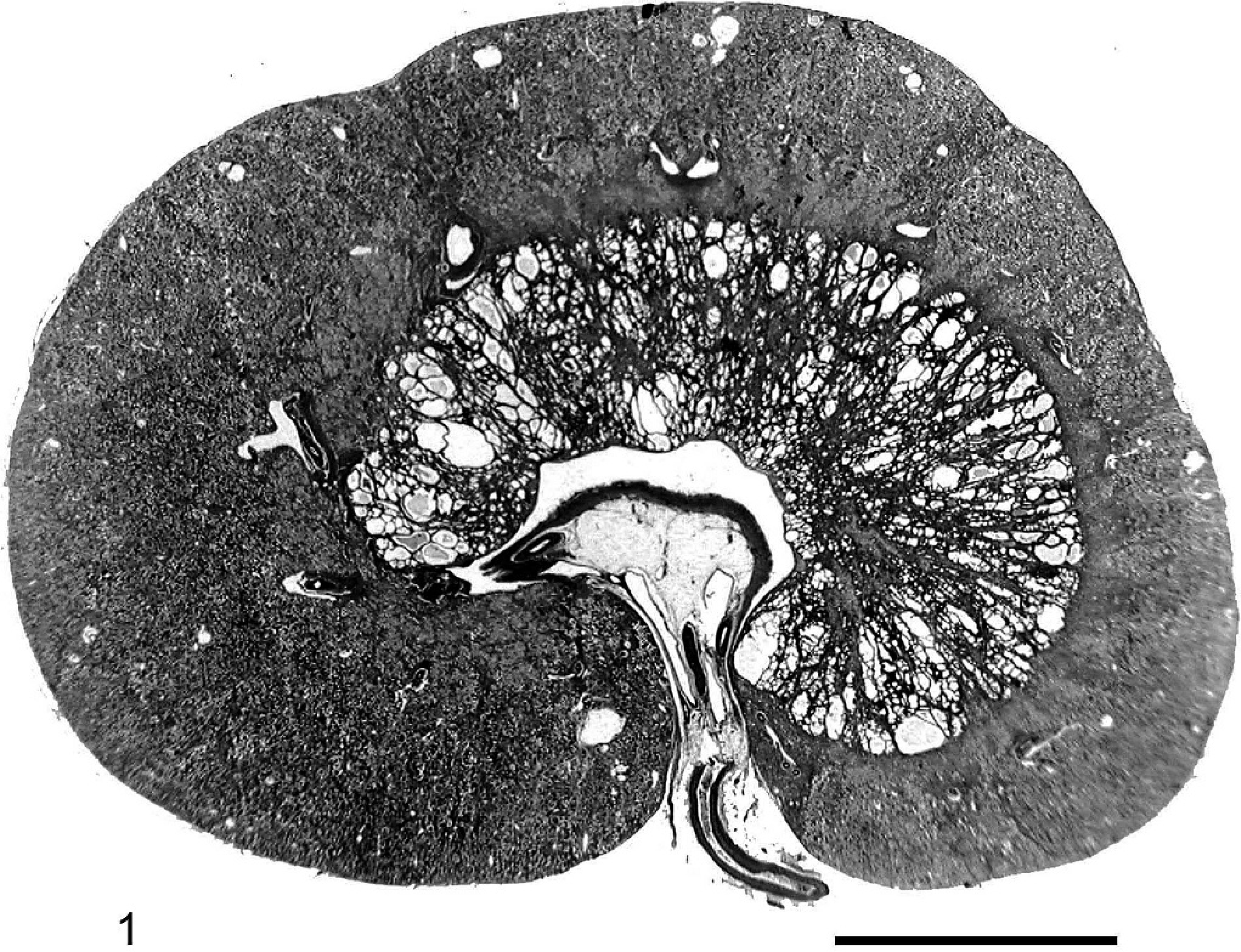
Kidney; 10-year-old dog. The cut surface shows various sized cysts from the inner to outer medulla. Dilation of renal pervis. HE. Bar = 1 cm.
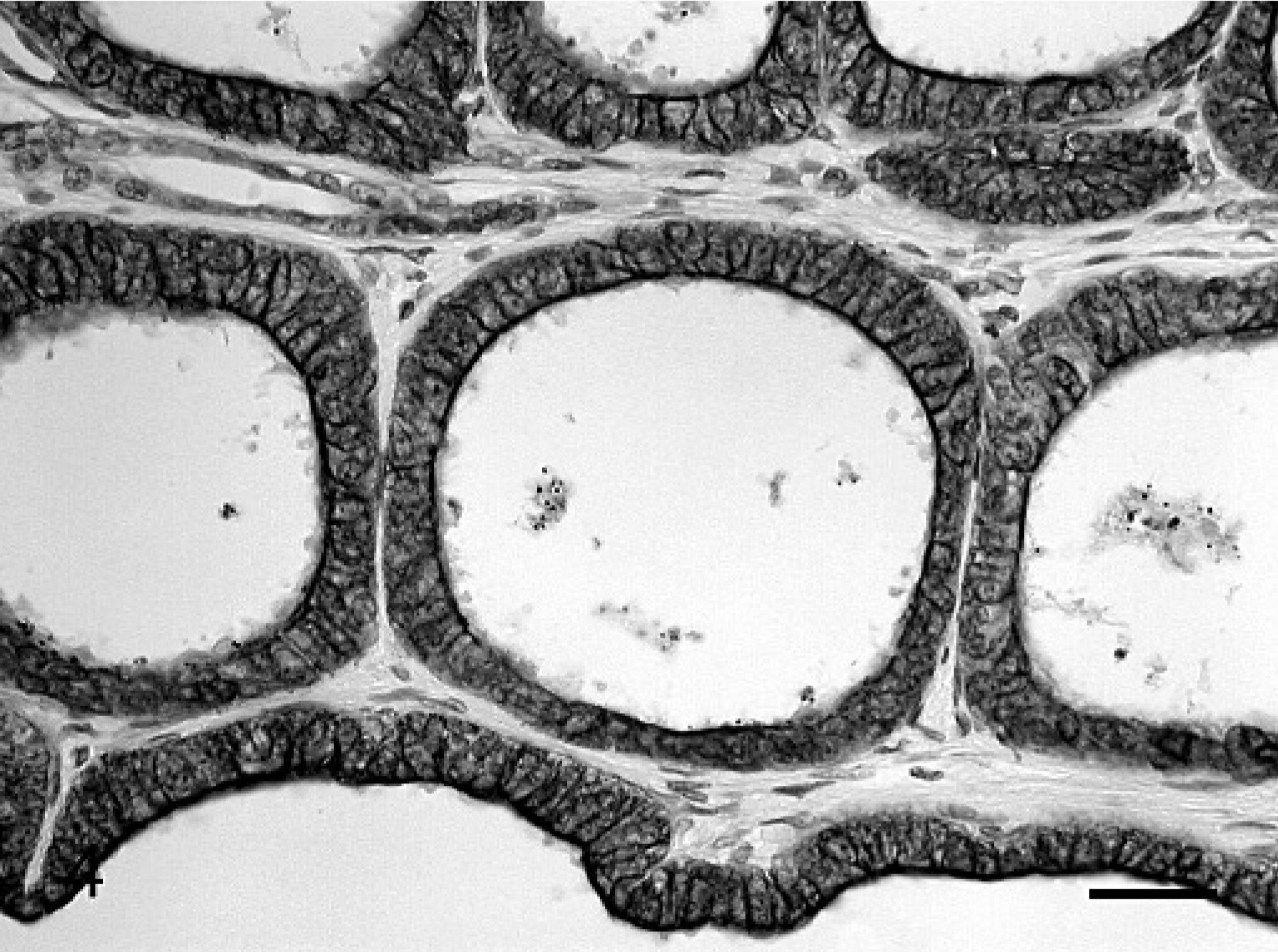
Histologically, almost all of these cysts were lined by monolayered or multilayered and columnar or cuboidal epithelium (Fig. 2). Large cysts seemed to be formed by the fusion of smaller cysts (Fig. 2). The lining cells of these cysts had round to oval nuclei and microvilli (Fig. 3). These structures were similar to canine epididymis. PAS-positive droplets and secretion were found in the lumens. Mitoses and atypia were not detected. Collagenous fiber increased in the medulla. In limited areas, dilation of glomeruli, glomerulosclerosis, and urolithiasis were also found.
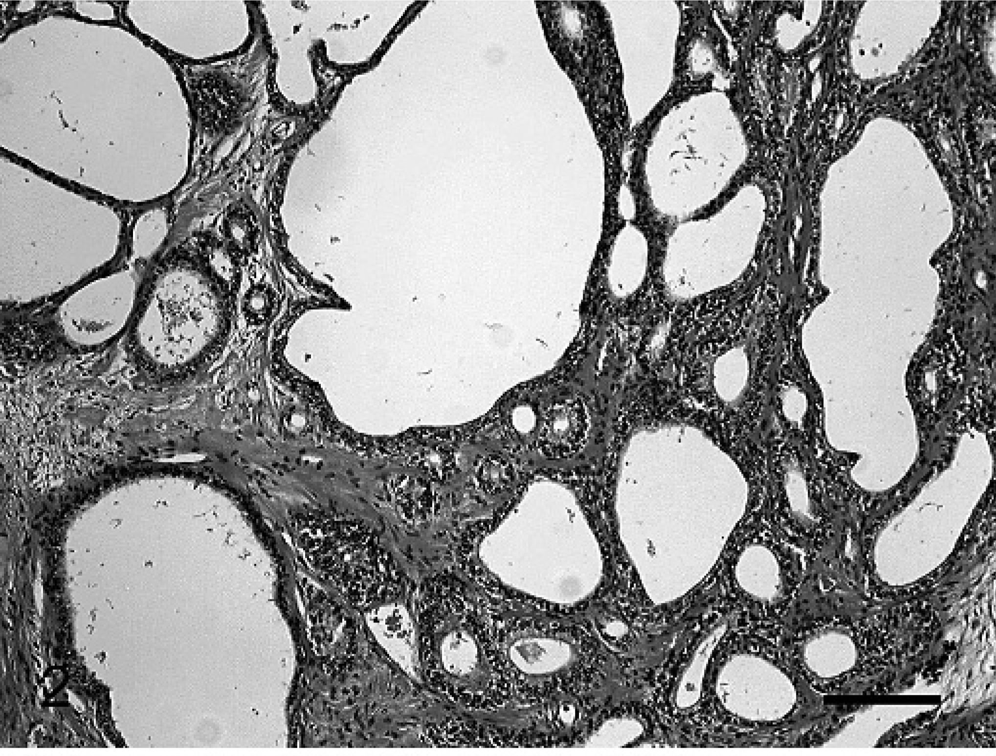
Kidney; 10-year-old dog. Cysts were lined by monolayered or multilayered cylindrical epithelium. Large cysts seemed to be formed by the fusion of smaller cysts. HE. Bar = 50 μm.
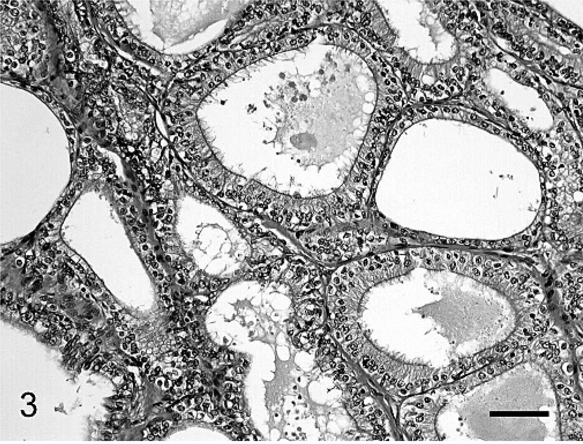
Kidney; 10-year-old dog. The lining cells of these cysts have round to oval nuclei and microvilli. These findings are similar to epididymis. HE. Bar = 30 μm.
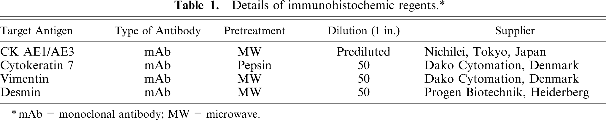
Contiguous sections were immunohistochemically examined by the avidin-biotin-peroxidase complex (ABC) procedure (Vectastain Elite ABC Kit; Vector Laboratories, Burlingame, CA). Primary antibodies and pretreatments used in this study are listed in Table 1. The blocking of endogenous peroxidase was done with H2O2 0.3% in methanol for 10 minutes. All sections were incubated with primary antibody at 4°C for 16 hours, with biotinated secondary antibody for 30 minutes at room temperature, and with avidin-peroxidase conjugate for 30 minutes. Staining was developed in a 0.05% 3,3′-diaminobenzidine solution. Kidneys and testes of 3 adult dogs were used as controls.
Details of immunohistochemic regents.∗
mAb = monoclonal antibody; MW = microwave.
All epithelial cells of the medullary cysts were stained with AE1/AE3 (Fig. 4, Table 2) but not stained with vimentin. CK7–positive cells were partially observed (Fig. 5, Table 2). Only a few desmin-positive cells were observed (Table 2). Comparing the present results with normal controls, the immunohistochemic appearance of these cysts was the same as the collecting duct.
Kidney; 10-year-old dog. All epithelial cells of the medullary cysts are stained with AE1/AE3. ABC complex method, Mayer's hematoxylin counterstain. Bar = 30 μm.
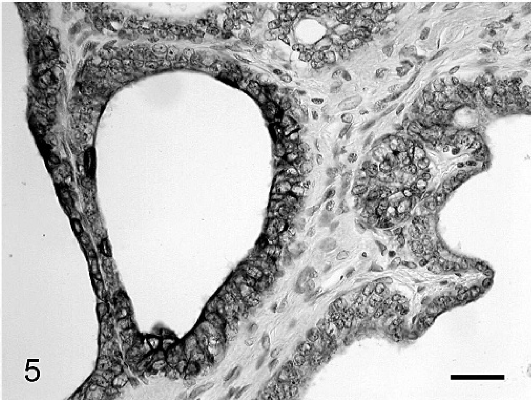
Kidney; 10-year-old dog. Epithelial cells of the medullary cysts are partially stained with CK7. ABC complex method, Mayer's hematoxylin. Bar = 30 μm.
Immunohistochemic labeling results.∗
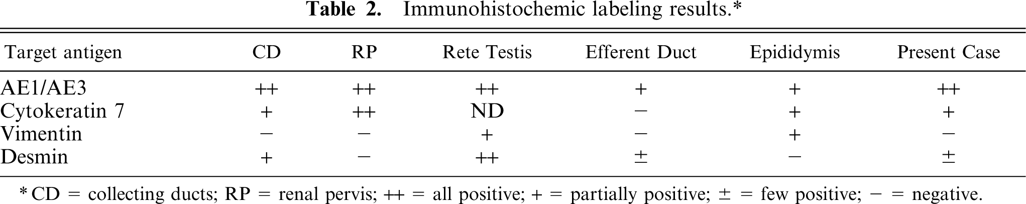
CD = collecting ducts; RP = renal pervis; ++ = all positive; + = partially positive; ± = few positive; − = negative.
The present case was diagnosed as medullary sponge kidney on the basis of the location of the cysts and symptoms. Ducts that resembled the epididymis seemed to be metanephric. The existence of this structure is one histopathologic criterion for the diagnosis of canine renal dysplasia. However, asynchronous differentiation of nephrons, which is the most common finding in canine renal dysplasia, 13 was not seen. Further, there was little difference in the cyst situation between canine renal dysplasia and the present case. The former cysts were found mainly in the outer medulla, 13 whereas the latter were widespread from the outer to the inner medulla. Besides the difference in the situation of the cysts, the presence or absence of morphologic anomaly in the cortex may decide the clinical outcome. The present case differs from previously reported canine renal dysplasia in terms of histologic and clinic findings. Histopathologically, cyst epithelium indicates the shape of the metanephric duct monolayered or multilayered and columnar or cuboidal epithelium, in partial agreement with the description of medullary sponge kidney in humans. 3, 15 Additionally, the opinion that medullary sponge kidney in humans occurs because of congenital morphologic anomaly 3 also agrees with the hypothesis that the present lesion occurred because of a remnant of the metanephric duct, either directly or indirectly. Immunohistochemic examinations in the present case were not similar to those for normal epididymis but rather like those for the collecting duct. A few cyst cells were positive for desmin. Desmin immunoreactivity was seen in part of the tubule cell between the distal tubule to the collecting duct in normal kidney. These results indicated that desmin-positive cells may have been derived from this area.
Renal dysplasia may be defined as a disorganized development of the renal parenchyma that is caused by defective interaction of ureteric bud and metanephric blastema. 2, 3, 13 The present case had a morphologic aberration of the collecting duct, but no aberration in the cortex, possibly indicating that these abnormal ducts induct normal cortical tubules. This dog presented only hyposthenuria without symptoms of chronic renal failure and seemed to reflect the location of the cyst mainly in the medulla-collecting duct rather than in the cortex.
To our knowledge, this is the first description of medullary sponge kidney in dog. There is no information about genetic background or developmental factors, but the present lesion may be congenital and occur in association with a remnant of the metanephric duct that is one of the most important findings of renal dysplasia. We should recognize this type of dysplasia-medullary sponge kidney in a dog as a new category of dysplasia in canine renal pathology, and this disease must be taken into consideration, especially for a dog presenting with hyposthenuria.
Footnotes
Acknowledgement
The work was supported by a grant-in-aid for the High Technological Research Center (Rakuno Gakuen University) from the Ministry of Education, Science, Sports, and Culture of Japan.