
Abstract
Necropsy of 2 white-tailed deer fawns who died acutely revealed diarrhea and melena in case No. 1 and no gross changes in case No. 2. Histologically, the livers of both deer displayed multifocal coagulative necrosis, with infiltrations of neutrophils, macrophages, and lymphocytes. By Warthin-Starry staining, bundles of filamentous bacteria were identified within hepatocytes at the periphery of the necrotic foci in case No. 1. There was multifocal myocardiocyte necrosis in case No. 1 and multifocal lymphoid necrosis of the Peyer's patches in case No. 2. Clostridium piliforme 16S ribosomal ribonucleic acid gene was detected in both livers by polymerase chain reaction (PCR) with C. piliforme-specific primers. The liver copper levels in both cases were normal to slightly elevated. The kidney copper level in case No. 2 was elevated. This represents the first published cases of Tyzzer's disease in deer, a novel use of PCR for the diagnosis of C piliforme infection, and a possible association between copper toxicosis and Tyzzer's disease.
Keywords
Tyzzer's disease is an enterohepatic infection caused by Clostridium piliforme, a large, intracellular, gram-variable, spore forming, anaerobic, filamentous, rod-shaped bacterium. 2, 4, 6 Affected animals are typically neonates and are often found dead in the absence of clinical disease. 3, 4 Common clinical signs, when present, may include diarrhea, depression, and anorexia, usually of short duration before resulting in death. 3, 4, 15 Typical postmortem findings include multiple 1- to 3-mm gray-white foci on the capsular and cut surfaces of the liver. 5, 7, 14, 15 Pale streaks through the myocardium and multifocal mucosal ulceration and hemorrhage in the ileum, the cecum, or the colon may be present in some species. 5, 15 Histologically, there is multifocal coagulative necrosis of the liver, with variable enteritis or myocardial necrosis. 5– 7, 14, 15 The proposed mode of transmission is fecal-oral, with the organism entering the portal circulation via the gastrointestinal tract and, subsequently, colonizing the liver. 4, 5 Tyzzer's disease has been reported in several laboratory, domestic, and wild species, although the disease has rarely been reported in ruminants. 2, 6, 7, 14, 15 This report represents a rare occurrence of Tyzzer's disease in a ruminant species and, to the best of the authors' knowledge, the first published cases of Tyzzer's disease in a cervid species. Furthermore, this represents a novel use of polymerase chain reaction (PCR) by using fresh-frozen tissue for the diagnosis of Tyzzer's disease and may represent an association between copper toxicosis and susceptibility to infection with C. piliforme.
A 5-day-old female (case No. 1) farm-raised white-tailed deer fawn (Odocoileus virginianus) developed acute lethargy, anorexia, and diarrhea. Despite supportive treatment, the fawn rapidly died and was submitted for necropsy. On postmortem examination, the perineal hair-coat was matted with wet feces. The ileum, the cecum, and the colon contained pasty black material consistent with melena. Portions of heart, lung, liver, spleen, kidney, and intestine collected for histologic examination were fixed in 10% neutral buffered formalin and processed routinely, embedded in paraffin, sectioned at 5 μm, and stained by using hematoxylin and eosin (HE). Additional paraffin-embedded sections of liver and kidney were later sectioned at 5 μm and stained by using Lindquist's rhodanine method, while sections of liver, heart, and intestine were stained by using the Warthin-Starry silver method. 9 A portion of the liver was frozen at -25°C and submitted for trace mineral and vitamin E evaluation, and PCR assay for C. piliforme. Additional tissues were submitted for bacteriology, electron microscopy for enteric viruses, and parasitology.
Six weeks later, a 4-day-old male (case No. 2) white-tailed deer fawn from the same farm died suddenly and was submitted for necropsy. No significant gross changes were detected on the postmortem examination. Portions of brain, heart, lung, liver, spleen, kidney, intestine, and skeletal muscle collected for histologic examination were fixed and processed as for case No. 1. A portion each of liver and kidney were frozen at -25°C. The frozen kidney was submitted for trace mineral evaluation, and the frozen liver was submitted for trace mineral and vitamin E evaluation, and for PCR assay for C. piliforme. Additional tissues were submitted for bacteriology, electron microscopy for enteric viruses, and parasitology.
Histologically, the livers of both deer were characterized by multifocal randomly distributed coagulative hepatocellular necrosis, with infiltrations of neutrophils, macrophages, and lymphocytes in the necrotic foci. Interlacing bundles of filamentous bacteria were identified within the cytoplasm of hepatocytes at the periphery of the necrotic foci by Warthin-Starry staining of the liver in case No. 1 but not in case No. 2 (Fig. 1). There was multifocal myocardiocyte necrosis characterized by cytoplasmic hypereosinophilia, myocardiofiber fragmentation, loss of cellular detail, and mineralization in case No. 1. The necrotic foci were infiltrated by small numbers of neutrophils, macrophages, and lymphocytes. No significant microscopic changes were detected in lung, spleen, kidney, or intestine of case No. 1. Although the gross postmortem findings in case No. 1 were suggestive of melena because of hemorrhagic enteritis, as seen in some rodents and rabbits, autolysis precluded more critical evaluation of the intestine in this case. There was multifocal lymphoid necrosis of the Peyer's patches in case No. 2. No significant microscopic changes were detected in brain, heart, lung, spleen, kidney, or skeletal muscle of case No. 2. Warthin-Starry staining was applied to sections of heart and intestine of both cases. Of these tissues, only within enterocytes of case No. 1 were bundles of intracytoplasmic filamentous bacteria detected; organisms were similar to those seen in the liver of case No. 1 (Fig. 2). Bacteriology, electron microscopy, and parasitology yielded negative results in both cases.
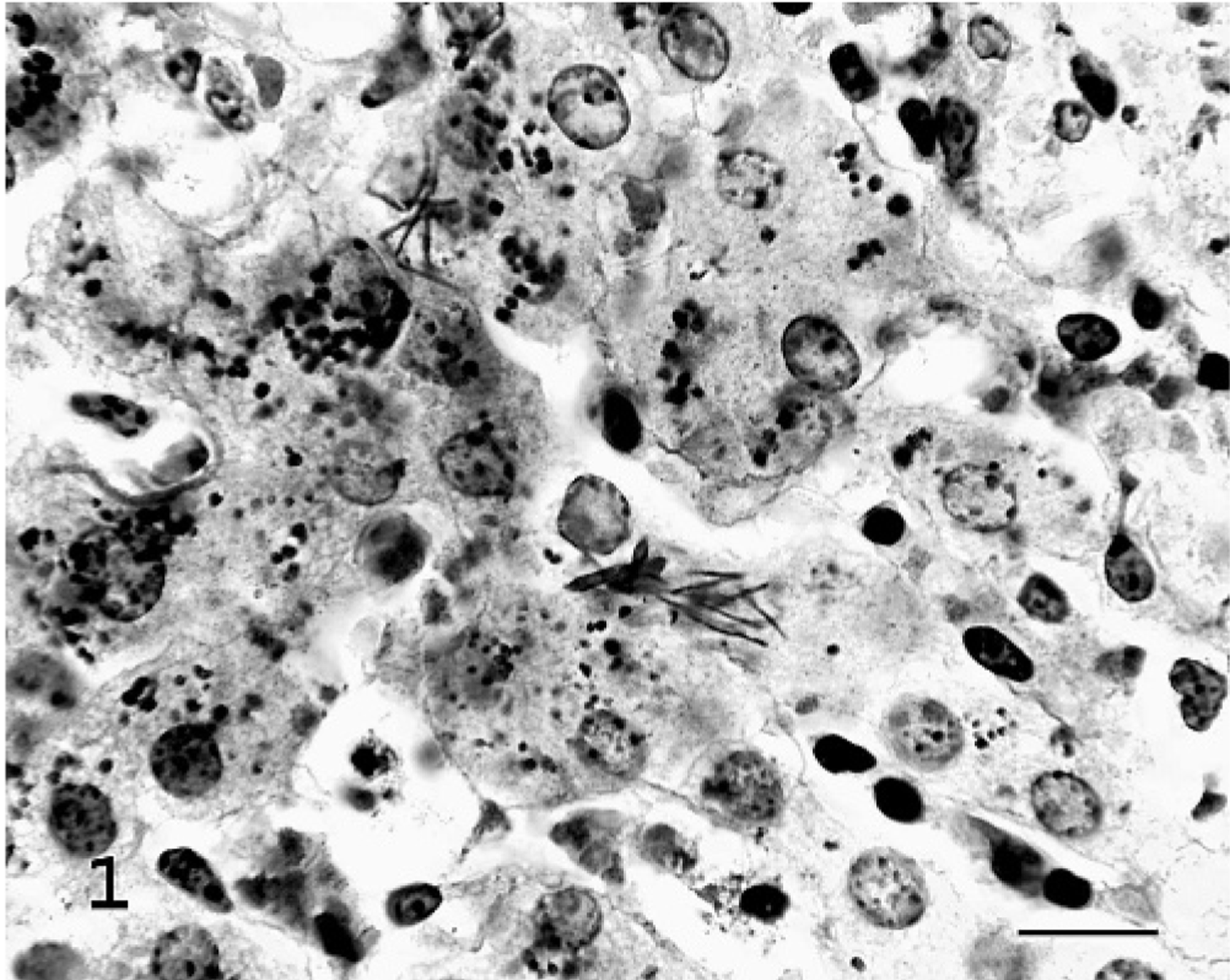
Liver; fawn No. 1. Interlacing bundles of filamentous bacteria within the cytoplasm of hepatocytes at the margin of a necrotic focus. Warthin-Starry method. Bar = 30 μm.
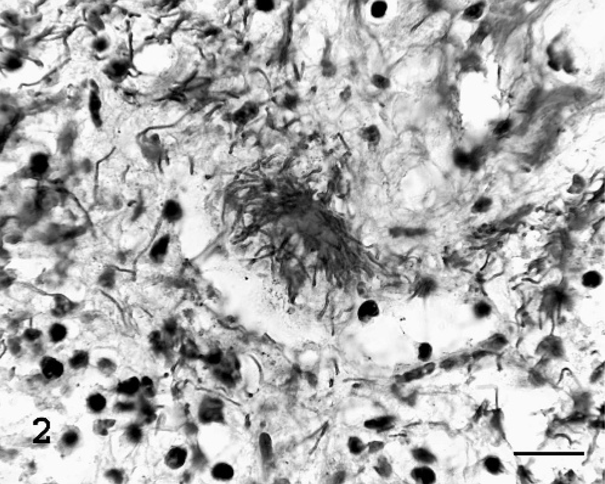
Jejunum; fawn No. 1. Interlacing bundles of filamentous bacteria within the cytoplasm of enterocytes. Warthin-Starry method. Bar = 30 μm.
Chromosomal deoxyribonucleic acid (DNA) from frozen liver samples from case No. 1, case No. 2, and a negative control deer liver was isolated by using a High Pure PCR Template Preparation Kit, following the protocol of the supplier (Roche Applied Science, Mannheim, Germany). The negative control liver was obtained from a white-tailed deer with no gross, microscopic, or bacteriologic evidence of disease. Purified C. piliforme DNA was used for a positive control. PCR detection of C. piliforme was performed in a 25-μl reaction mixture that contained 200 ng of each DNA template, 500 nM each of forward and reverse primers, and a PuReTaq Ready-To-Go PCR bead (Amersham Biosciences, Buckinghamshire, England). The sequences of the 16S ribosomal ribonucleic acid (rRNA) gene-based primers were as follows: forward primer, 5′-ACC ATT GAC AGC CTA CGT AA-3′, reverse primer, 5′-GTC TCG CTT CAC TTT GTT GTA-3′. 13 Thermocycling conditions were as follows: 35 cycles of 94°C for 1 minute, 53°C for 1 minute, and 72°C for 1 minute, followed by incubation at 72°C for 7 minutes. PCR products were electrophoresed on a 2.5% agarose gel and were visualized by staining with ethidium bromide.
PCR products of the expected size (∼270 bp) were extracted from the agarose gel by using a QIAquick Gel Extraction Kit according to the instruction of the supplier (Qiagen, Valencia, California). Subsequently, the eluted DNA solution was dried and resuspended in sterile distilled water. Twenty ng of the purified PCR products were sequenced by using the ABI Prism 310 sequencer (Applied Biosystems, Foster City, California). For the samples that did not produce detectable C. piliforme–specific product in the first-round PCR, the agarose gel bands that corresponded to the PCR product amplified from the C. piliforme DNA control were excised, and the DNA was extracted from these bands by using a QIAquick Gel Extraction Kit (Qiagen). The resulting DNA samples were dissolved in 10 μl of sterile distilled water, 2 μl of which was used to perform a second-round (nested) PCR as described above.
Multiple amplicons were produced by a first-round PCR from all 3 liver samples (Fig. 3A). Only liver DNA extracted from case No. 1 produced an abundant amplicon of a size similar to that of the control C. piliforme DNA template (∼270 bp). This PCR product and a slightly larger product were purified from the gel and were sequenced. The nucleotide sequence of the 270 bp product had 97% sequence similarity to the corresponding region of the C. piliforme 16S rRNA gene deposited in the GenBank database by various sources. The larger PCR product did not produce a readable sequence, indicating that the purified DNA template contained more than 1 PCR product.
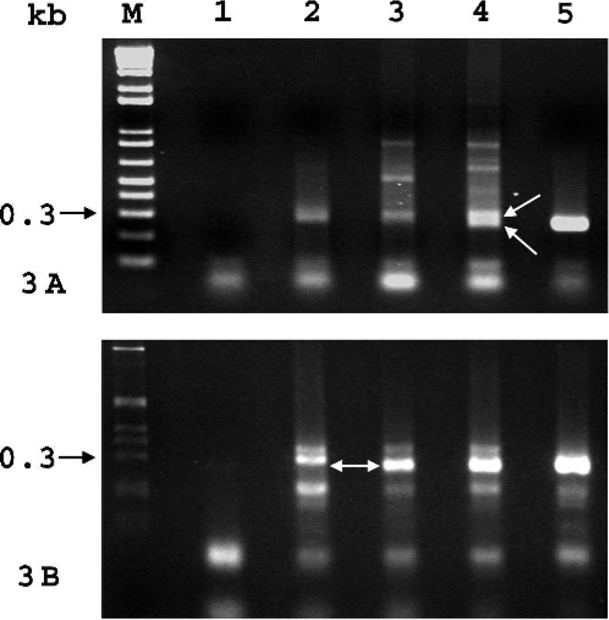
16s rRNA gene-based PCR detection of C. piliforme.
To investigate whether liver samples yielding negative first-round PCR results for C. piliforme were because of a low number of organisms present in the samples, the DNA purified from the agarose gel bands corresponding to the size of the C. piliforme–positive control product were subjected to a second-round PCR with the same primers (Fig. 3B). A 270-bp DNA fragment was amplified from case No. 2, indicating that case No. 2 was also infected with C. piliforme but at a relatively low level. In contrast, a similar PCR product was not produced from the liver of an unaffected white-tailed deer used as a negative control.
Frozen liver samples from each case and frozen kidney from case No. 2 were submitted for nutritional and trace mineral analysis by inductively coupled plasma–mass spectrometry (ICP-MS). After overnight digestion with 70% nitric acid at 70°C, sample digests were diluted with 18 MΩ cm double-deionized water to a final volume of 10 ml after addition of 5 internal standards. Before analysis with an Elan 6100 ICP-MS (Perkin Elmer, Inc., Wellesley, Massachusetts), a standard curve was generated and a standard reference material (1577b bovine liver), with certified mineral values from the National Institute of Standards and Technology, was analyzed. All final concentrations, including arsenic, cadmium, calcium, cobalt, copper, iron, lead, magnesium, manganese, molybdenum, phosphorus, selenium, and zinc, were reported in parts per million on a wet-weight basis.
In addition, vitamin E levels were evaluated on the frozen liver samples of each case by liquid chromatography and fluorescence spectrometry. Proteins were precipitated with ethanol that contained 30 ppm butylated hydroxytoluene and delta tocopherol (internal standard), followed by extraction with hexane. Samples were analyzed by using a Thermo SpectraSystem P4000 high-pressure liquid-chromatography–fluorescence-spectrometry system (Thermo Electron Corp., Waltham, Massachusetts) with a 150 × 3-mm 5-μm Betasil C18 reverse-phase column (Thermo Electron). A control serum and a serum spike were included with each run. Four standards were used to construct a calibration curve by using delta tocopherol. Vitamin E as alpha-tocopherol concentrations were reported in parts per million on a wet-weight basis.
The liver copper levels were 507 and 600 ppm on a wet-weight basis for case Nos. 1 and 2, respectively. In addition, kidney copper for case No. 2 was 222 ppm on a wet-weight basis. Kidney from case No. 1 was not available for testing. Reported normal liver copper values for neonatal cervids range from 100 to 1480 ppm on a wet-weight basis (after conversion, assuming 20% liver dry matter). 8, 11, 12 Reported normal kidney copper values are 7 to 20 ppm on a wet-weight basis, with little reported difference in fetal levels versus adult levels. 1, 11 Sections of liver from both case No. 1 and case No. 2 displayed moderate staining for copper within hepatocytes by the rhodanine method. The kidney from case No. 1 showed no rhodanine staining for copper, whereas that of case No. 2 showed moderate staining within the renal tubular epithelial cells. Although these liver copper levels appear high for most domestic species, it is possible that they are within normal limits for neonatal deer, as suggested in several published reports. 8, 11, 12 The elevated kidney copper and positive copper staining in the kidney, however, are highly suggestive of copper toxicosis. It is well documented that newly regenerating hepatocytes retain very little copper and that total liver copper diminishes as copper escapes from necrotic hepatocytes. 10 Thus, in copper toxicosis, the liver copper level may be nearly normal, while copper levels in other tissues, such as the kidneys, are elevated. 10 All other mineral levels and vitamin E levels in both cases were within reported normal ranges for neonatal white-tailed deer.
The diagnosis of Tyzzer's disease is typically based on histologic findings by staining with HE and Warthin-Starry methods, in addition to suggestive clinical history or gross postmortem findings. This report represents a novel use of nested PCR by using fresh-frozen tissues for confirmation of C. piliforme infection. In addition, this may represent an association between copper toxicosis and Tyzzer's disease. The authors suggest that hepatocellular injury because of excessive lysosomal copper accumulation may have led to hepatocellular necrosis and release of copper into the bloodstream. This copper may have been absorbed by renal tubular epithelial cells and caused elevated kidney copper levels. In the presence of C. piliforme passing through the gastrointestinal tract, the hepatocellular damage may have provided a nidus for infection with C. piliforme. This is consistent with the common association of Tyzzer's disease with a primary stressor, as this disease is frequently believed to occur secondary to immunocompromise from nutritional, environmental, or infectious causes, or from treatment with corticosteroids. 2, 3, 7 Finally, this represents the first report of Tyzzer's disease in a cervid species. While the histologic changes in these cases are similar to those of other species, the gross postmortem changes are variable. Deer may develop no gross changes in the liver with C. piliforme infection and may instead develop gross changes suggestive of hemorrhagic typhlitis or colitis, as seen in some rabbits and rodents.
Footnotes
Acknowledgements
We thank Dr. Robert Livingston (University of Missouri—Columbia) for providing C. piliforme DNA and the 16S rRNA gene-based primers; Dr. Robert Van Saun (The Pennsylvania State University); Carol Buckley, and Margie Cummings (University of Pennsylvania) for toxicology assays; and Vivian Ng (Massachusetts Institute of Technology) for molecular assays.