
Abstract
Systemic granulomatous disease involving the spleen, heart, lymph nodes, omentum, liver, kidney, lung, mediastinum, and salivary glands developed in an 8-year-old Rottweiler. The dog also had sialometaplasia of both submandibular salivary glands. Bartonella henselae and B. vinsonii subsp. berkhoffii DNA was amplified from the salivary gland by polymerase chain reaction analysis. Bartonellae may be the cause of this systemic disease, but to the authors' knowledge, involvement of omentum, mediastinum, and salivary glands has not previously been reported in association with Bartonella infection. Bartonellae should be considered potential causes of sialometaplasia.
Bartonella spp. are gram-negative, intracellular, bacterial rods that induce disease in a wide variety of animal species. The disease is transmitted through fleas and ticks. The bacteria can be found in many healthy animals that may act as carriers. B. vinsonii subsp. berkhoffii and B. clarridgeiae are the species most often isolated from dogs, and they have induced uveitis, endocarditis, rhinitis, and granulomatous disease in liver, lymph nodes, heart, and spleen. 1, 3– 6
Sialometaplasia is a rare disease of unknown cause in dogs and cats. The disease in dogs usually affects one submandibular salivary gland and induces infarction, inflammation, and squamous metaplasia of the glandular epithelium and ducts. 2
An 8-year-old, spayed female Rottweiler presented with swollen, firm, submandibular salivary glands; subcutaneous edema of the face, thorax, and peripheral limbs; and anorexia, coughing, and lethargy of 1 week's duration. Laboratory tests indicated leukocytosis, neutrophilia, lymphopenia, mild hypoalbuminemia, hyperglobulinemia, and increased urinary protein-to-creatinine ratio. Thoracic radiography and ultrasonography revealed a consolidated right middle lung lobe and an enlarged hilar lymph node. Cytologic examination of lung aspirate and bronchoalveolar lavage specimens revealed granulomatous inflammation. Results of bacterial and fungal culture of the bronchoalveolar lavage specimen and an aspirate of the salivary gland were negative.
Biopsy of one submandibular salivary gland revealed granulomatous sialoadenitis, infarction, and metaplasia of the glandular and ductal epithelium (Fig. 1), and a diagnosis of sialometaplasia was made. The dog had no apparent response to antibiotic therapy, but had partial response to phenobarbital therapy. The dog was euthanized because of continued systemic disease, with a suspected poor prognosis for recovery.
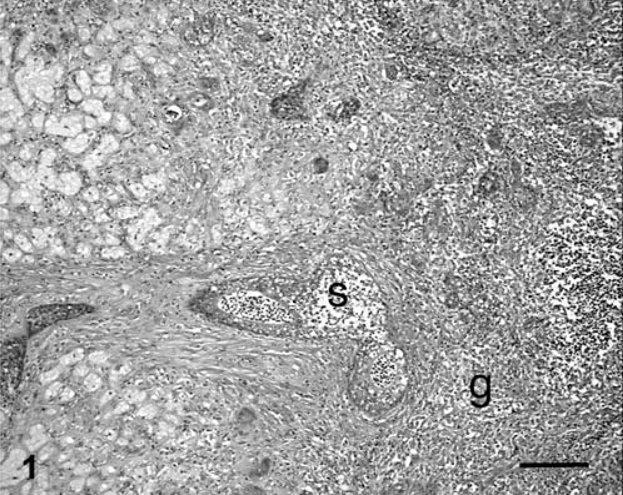
Salivary gland; dog. Notice infarction, squamous metaplasia (s), and granulomatous sialoadenitis (g). HE. Bar = 100 µm.
Necropsy revealed large, firm, submandibular salivary glands that were hemorrhagic and necrotic. A multilobular, firm, hemorrhagic, necrotic mass also was observed in the mediastinum ventral to the esophagus, extending from the thoracic inlet to the heart, and further extending into the right cranial lung lobe affecting 75% of the lobe. The hilar lymph nodes were encased by the fibrous mass. The heart contained numerous white nodules throughout 40% of both ventricles.
Microscopically, lesions in the salivary glands, lung, mediastinum, hilar lymph node, and heart contained granulomatous to pyogranulomatous inflammation with fibrosis, hemorrhage, and necrosis (Fig. 2). The liver, kidney, omentum, and spleen also had mild, multifocal, granulomatous inflammation. A systemic granulomatous disease of probable infectious cause was the final diagnosis. Results of Gram and periodic acid–Schiff staining of the tissues were negative for bacteria and fungi, respectively.
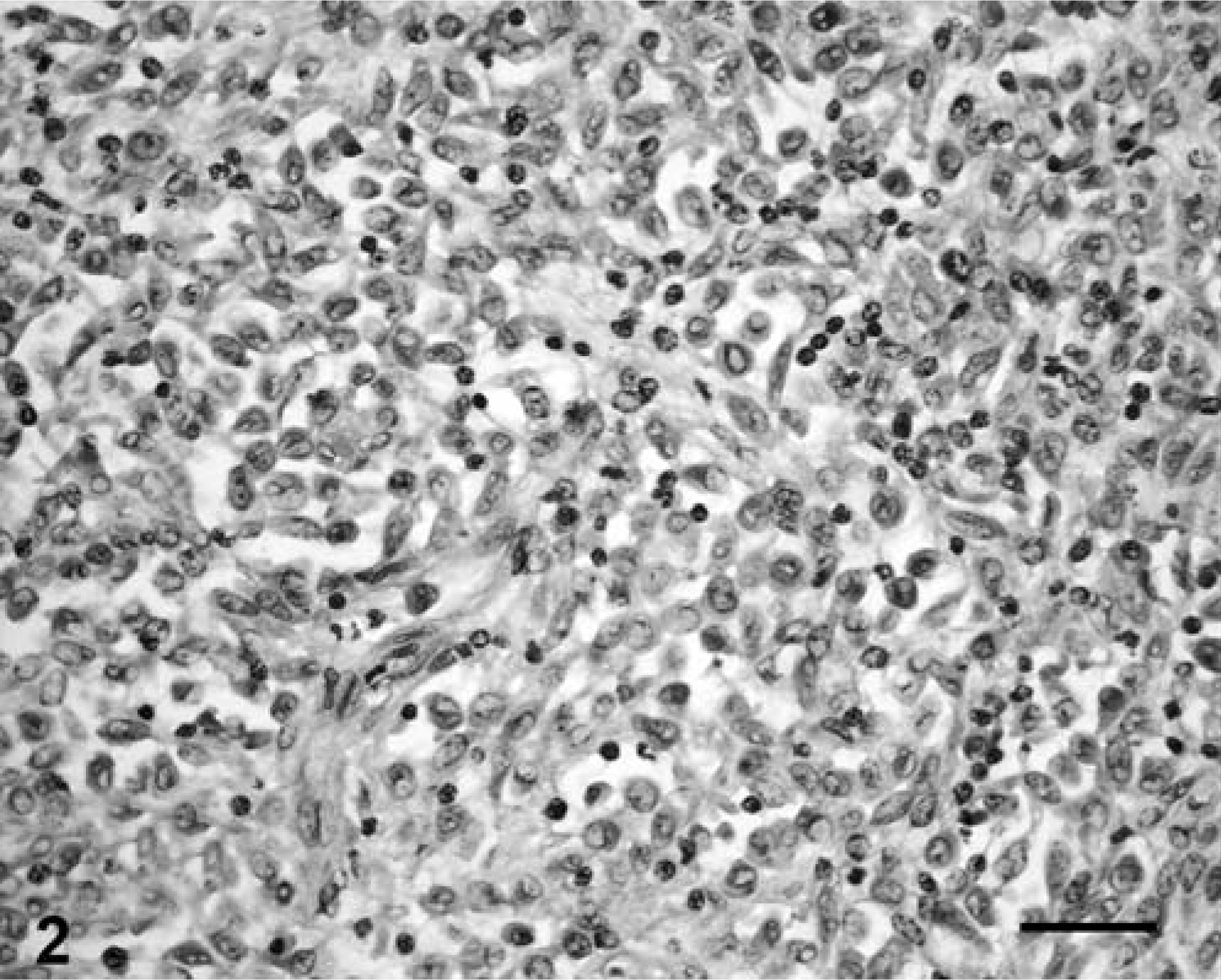
Lymph node; dog. Notice granulomatous inflammation typical of that seen in all affected organs. HE. Bar = 25 µm.
Real-time polymerase chain reaction (PCR) analysis was performed on DNA extracted from paraffin-embedded salivary gland tissue. Bartonella genus screening of the intergenic transcribed sequence region was done by PCR analysis using a scorpion 321 fluorescent probe (Fisher Scientific, Pittsburgh, PA). Bartonella species identification was done using Taqman fluorescent probes for B. henselae, B. quintana, and B. vinsonii subsp. berkhoffii with real-time PCR analysis (SmartCycle II system, Cepheid, Sunnyvale, CA). DNA matching B. henselae and B. vinsonii subsp. berkhoffii was identified.
Immunohistochemical analysis was done on specimens from the mediastinal mass using a mouse anticat scratch antibody (1 ° 100 dilution; Biocare Medical) that detects B. henselae. The ENVISION horseradish peroxidase (HRP) detection system (Dako, Carpinteria, CA) was used with diaminobenzedine as chromagen. The tissue was negative for B. henselae.
Bartonella infection in dogs has been reported to cause systemic granulomatous disease in liver, lung, heart, spleen, lymph nodes, eyes, and endocardium. 1, 3– 6 The similarity of the lesions in this dog to those reported for Bartonella spp. prompted the search for bartonellae. The presence of Bartonella DNA in the tissues of this case does not definitively establish it as the cause of the lesions, but the association of the organism with lesions known to be caused by Bartonella spp. strongly supports bartonellae as the cause. This may be a case of dual infection with two Bartonella species. B. vinsonii subsp. berkhoffii is the most common Bartonella species infecting dogs, whereas B. henselae is infrequently found in dogs. 1, 3
This case is unusual in that, to our knowledge, involvement of the mediastinum, omentum, kidneys, and salivary glands has not been previously associated with Bartonella infection. The involvement of the salivary glands is particularly interesting as this organ is uncommonly affected by disease in dogs. The lesions in the salivary glands were consistent with sialometaplasia, a rare disease of unknown cause in the dog. The disease causes infarction, with squamous metaplasia of the glandular and ductal epithelium. Trauma and infection have been suggested as possible causes of sialometaplasia. 2 The infarction is thought to be vascular in origin, caused by trauma or inflammation of the blood vessels. The findings in this case support infectious disease as a cause of sialometaplasia, and Bartonella infection should be considered a possible cause of sialometaplasia in dogs.