
Abstract
Chronic wasting disease (CWD), a transmissible spongiform encephalopathy (TSE) of deer and elk, is one of a group of fatal, neurologic diseases that affect several mammalian species, including human beings. infection by the causative agent induces accumulations of an abnormal form of prion protein (PrPres) in nervous and lymphoid tissues. This report documents the presence of PrPres within ectopic lymphoid follicles in a kidney of a white-tailed deer that had been experimentally inoculated by the intracerebral route with CWD 10 months previously. The deer was nonclinical, but spongiform lesions characteristic of TSE were detected in tissues of the central nervous system (CNS) and PrPres was seen in CNS and in lymphoid tissues by immunohistochemistry. The demonstration of PrPres in lymphoid tissue in the kidney of this deer corroborates a recently published finding of PrPres in lymphoid follicles of organs other than CNS and lymphoid tissues in laboratory animals with TSE (scrapie).
Chronic wasting disease (CWD) is a fatal transmissible spongiform encephalopathy (TSE) that has been identified in captive and free-ranging cervids. 7, 8 CWD has been experimentally transmitted by intracerebral inoculation of infected brain from mule deer into a variety of animal species, including a goat and cattle. 1, 2, 7, 8
Cervids affected with CWD show accumulation of an abnormal form of prion protein (PrPres) in tissues of the central nervous system (CNS) and lymphoid system and not in other tissues or organs. In this article, we document the presence of PrPres in ectopic lymphoid tissue of kidney in a white-tailed deer experimentally inoculated with CWD.
The subject was part of an experiment to evaluate pathogenicity in white-tailed deer of CWD isolates from elk, mule deer, and white-tailed deer. 2 All inoculated animals were housed in a biosafety level-2 containment facility at the National Animal Disease Center (NADC), Ames, Iowa. Personnel wore protective clothing while in the isolation facility and showered when leaving the facility. The inoculum was obtained from a pool of brains from CWD-positive white-tailed deer, all of which were positive for PrPres by immunohistochemistry (IHC) and by Western blot. The material was ground in a mechanical grinder, gentamicin was added at 100 µg/ml, and the final concentration of 10% (wt/vol) was made with physiologic buffered saline.
The deer was inoculated intracerebrally at 6 months of age with 1 ml of inoculum. The method of inoculation has been described previously. 4 The clinically healthy deer was euthanatized at 10.3 months postinoculation (PI) to attempt detection of PrPres. A complete necropsy was conducted within 1 hour after euthanasia. Representative samples of lung, liver, kidney, spleen, salivary gland, thyroid gland, reticulum, rumen, omasum, abomasum, intestines (ileum, colon), adrenal gland, pancreas, urinary bladder, lymph nodes (retropharyngeal, prescapular, mesenteric, popliteal), tonsil, striated muscles (heart, tongue, masseter, diaphragm), eye, sciatic nerve, trigeminal ganglion, pituitary gland, spinal cord (cervical, thoracic, lumbar), and one half of the sagitally cut brain were immersion fixed in 10% neutral buffered formalin. Tissues were embedded in paraffin wax and sectioned at 5 µm. The sections were stained with hematoxylin and eosin (HE) and immunolabeled by an IHC method for detection of PrPres, as described previously. 4
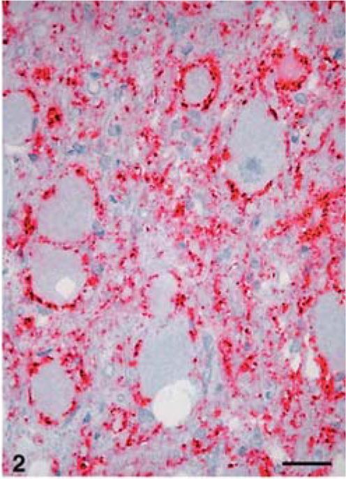
The carcass was in good nutritional state and no significant gross lesions were seen. Microscopically, lesions of spongiform encephalopathy (vacuolation of neuronal perikarya and neuropil) were seen only in the obex area of the medulla (Fig. 1). In the renal pelvis of the kidney section, there were three distinct lymphoid follicles that were located subjacent to the uroepithelial layer (Fig. 3) No other significant lesions were seen.
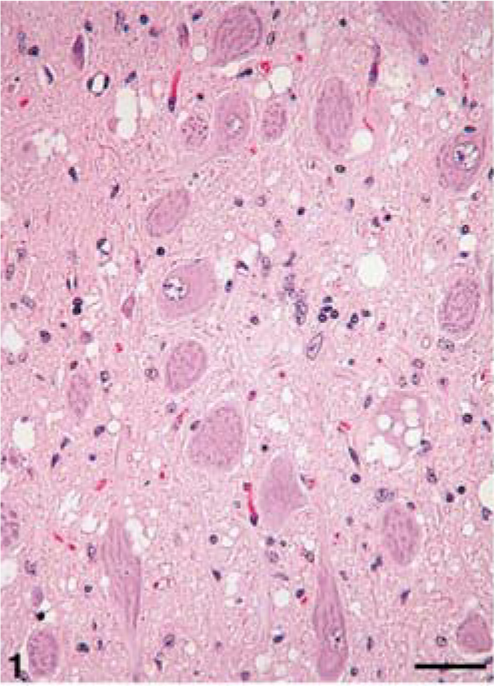
Brain; brainstem (obex) of white-tailed deer. Spongiform encephalopathy characterized by variable sized clear vacuoles is present in neuropil as well as within one neuron. HE. Bar = 70 µm.
Kidney; renal pelvis, and medulla (KM) of white-tailed deer. Note the presence of two lymphoid follicles subjacent to the uroepithelial layer. HE. Bar = 140 µm.
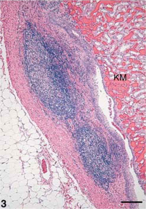
IHC revealed PrPres in brain (cerebrum, cerebellum, midbrain, and brainstem, Fig. 2), spinal cord (cervical), tonsils (pharyngeal and palatine), Peyer's patches in the ileum and in lymph nodes (retropharyngeal, prescapular, mesenteric, and popliteal). It was also observed in the myenteric plexus of small intestines. In the brain, the immunostaining was diffusely distributed throughout gray matter neuropil in all parts of the CNS, including both granular and molecular layers of cerebellum. The immunolabeling pattern was primarily punctate and granular, with some small aggregates and only a few accumulations large enough to be described as plaques. Some neurons contained large immunolabeled granules in the perikaryon, but more commonly, the location of PrPres was perineuronal, forming a ring around the cell (Fig. 2).
Brain; brainstem (obex) of white-tailed deer. There is perineuronal PrPres immunolabeling. Diffuse immunolabeling is also present in the neuropil. Stained for PrPres by IHC. Bar = 50 µm.
PrPres was detected in pars nervosa of the pituitary gland. However, neither the Gasserian (trigeminal) ganglia nor the retina showed immunolabeling for PrPres.
Immunolabeling for PrPres by IHC was detected in two of three ectopic lymphoid follicles in the renal hilus. The staining was within the central area of each follicle (Fig. 4) and resembled the immunolabeling that was observed elsewhere in lymph nodes.
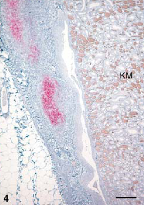
Kidney; renal pelvis, and medulla (KM) of white-tailed deer. Note the presence of PrPres immunolabeling within the lymphoid follicles subjacent to the uroepithelial layer. Immunolabeled for PrPres by IHC. Bar = 140 µm.
CWD is a transmissible spongiform encephalopathy and, like all other TSEs, it is characterized by a long incubation period, which in deer is seldom less than 18 months. 7, 8 In cervids, clinical CWD is characterized by emaciation, changes in behavior, and excessive salivation. 7, 8 Clinical signs of CWD were not observed in the presently described case. However, the morphologic lesions of SE were quite obvious at this early time of death (10.3 months PI). Also, PrPres was present not only in the brain, but also in lymphoid tissues (tonsils, spleen, various lymph nodes, and Peyer's patches). This is interesting because the deer was inoculated intracerebrally and yet PrPres was detected extensively in peripheral tissues of the carcass. This suggests that, as long established, after intracerebral injections, the inoculum gained rapid access to the circulatory system. 6
Sheep with scrapie and cervids with CWD have accumulations of PrPres in tissues of the CNS and lymphatic systems. The findings in the present case confirm previous reports of early involvement in the obex region of CNS and in lymphoid tissues of white-tailed deer with CWD. 7, 8
Recently, PrPres was also demonstrated in inflammatory lymphoid tissues in kidney, pancreas, and liver of mice with scrapie. 5 These mice were experimentally infected with chronic inflammatory diseases that had induced formation of lymphoid follicles within the parenchymatous organs. Similar to the presently described cervid case, the PrPres in mice was present within the lymphoid follicles of the inflammatory tissue. Authors of the mouse study concluded that formation of lymphoid follicles due to a variety of chronic inflammatory diseases resulted in PrPres accumulation in otherwise prion-free organs. In the presently described cervid, because there was no other morphological evidence of inflammation, it was not possible to conclude whether the ectopic lymphoid follicles were the result of chronic inflammation that had partially resolved.
In the mouse study, 5 the authors also stated that knowledge of PrPres distribution within infected hosts is vital to considerations of protection against the spread of TSEs to humans. However, at this time, BSE is the only animal TSE implicated as a zoonosis, and human cases are considered to have resulted principally from consumption of infected CNS tissues.
As previously documented, 3 PrPres was not observed by IHC in striated muscles (heart, tongue, masseter, diaphragm) of the experimental deer. This observation is in agreement with our previous findings in which striated muscle tissues from 20 experimental animals (cattle, sheep, elk, and raccoons) were examined for PrPres by IHC. In these animals, all of which had developed a TSE after experimental inoculation, PrPres was found by IHC in the CNS, but not in muscle tissues. 3