
Abstract
Deciduoid mesothelioma is a rare variant of epithelial mesothelioma, up to now only described in human pathology, which bears remarkable cytomorphologic resemblance to the endometrium of pregnancy, termed decidua. A case of peritoneal mesothelioma with deciduoid features in a 10-year-old, female dog is reported. Multiple whitish-gray nodules (1-5 mm in diameter) in parietal peritoneum and mesentery were histologically composed of large, proliferating, polygonal or ovoid cells with an abundant eosinophilic, glassy cytoplasm. Immunohistochemical evaluation indicated that the neoplastic cells coexpressed cytokeratin and vimentin with strong and diffuse cytoplasmic staining, and ultrastructural analysis showed long and slender mesothelial-type microvilli; these findings confirmed the mesothelial origin of the tumor.
Mesotheliomas (MMs) are rare tumors derived from the mesothelial cell lining of serous membranes and have been reported in dogs, cats, goats, horses, rats, hamster, and cattle. 1, 7 In dogs, like in humans, the pleura is the main site of MM development after the pericardial and the peritoneal cavity. The average age of onset is 8 years, and according to some reports, the tumor is more common in male dogs, although there does not seem to be any strong breed predilection. 7 In humans, the development of mesothelioma after exposure to asbestos is well documented, whereas the association between asbestos fibers, and mesothelioma has not yet been definitely confirmed in animals. Ferruginous bodies, suggestive of asbestos exposure, have been found in the lungs of some urban dogs with mesothelioma, and associations have been made between mesothelioma in dogs and exposure of owners to asbestos. 5, 6 Exposure to pesticide is a cofactor that apparently increases the risk of mesothelioma. 5
The wide range of morphologic appearances is one of the main characteristics of mesothelioma. MMs are classically classified, according to the histologic growth pattern, as epithelial or sarcomatoid or biphasic, but several subtypes have been described in human pathology, such as lymphohistiocytoid, desmoplastic, microcystic, clear cell, signet ring, small cell, and deciduoid. 8, 9 In dogs, only two uncommon variants (cystic and sclerosing) have also been described, all in the peritoneal localization. 2– 4 To our knowledge, in domestic animals, the deciduoid variant of mesothelioma has not yet been reported.
A 10-year-old, female, crossbred dog was admitted to a private clinic (Clinica veterinaria Avoni Ivano, Imola, Bologna) with a 1-month history of weight loss and acute, severe abdominal distension. At clinical examination, the patient showed marked cachexia and evidence of ascites. Ultrasonography confirmed the widespread abdominal effusion and revealed the presence of a 3.2- × 1.6-cm mass in the mesentery, cranially to the urinary bladder, associated with a diffuse mesenteric thickening. At the owner's request, further diagnostic investigations were not performed, and the dog was euthanatized. Representative tissue samples of the mass and the mesentery were collected by the practitioner immediately after death and fixed in 10% formalin for routine histologic examination. Paraffin sections, 4 µm thick, were prepared and stained with hematoxylin and eosin (HE) and Periodic acid-Schiff (PAS).
For immunohistochemical analysis, the following antibodies were applied in appropriate dilutions on samples sections: pancytokeratin (Dako, Glostrup, Denmark), cytokeratin AE1/AE3 (Dako), cytokeratin 5/6 (Dako), vimentin (Dako), anticalretinin (Dako), smooth-muscle actin (Dako), desmin (Dako), S-100 protein (Dako), and CD117 (Dako), using the labeled streptavidin–biotin method (LSAB™ kit, Dako). For transmission electron microscopy (TEM), small pieces of formalin-fixed tissue were twice rinsed in 0.1 M cacodylate buffer, postfixed in 1% osmium tetroxide, and epoxy resin–embedded. Ultrathin sections were double-stained with uranyl acetate and lead citrate and ultrastructurally examined.
Grossly, the samples were characterized by nodular bosselated surface, with disseminated, multifocal-to-coalescent, 1–5-mm diameter, whitish-gray nodules (Fig. 1). At microscopic examination, the tissue was composed of large, epithelial-like, rounded, ovoid or polygonal cells arranged in solid sheets and occasionally in glandular and papillary structures (Fig. 2). Mild, mixed-inflammatory infiltrates were seen within the sheets of tumor cells. The neoplastic cells had abundant eosinophilic glassy cytoplasm with well-defined cytoplasmic borders. The cytoplasm of some cells had an eccentric outer pale zone, sharply demarcated from a central glassy perinuclear region, and there were occasional large vacuoles (Fig. 3). The nuclei were large, round or oval vesicular with a well-defined, and occasionally indented, nuclear membrane and prominent single nucleoli; cells with more than one nucleus were occasionally present. At high magnification (400×), the cell surface exhibited a thick, brush-like border; the mitotic activity was low (one–two mitoses per 10 400× fields). The PAS reaction stained faintly the cell boundaries, but not the cytoplasm or the intracytoplasmic vacuoles.
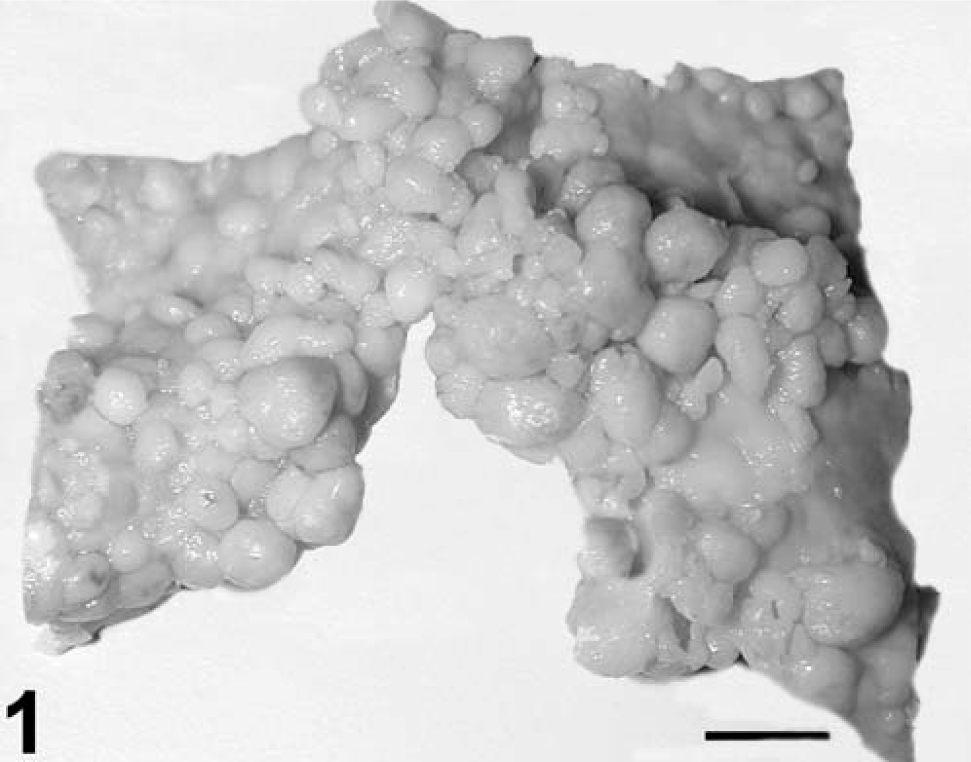
Mesentery; canine deciduoid mesothelioma. A formalin fixed small specimen showed multifocal to coalescent whitish-gray nodules on the serosa. Bar = 5 mm.
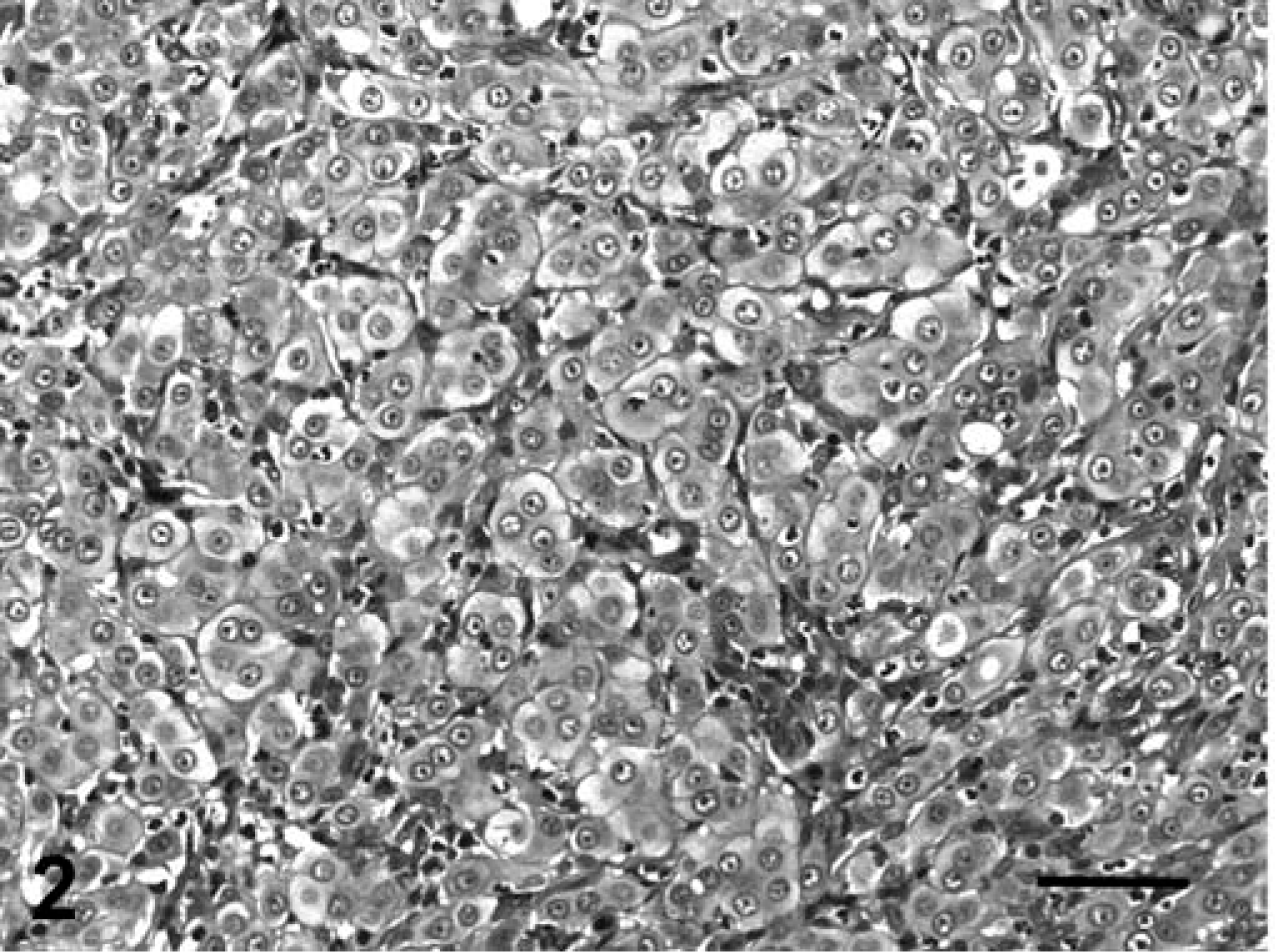
Mesentery; canine deciduoid mesothelioma. Confluent sheets of large, round cells with abundant eosinophilic cytoplasm and well-defined cell borders. HE stain, Bar = 50 µm.
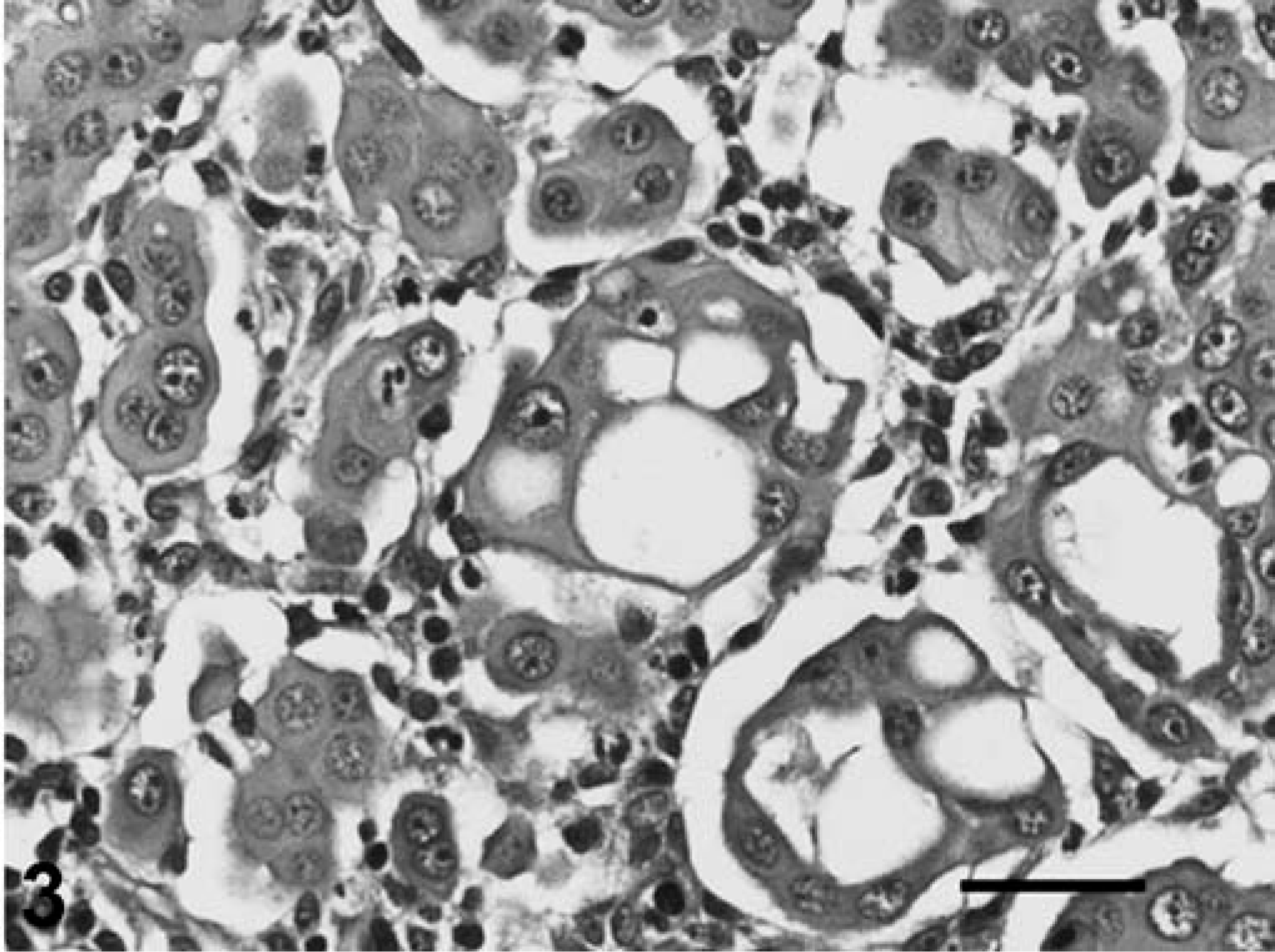
Mesentery; canine deciduoid mesothelioma. Large intracytoplasmic vacuoles were occasionally present within tumor cells. HE stain, Bar = 20 µm.
Immunohistochemistry showed strong, diffuse, cytoplasmic staining of neoplastic cells for pancytokeratin and cytokeratin AE1/AE3 (Fig. 4a) and focal, cytoplasmic staining in scattered cells for cytokeratin 5/6. Tumor cells also stained intensely positive for vimentin (Fig. 4b), whereas all other markers were negative.
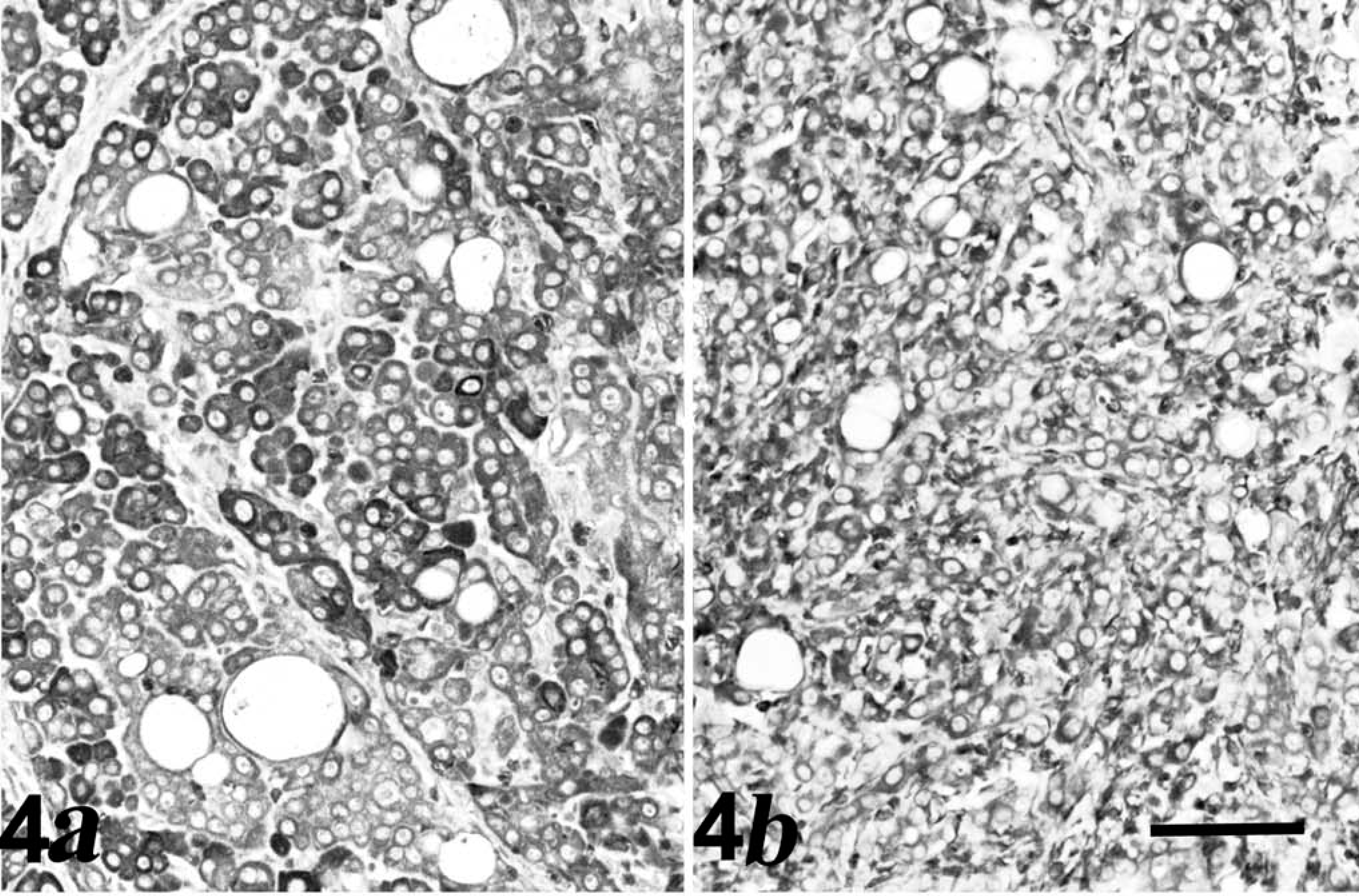
Mesentery; canine deciduoid mesothelioma. The neoplastic cells were strongly immunoreactive for Cytokeratin.
Electron microscopy showed closely apposed large and polygonal tumor cells, with regular oval nuclei, joined by desmosomes with tonofibril tails. The cytoplasm contained intermediate filaments, some forming tonofibrils and others in more dispersed arrangements, and a variety of organelles, which included clusters of mitochondria, some cisternae of rough endoplasmic reticulum and focally prominent, smooth endoplasmic reticulum. Numerous, delicate, elongated slender microvilli without rootlets were noted on the cell surface; scattered cells contained intracytoplasmic vacuoles hemmed by a membrane with the same type of microvilli (intracytoplasmic lumina) (Fig. 5).
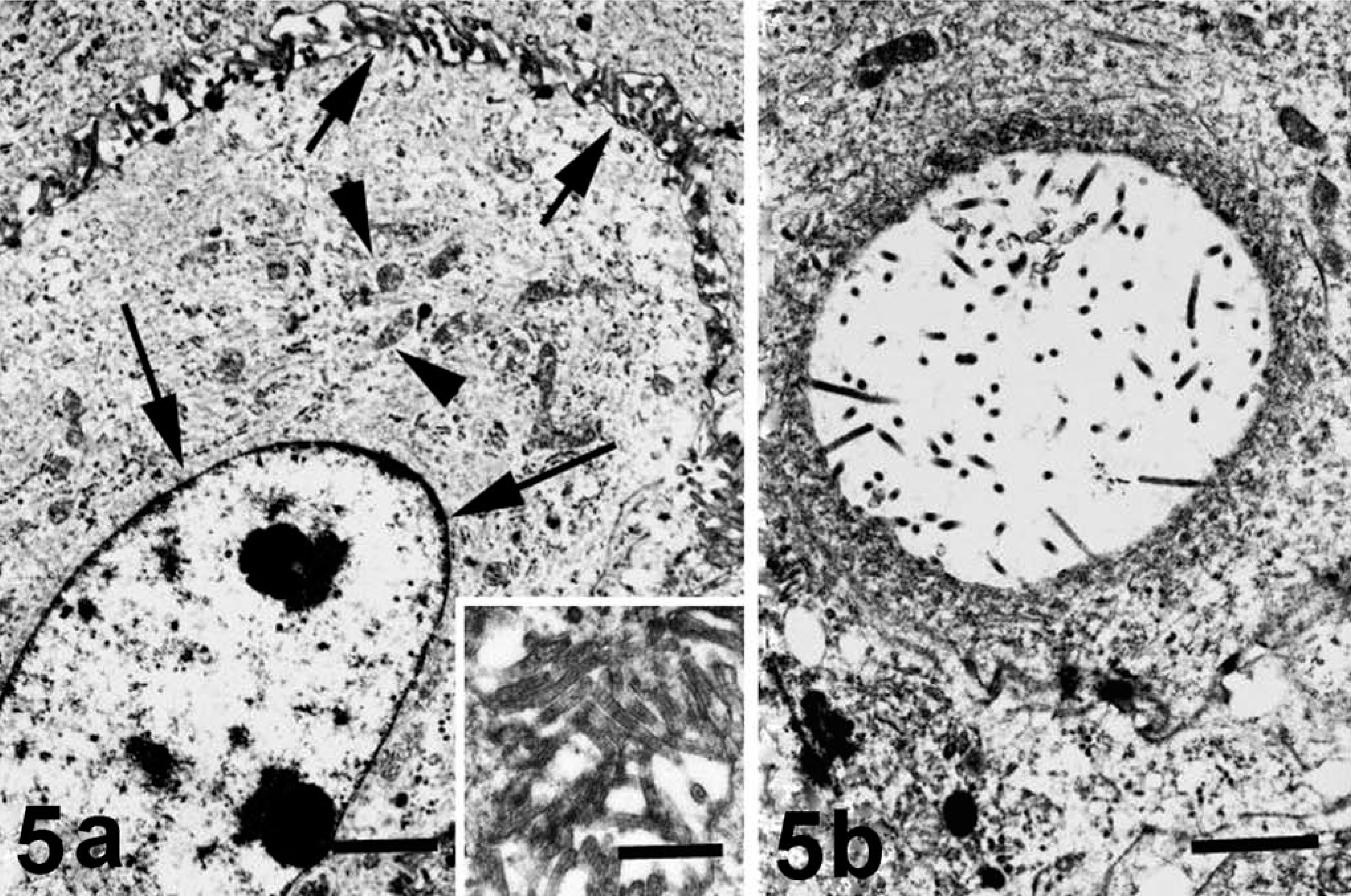
Electron micrograph, mesentery; canine deciduoid mesothelioma.
The gross and histologic findings suggested the diagnosis of peritoneal mesothelioma. Alternative diagnoses, according to the histologic features of large epithelial-like eosinophilic cells, included alveolar rhabdomyosarcoma or peritoneal metastases of hepatocellular carcinoma or hepatoid perianal gland adenocarcinoma. Nevertheless, anamnestic and postmortem findings did not support the latter hypotheses, which were also excluded by immunohistochemistry (coexpression of cytokeratin and vimentin, negativity to other markers) and electron microscopy (mesothelial-type microvilli). The close morphologic resemblance of the tumor structure with the deciduoid variant of human mesothelioma suggested a definitive diagnosis of deciduoid peritoneal mesothelioma, a recently characterized unusual morphologic variant of epithelial mesothelioma, with remarkable cytomorphologic resemblance to decidua or decidualized tissue. Decidua is the epithelial lining of the endometrium, which is formed after the blastocyst is implanted onto the uterus, and which, in some mammals (human, dog, cat), is histologically composed by large round or polygonal “epithelioid” stromal cells, with a large, vesicular nucleus (decidual cells). 8, 10
Human deciduoid mesothelioma (DM) has been reported especially in the peritoneum but also in the pleural cavities and pericardium. 8– 11 The first case of deciduoid features in malignant mesothelioma was described in 1985 by Talerman et al. 13 as a “sheet-like proliferation of large rounded, ovoid, or polygonal cells with pale to bright, eosinophilic, slightly foamy or vacuolated cytoplasm containing a large round or oval vesicular nucleus with a single prominent nucleolus and occasionally binucleated forms.” Thirty-one cases of DM has been described in humans, and it appears that the so-called DM has some clinical and pathologic features that are dissimilar to mesothelioma in general, such as the predilection to the peritoneum, a female preponderance, and the occurrence in juvenile patients. 12 Nevertheless, it has been demonstrated that the resemblance of DM to decidua is only based on morphologic similarities and not on the same embryologic origin. 11
In the described cases of human DM the immunohistochemical profile is similar to that of classic mesothelioma, with coexpression of cytokeratin and vimentin, which has also been demonstrated in canine mesothelioma. 2, 4 According to our results, canine DM shows the same twofold immunoreactivity of neoplastic cells, which is rarely observed in canine tumors, except for renal and highly anaplastic carcinomas, Sertoli cell tumors, thyroid tumors, and synovial sarcoma. Calretinin, a calcium-binding protein expressed in human mesothelial cells, is used as a diagnostic marker of mesothelioma. 9 In dogs, it is not constantly expressed in normal mesothelial cells (personal observation), and its expression has never been observed in canine mesotheliom, 4 nor in this case. TEM can be useful in the diagnosis of MM by the demonstration of mesothelial-type microvilli in the cell surface, which helps to differentiate mesothelioma from other tumors.
The etiology of deciduoid mesothelioma seems not to be related to asbestos exposure, 9 except for very few cases. 11 The majority of reported cases of DM involved young females; this raised the possibility that the tumor could be induced or at least stimulated by sex hormones, but this hypothesis was opposed by the absence of immunoreactivity for hormonal receptors in the neoplastic cells. 8
After due consideration, on the basis of histochemical, immunohistochemical and ultrastructural findings, this article appears to be the first report of peritoneal deciduoid mesothelioma in the dog. This peculiar histologic type of mesothelioma needs, therefore, to be considered as a first step for the identification of additional cases with the ultimate objective of understanding if, indeed, this is a unique clinical and pathologic entity or just another morphologic variant of mesothelioma.
Footnotes
Acknowledgements
We are grateful to Dr. Paola Preda for technical assistance in electron microscopy observations.