
Abstract
Regional suppurative meningoencephalitis and ventriculitis of variable chronicity was diagnosed in three young dogs residing in Colorado. Grass awns were grossly identified in the right occipital cortex of one dog and in the right lateral ventricle of another. Intralesional plant material was microscopically evident in the dura mater overlying the right occipital cortex of the third dog. One grass awn was identified as a floret of Hordeum jabatum. In each case, aerobic culture of brain tissue identified multiple isolates of bacteria. The dogs presented with clinically variable, rapidly progressive neurologic dysfunction, including tetraplegia, depressed mentation, and episodic extensor rigidity, ataxia, circling, stupor, vocalization, and head-pressing. Encephalitis due to bacteria introduced from migrating plant foreign material is a potential sequela of intranasal, periocular, or pharyngeal foreign bodies.
Plant material, especially the grass awn, is a common cause of foreign body-related disease in animals. The grass awn is especially adept at migration as its sharp, pointed anterior and proximally directed, barbed posterior facilitate cutaneous or mucosal penetration and progressive forward movement. Grass awn migration most commonly occurs in the subcutis and external otic canal, but also affects the conjunctiva, nasal sinuses, and oral cavity with relative frequency. 3 Subcutaneous abscesses associated with awn migrations often involve the interdigital web, head, face, neck, flank, and costochondral areas. 3, 6 Penetration of the bronchial or esophageal mucosa may occur subsequent to inhalation or ingestion of an awn, resulting in lobar pneumonia, chronic pleuritis, or pyothorax. 6, 7 Inhaled awns that are deflected by the diaphragm may migrate along the crura and eventually cause osteomyelitis and discospondylitis of the rostral lumbar vertebrae. 9, 13
Migrating foreign bodies affecting the central nervous system (CNS) have rarely been documented in domestic animals. Intracranial foreign bodies reported in veterinary literature are scant, including two cases of encephalitis in the dog due to a migrating porcupine quill and a sewing needle. 5, 11 There is a single report of feline encephalitis due to intralesional plant material. 1 Here, we describe three dogs in which presence of intracranial plant material resulted in suppurative meningoencephalitis and ventriculitis associated with intralesional bacteria.
Dog No. 1, a 5-month-old, spayed female terrier-crossbreed, presented with acute onset of tetraparesis and depressed mentation. Three months prior to presentation, the dog had been diagnosed with bilateral mild chronic conjunctivitis. Neurologic examination revealed bilateral ventral strabismus and decreased conscious proprioception in the left pelvic limb. Abnormalities were not identified on a complete blood count (CBC), serum biochemistry panel, or urinalysis. While hospitalized, the dog became recumbent, with limb extensor rigidity, obtundation, tachycardia (300 beats/min), and hyperthermia (rectal temperature of 40.3°C [104.6°F]). Dog No. 1 died within 9 hours of presentation.
At necropsy, meningeal hemorrhage extended along the caudal portion of the cerebellum and brain stem. The right lateral and third ventricles were filled with thick, yellow-green, mucinous exudate (Fig. 1). Within the exudate was a 1.25-cm × 1-mm linear plant foreign body which, when examined under a dissecting microscope, had a green fibrillar internal structure. On the ventral surface of the left olfactory tract was a 2-mm area of red-brown discoloration, interpreted as hemorrhage or hemosiderosis associated with foreign body migration. Other gross lesions were not identified. Histologic examination of the lateral ventricle and parietal cortex revealed severe acute suppurative meningoencephalitis and ventriculitis. The ventricle was expanded with luminal sheets of viable and degenerate neutrophils, which effaced the ependyma and regionally extended into periventricular white matter, parietal cortex, and meninges. Several colonies of rod-shaped bacteria were evident within the inflammatory focus. The adjacent parenchyma contained perivascular cuffing by mononuclear leukocytes, perivascular edema, multifocal areas of hemorrhage, and regional rarefaction. Aerobic culture of the ventricular exudate yielded moderate growth of coagulase-negative Staphylococcus sp, hemolytic Escherichia coli, Pasteurella multocida, and S. aureus.
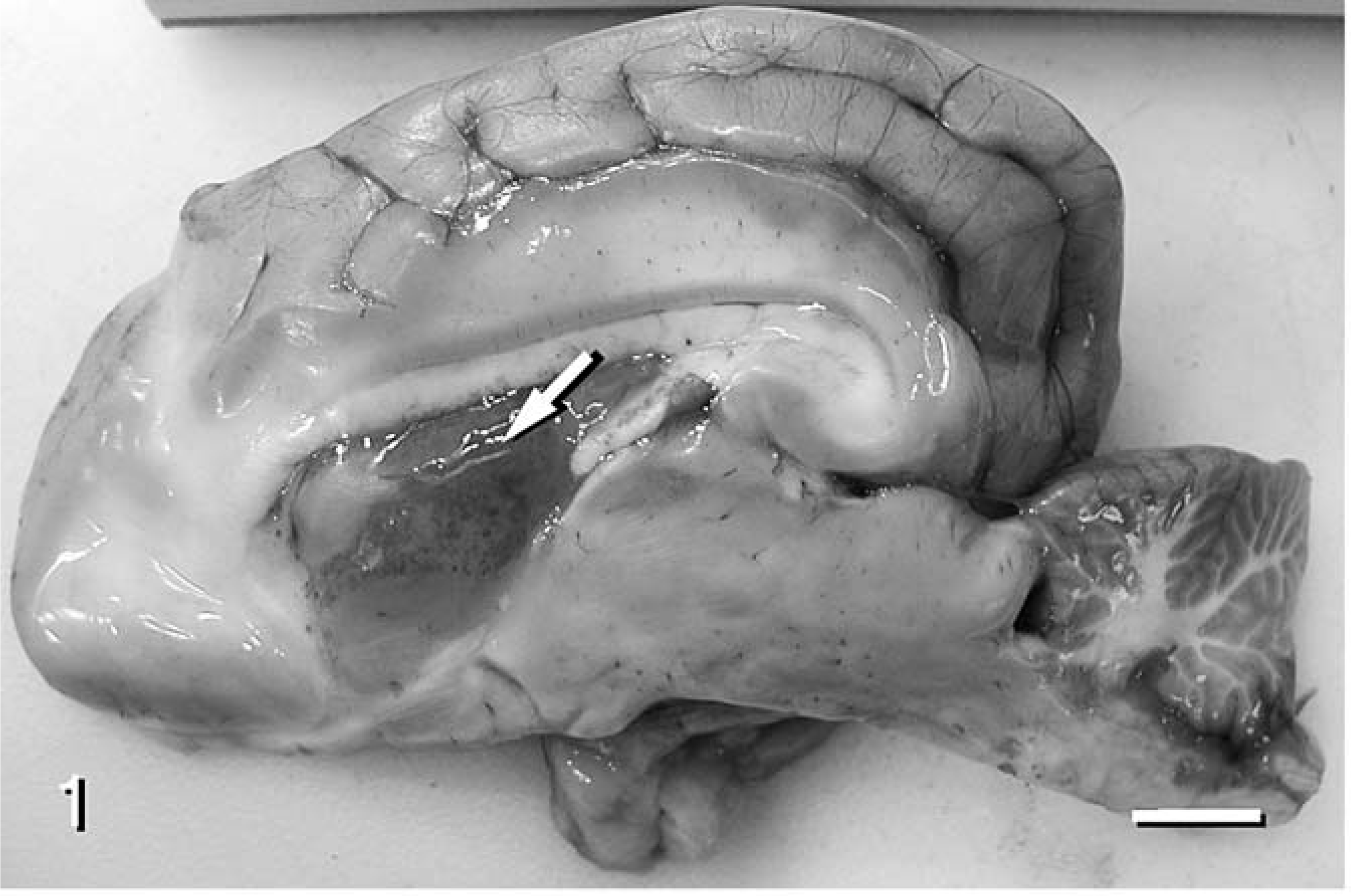
Brain; dog No. 1. The choroid plexus of the right lateral ventricle is congested with petechial hemorrhages. Mucinous exudate fills the lumen of the ventricle and contains a plant foreign body (arrow). Hemorrhage extends along the meninges of the dorsal aspect of the medulla oblongata. Bar = 1 cm.
Dog No. 2, a 4-month-old, reproductively intact male Great Dane, presented with acute onset of tetraparesis, depressed mentation, and tonic-clonic muscular contractions. The dog was treated with IV-administered fluids, dextrose, penicillin, and Nembutal, with little response. While hospitalized, episodes of stupor, vocalization, aggression, and unconsciousness occurred. Dog No. 2 died 46 hours following presentation.
At necropsy, a grass awn spanned the ventrolateral aspect of the right occipital lobe, internal capsule, and rostral horn of the lateral ventricle (Fig. 2). The grass awn was composed of a fusiform grain sheath that contained two closely packed seeds with three 1.5-cm radiating stylets. The surrounding parenchyma was cavitated with malacia and centrally contained fibrinopurulent exudate. A plaque of fibrinopurulent exudate surrounded the penetrated cortical surface. The third ventricle was moderately distended and internally coated with fibrinous exudate. Additional gross lesions were not identified. On the basis of morphologic characteristics, the grass awn was identified at the Colorado State University Weed Research Laboratory as a floret of Hordeum jabatum, commonly known as foxtail barley or squirrel tail. Within the parenchyma surrounding the grass awn, severe subacute fibrinosuppurative meningoencephalitis and ventriculitis were apparent microscopically. Numerous viable and degenerate neutrophils, admixed with necrotic cell debris, abundant fibrin, few gitter cells, and rare coccal bacteria extended throughout the clefted malacic area and overlying meninges, and regionally eroded the ependyma of the lateral ventricle. Throughout the adjacent tissue, rarefaction of cerebral parenchyma, pronounced mononuclear perivascular cuffing, perivascular proteinaceous fluid exudation, mild perivascular fibroplasia, and multifocal areas of hemorrhage were evident. There also was severe, diffuse, lymphocytic meningitis of the entire brain and rostral portion of the spinal cord. Aerobic culture of the right olfactory bulb yielded a mixed growth of β-hemolytic Streptococcus sp. and E. coli.
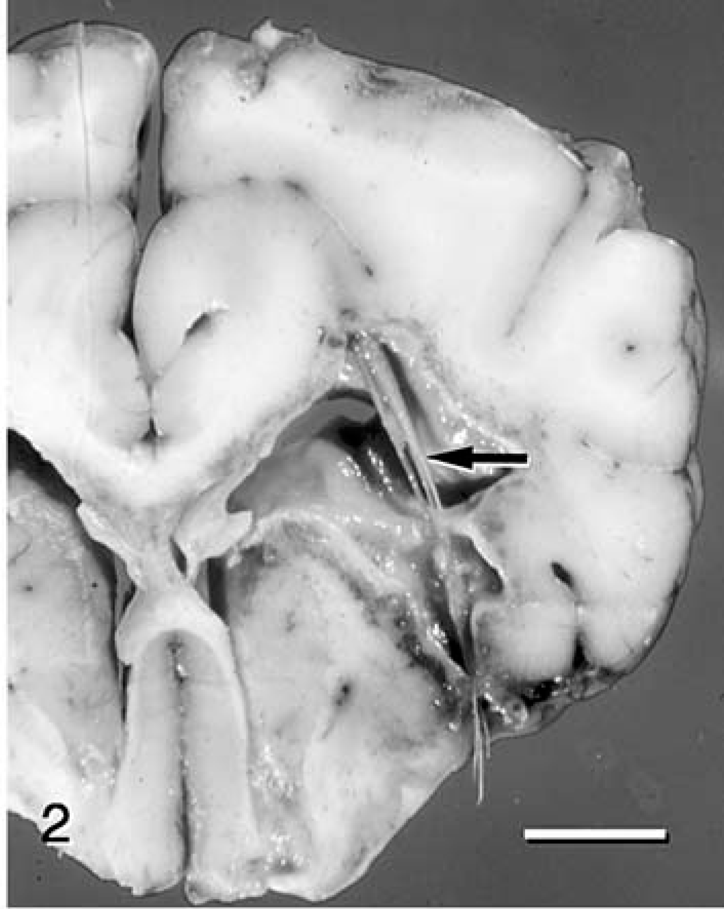
Brain; dog No. 2. A grass awn spans the ventral right occipital lobe and the lateral ventricle. Cortex surrounding the awn and periventricular white matter are affected with malacia. Bar = 5 mm.
Dog No. 3, a 4-year-old, reproductively intact female Brittany Spaniel, was referred to Colorado State University Veterinary Teaching Hospital because of ataxia, depressed mentation, and head pressing of one day's duration. One month prior to referral, a similar episode had been observed that appeared to improve in response to administration of carprofen. Neurologic examination revealed depressed mentation, mild ataxia, right-sided facial hyperesthesia, ventrolateral strabismus of the right eye, delayed menace response of the left eye, delayed sensory trigeminal nerve and facial nerve responses on the left side, and decreased conscious proprioception in the left pelvic and thoracic limbs. These deficits were suggestive of a right-sided cerebral lesion. While hospitalized, intermittent episodes of circling to the right, head pressing, and pronounced ataxia were observed. Results of a CBC and serum biochemistry panel were unremarkable. Examination of a cerebral spinal fluid (CSF) sample obtained from the cerebellomedullary cistern suggested mixed inflammation, with increased protein concentration (90 mg/dl), and pleocytosis (8 erythrocytes/µl, 10 nucleated cells/µl, 63% nondegenerate neutrophils, 17% large mononuclear cells, 18% lymphocytes). The CSF titers for Toxoplasma gondii IgM and IgG, Rocky Mountain spotted fever IgG, and Ehrlichia canis IgG were negative (each < 1:10). Titers for canine distemper virus (CDV) IgG were slightly increased in CSF (1:16) and serum (1:128). Titers for CDV IgM were negative in CSF (<1:2), and were increased in serum (1:32). Dog No. 3 was discharged with a diagnosis of CNS inflammatory disease, and was prescribed oral prednisone therapy.
Dog No. 3 re-presented 4 days later because of progressive signs of neurologic dysfunction. Additional signs included obtundation, marked ataxia, conscious proprioceptive deficits in all limbs, bilateral head and facial hyperpathia, and intermittent vocalization. A serum biochemistry panel indicated hyperglycemia (160 mg/dl), hyperalbuminemia (4.3 g/dl), hypochloremia (102 mEq/L), and a titrational metabolic acidosis (anion gap of 33, bicarbonate concentration of 14.2 mEq/L). Results of a CBC and urinalysis were within normal limits. The dog was hospitalized for further treatment, including intravenous administration of mannitol (single dose of 1 g/kg of body weight given over 15 minutes) and cytosine arabinoside, and oral administration of doxycycline and prednisone. Seizure-like behavior (stupor, facial muscle fasciculation, head turning to the left, limb rigidity, and opisthotonus) was intermittently observed.
Due to progression of clinical signs of disease despite medical therapy, dog No. 3 was euthanatized 36 hours later. At necropsy, a poorly delineated, 2.5-cm-diameter, dark-tan, friable mass spanned the right occipital lobe, from the caudal cortical surface to the floor of the lateral ventricle. Dura mater adhered to the surface of the mass, which had distorted and flattened gyral architecture. Within the mass were central areas of cavitation, which contained thin, clear, green-tan fluid. The lumen of the lateral ventricle was regionally attenuated by the mass. Other gross lesions were not identified. Histologic examination of the right caudal occipital lobe revealed a regionally extensive area of severe chronic suppurative meningoencephalitis and ventriculitis. Coalescing aggregates of viable and degenerate neutrophils, admixed with necrotic cell debris and few gitter cells extended throughout the occipital cortex and subcortical and periventricular white matter, and effaced the ependyma of the lateral ventricle. The inner aspect of the overlying dura mater was expanded with fibrous connective tissue that centrally contained a 60-µm profile of birefringent multicellular foreign body consistent with plant material (Fig. 3). Small colonies of rod-shaped bacteria were sparse within areas of inflammation. Parenchyma affected with suppurative inflammation was distorted with rarefaction, cavitation, and stromal collapse. Mild spongiosis of the neuropil affected the marginating gray matter, which contained few necrotic neurons and hypertrophic astrocytes. Vessels in adjacent parenchyma were lined by hypertrophic endothelium and often were cuffed by mononuclear leukocytes within proteinaceous fluid. Aerobic culture of the caudal occipital lobe yielded light growth of Corynebacterium and Micrococcus spp.
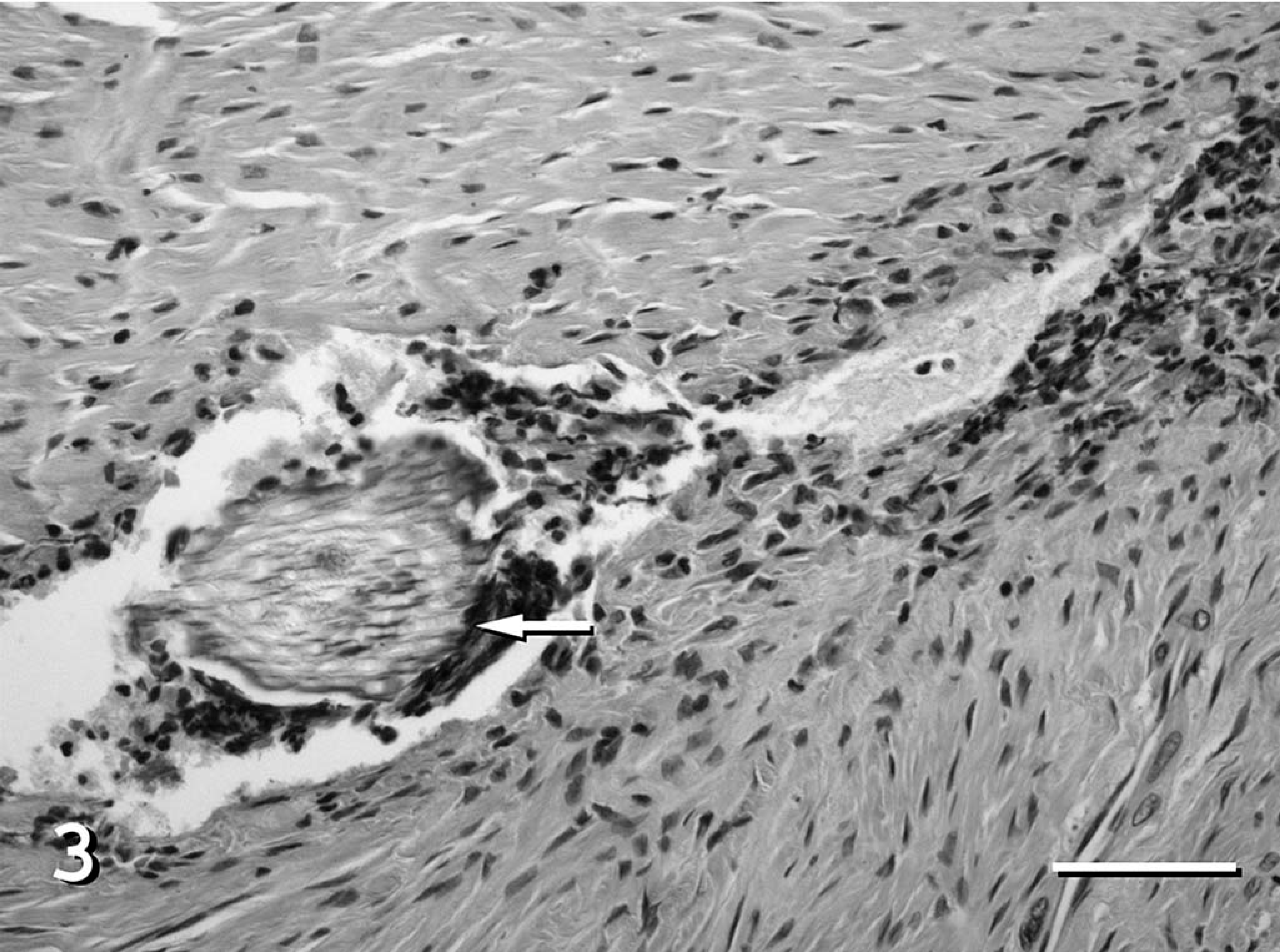
Dura mater; dog No. 3. A refractile plant foreign body (arrow) is situated within the thick and fibrotic dura. Necrotic cell debris, degenerate neutrophils, lymphocytes, and plasma cells surround the foreign material. HE. Bar = 50 µm.
Focal suppurative encephalitis often develops secondary to local extension of adjacent septic foci, such as otitis interna, or from hematogenous bacterial implantation, as with valvular endocarditis or other causes of bacteremia. Migratory foreign material may introduce bacterial agents; however, intracranial foreign bodies are rare in domestic animals. Additionally, to the authors' knowledge, there have not been reported cases of canine encephalitis involving foreign bodies consisting of plant material.
Migration routes for reported cases of foreign bodies that have penetrated the CNS of domestic animals usually are obscure. A migratory tract was not grossly evident in a cat affected with suppurative encephalitis with intralesional plant material. 1 In that case, plant material was recognized microscopically and nasal migration with penetration of the cribriform plate was considered as the cat was affected with chronic rhinitis. Plant material was identified in a granuloma removed from the rostral portion of the cervical vertebral canal of a mixed-breed dog, 10 and in the rostral cervical epidural fat of a Labrador Retriever that had suffered a pharyngeal stick injury 5 months earlier. 4 A migration tract was not evident in either case; however, in both cases, the plant material was suspected to have perforated the pharyngeal soft tissues and entered the vertebral canal through the atlanto-occipital joint. A porcupine quill was associated with granulomatous encephalitis in an adult Saint Bernard. 5 The foreign body was situated vertically in an occipital lobe and was suspected to have entered the cranium through the foramen magnum. A brain abscess associated with a sewing needle was reported in a 12-year-old dog. 11 In that case, the foreign body penetrated the oropharyngeal mucosa through pharyngeal soft tissue and the base of the left temporal wing of the basisphenoid bone, extending into the base of the brain in the left pyriform region.
In the cases described herein, definitive foreign body migratory tracts for intracranial penetration were not grossly evident. The discoloration of the optic tract surface in dog No. 1, and the preceding 3-month history of conjunctivitis, may indicate a periocular pathway. Intracranial penetration is not an uncommon complication of periorbital wooden foreign bodies in humans. 12 The positioning of the awn in dog No. 2 suggests penetration of the ventral aspect of the cranium, possibly involving transmigration of the cribriform plate. The migration route in dog No. 3 may have involved the occipital foramen, as the plant material was localized in close proximity within the dura overlying the caudal right occipital lobe. Although positioning of the plant material may provide insight for a potential entry site, gravity and the circulation of CSF may influence the migration of intracranial foreign bodies. 8
The type of intralesional plant material in dog No. 3 could not be determined; however, the morphologic characteristics of the material found in dog Nos. 1 and 2 were consistent with grass awn. The bacteria identified microscopically and isolated from the encephalitic foci were likely introduced by the migratory plant material. The suppurative infiltrate was probably a response to both elements, although bacteria may be more pyogenic than is migratory foreign material. Streptococcus sp, S. aureus, P. multocida, Actinomyces sp., and Nocardia sp. are frequently isolated from inflammatory lesions associated with grass awn migration. 3, 6 Many of these isolates are common commensal organisms of the respiratory or integumentary system, suggesting potential modes of introduction into the brain. As was exemplified in these cases and is typical of disease mediated by other foreign bodies, multiple bacterial species are often isolated from grass awn-related lesions. Culture of CSF may have been useful for antemortem diagnosis of bacterial meningoencephalitis, but in any case, was not done. CSF culture is frequently unrewarding even with substantial CNS bacterial infection. 2, 14 Proposed reasons include bacterial sequestration within an abscess and low absolute numbers of organisms needed to incite encephalitis and lytic necrosis. Although unlikely, culture of a CSF specimen that yields multiple isolates may support involvement of a CNS foreign body. If the source of bacterial meningoencephalitis cannot be determined, concurrent productive blood culture results could indicate hematogenous origin.
Encephalitis due to bacteria introduced from migrating plant material is an uncommon syndrome in dogs that presents a dilemma for antemortem diagnosis. The presence of foreign material in the CNS without an apparent migration tract is clinically and pathologically confounding. This fatal condition is a possible consequence of embedded intranasal, periocular, or pharyngeal plant material. Cases of canine regional suppurative encephalitis in which a primary cause or septic focus cannot be identified should be closely examined for inconspicuous intralesional plant material.