
Abstract
A 1-year-old, spayed, female, mixed-breed dog had two reddish-purple cutaneous lesions, one on the right dorsal antebrachium and the other on the right shoulder. The lesions consisted of approximately 13 x 3 cm and 15 x 10 cm, irregular, patchy regions of 0.5-3.0 cm, circular, sometimes raised, reddish-purple swellings resembling ecchymoses. The lesion on the antebrachium had been noticed since the dog was adopted at 6 months of age and appeared to have increased in size over an 11-week period, at which time skin punch biopsy revealed an infiltrative pattern of well-differentiated blood vessels leading to an interpretation that the lesion was a well-differentiated hemangiosarcoma. The second lesion was revealed when the dog had its fur shaved in that area during surgical preparation to excise the antebrachial lesion. No other skin lesions were found on the dog. Microscopically, there was a widely disseminated and infiltrative-like pattern of benign-appearing small blood vessels, which were throughout the superficial and deep dermis and subcutis. Although the disseminated nature suggested malignancy, the histologic appearance of well-differentiated small blood vessels and nonprogressive clinical features indicate that the lesions were benign. The dog has been followed for 6 years and to date has no evidence of progression of the antebrachial lesion or shoulder lesion. To the authors' knowledge, this is the first report of a congenital angiomatosis-like lesion in a young dog, with extensive involvement of the forelimb.
Keywords
Hemangiomas and hemangiosarcomas are neoplasms of vascular endothelium that commonly occur in dogs and rarely in other domestic animals. 3 In humans, hemangiomas are common, whereas hemangiosarcomas are rare. Hemangiomas are benign and are usually solitary masses in the dermis or subcutis. On the basis of the size of the vascular channels, these tumors are classified as cavernous or capillary hemangiomas. 5 Hemangiosarcoma is the malignant counterpart of hemangioma and most commonly presents as a multicentric disease involving the right auricle, spleen, liver, and lungs of dogs. 3 Cutaneous involvement of hemangiosarcomas can be solitary or, sometimes, part of the multicentric syndrome. 3, 4 Some dermal hemangiomas and hemangiosarcomas appear to be the result of chronic solar irradiation. 4
A 1-year-old, spayed, female, mixed-breed dog had two reddish-purple, discolored cutaneous lesions, one on the right dorsal antebrachium over the carpus and the other on the right shoulder (Fig. 1). The lesions consisted of approximately 13 × 3 cm and 15 × 10 cm, irregular, patchy regions of 0.5–3.0 cm, circular, sometimes raised, reddish-purple swellings resembling ecchymoses (Fig. 1). The lesion on the antebrachium had been noticed since the dog was adopted at 6 months of age, and the dog licked the area frequently leading to an initial diagnosis of acral lick granuloma. The lesion remained despite therapy with Cephalexin (22 mg/kg) twice a day, intralesional Depo Medrol, bandaging, and topical dimethyl sulfoxide mixed with flunixin and slowly enlarged over an 11-week period. Results of skin scrape and fungal culture were negative for infectious agents. Two skin punch biopsy specimens at this time revealed an infiltrative pattern of well-differentiated blood vessels leading to an interpretation that the lesion might represent a well-differentiated, infiltrating hemangiosarcoma. The decision to attempt complete and wide surgical excision of the antebrachial lesion was based on the potential malignancy of the lesion as well as associated chronic irritation. The shoulder lesion (Fig. 1) was revealed when the dog had its fur shaved in that area during surgical preparation. No other skin lesions were found on the dog after a thorough examination of the nonshaved remaining areas of the body. A 13 × 3 × 0.6-cm elliptical wedge from the right front foreleg and a single skin punch biopsy from the right shoulder were submitted for histopathologic examination. On cut section of the biopsy specimens, there was dark purple discoloration of the dermis and subcutis. The biopsy samples were immediately fixed in a 10% solution of phosphate-buffered formalin for approximately 48 hours before being routinely processed and stained with hematoxylin and eosin for light microscopic examination. To further characterize the lesions, additional sections from paraffin-embedded tissues were prepared and stained with Masson's trichrome stain for collagen, and immunohistochemical stain for factor-VIII–related antigen for vascular endothelium and desmin and smooth muscle actin for smooth muscle cells. Immunohistochemical staining of formalin-fixed, paraffin-embedded tissue sections was performed using Ventana NexES system (Ventana Medical System, Tuscon, AZ) and I-View DAB detection system (Ventana Medical System), according to the manufacturer's instructions. Primary antibodies used were factor-VIII–related antigen (Biogenex, San Ramon, CA), desmin (Ventana Medical System), and smooth muscle actin (Cell Marque, Hot Springs, AR).
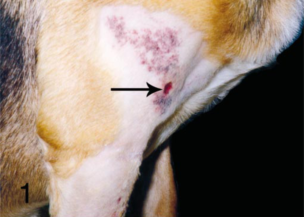
Skin; mixed-breed dog. Multiple, red, discolored lesions on the foreleg with punch biopsy site (arrow).
Histologically, there were multiple irregular capillaries and endothelial-lined cavernous blood vessels scattered throughout and infiltrating the superficial and deep dermis, surrounding adnexa, and extending into subcutaneous adipose tissue and muscle (Fig. 2). The proliferative blood vessels were filled with blood and were lined by a single layer of well-differentiated flattened endothelial cells (Figs. 3, 4). In some areas, occasional plump endothelial cells lined trabecular structures, which were often forming vascular channels (Fig. 5). The majority of the proliferative vascular channels were filled with erythrocytes, and some blood vessels contained fibrin thrombi, which partially or entirely occluded the lumina. Endothelial cells of proliferative blood vessels were supported by a single to double layer of smooth muscle cells, which were demonstrated by immunohistochemical staining of smooth muscle actin (Fig. 6) and desmin (not shown). Trichrome stain revealed collagenous matrix supporting and separating individual blood vessels. Occasional mild to moderate infiltration of neutrophils was present in the dermis adjacent to or in the proliferative vessels. Regionally, the epidermis was raised, moderately hyperplastic, often covered by hyperkeratotic crusts, and had rete pegs separating the blood vessels.
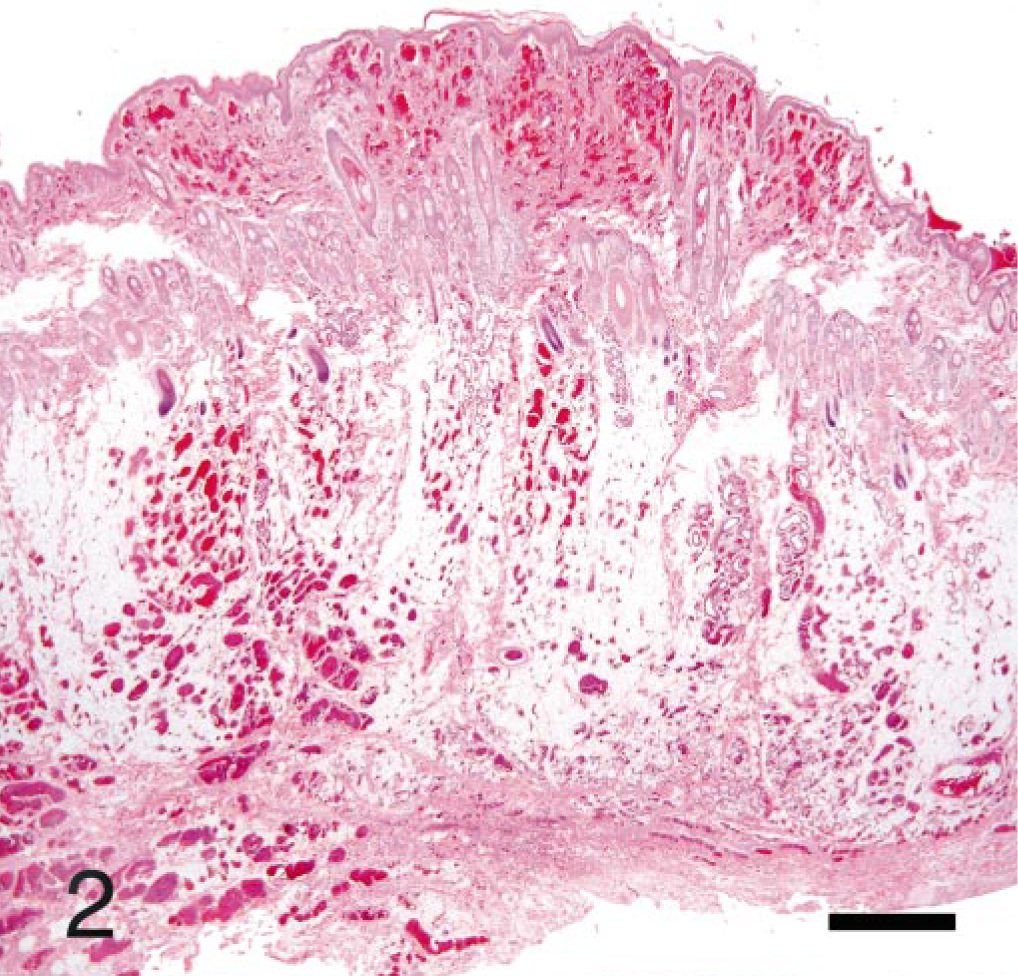
Skin from right shoulder; mixed-breed dog. Nonencapsulated, poorly demarcated, proliferative small caliber vascular channels in the superficial and deep dermis extending to the underlying subcutis. Note the infiltrative-like nature. HE. Bar = 600 μm.
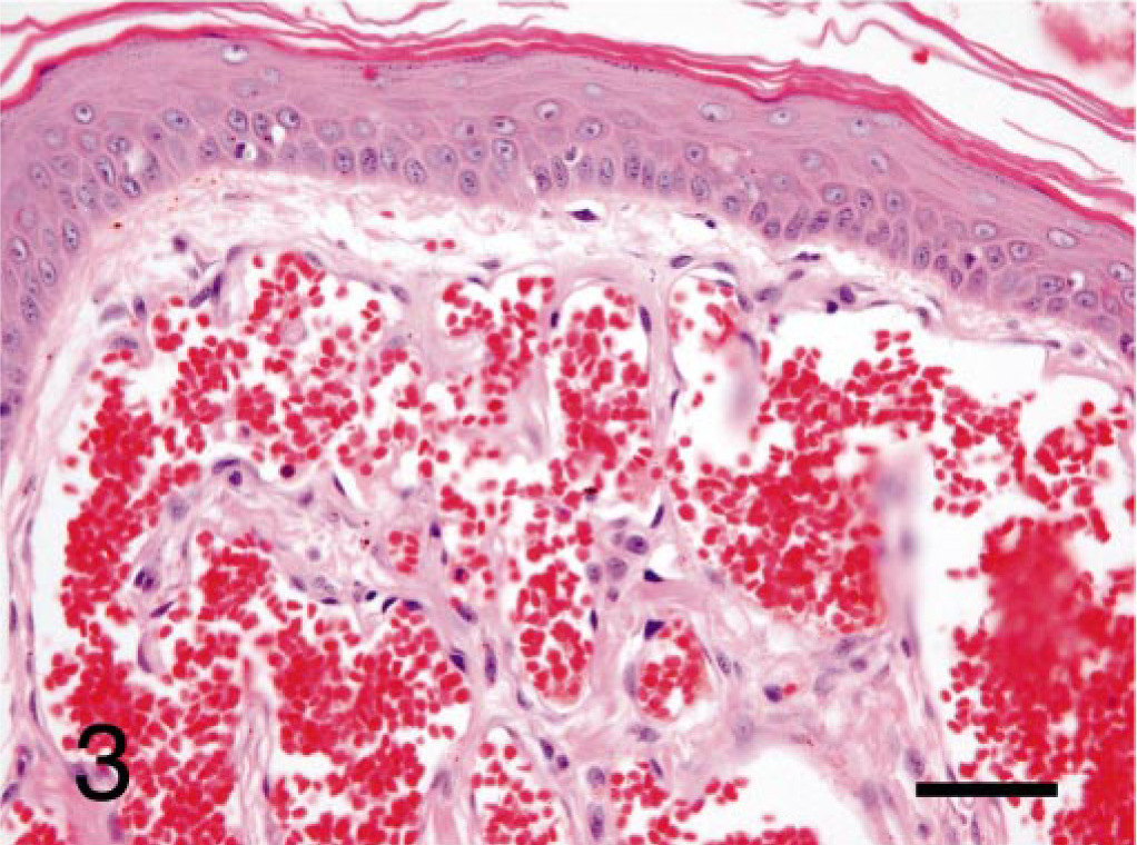
Skin from right shoulder; mixed-breed dog. Proliferative small caliber vascular channels lined by flattened endothelial cells are replacing normal architecture of the superficial dermis. Vascular channels are filled with erythrocytes. HE. Bar = 100 μm.
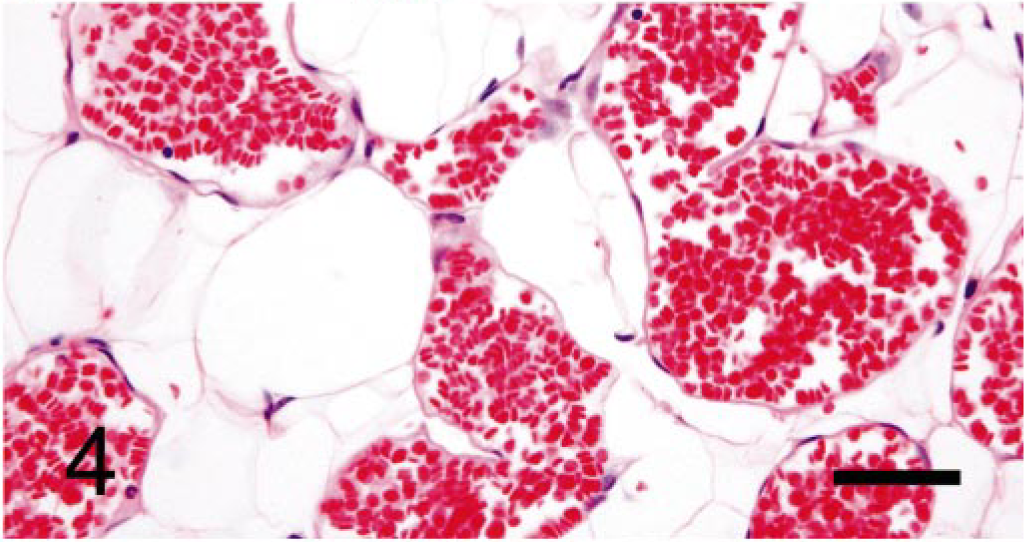
Skin from right shoulder; mixed-breed dog. Proliferative small caliber vascular channels lined by flattened endothelial cells are diffusely scattered in mature adipose tissue of the subcutis. HE. Bar = 100 μm.
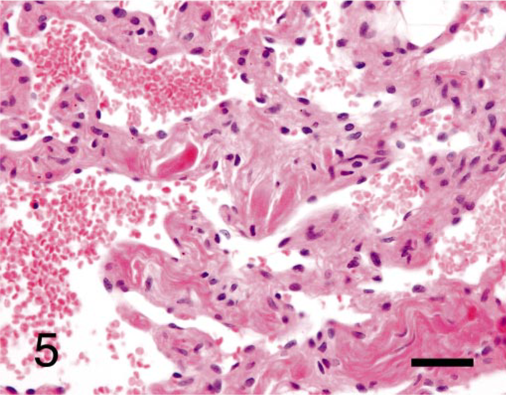
Skin from right shoulder; mixed-breed dog. Note that some blind-ended trabeculae lined by plump endothelial cells are forming vascular channels, which contain erythrocytes. HE. Bar = 80 μm.
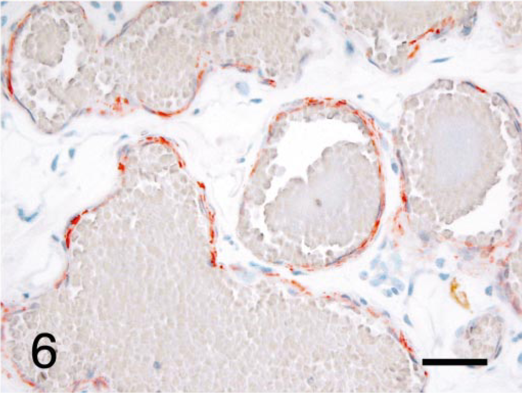
Skin from right shoulder; mixed-breed dog. Endothelial cells of small caliber vascular channels are supported by one or two layers of smooth muscle cells, which are positively labeled with smooth muscle actin antibody. Ventana NexES system with I-View DAB kit. Bar = 100 μm.
On the basis of the histopathologic findings, the lesions in the excisional biopsy from the antebrachium and punch biopsy of the shoulder were diagnosed as hemangiomas, cavernous-type, dermal, and subcutaneous. We believed these to be benign lesions, although the multicentric distribution and widely disseminated and infiltrative nature raised some concern and uncertainty about the biological potential of these lesions. Such an infiltrative and disseminated pattern of benign-appearing blood vessels has not been described in the dog. The benign nature of the lesions was supported by the organized structure of the vessels demonstrated by the immunohistochemical and collagen stains. The vessels were typically lined by a layer of well-differentiated flattened endothelium, with support by single to double layer of smooth muscle cells that were further supported by collagenous matrix. Now, almost 6 years after the excision of the antebrachial lesion, the dog remains healthy, and the surgical sites have healed completely, with no progression of the dorsal antebrachial lesion or shoulder lesions beyond the original site. There was slight enlargement and thickening of the lesions since the initial presentation may have been related to vascular congestion, chronic inflammation from self-induced trauma, and the occurrence of multifocal thrombosis. A few, similar appearing 0.5–1 cm, small, firm nodules exist at the area of the surgical site on the antebrachium.
The clinical and microscopic features of the skin lesions in this dog are consistent with benign vascular proliferations and are similar regarding age of onset, locations of the lesion, gross and histopathologic findings, and biological prognosis to that described as angiomatosis in children. 2, 11 In humans, more than half of the cases occur in the lower extremities, followed by the chest wall, abdomen, and upper extremity. 11 Human angiomatosis is a diffuse form of hemangioma that affects a large segment of the body in a contiguous fashion, either by vertical extension to involve multiple tissue planes or by crossing muscle compartments to involve similar tissue types. 2, 11 The occurrence in childhood and young animals suggests a possible dysontogenic origin, but the endothelial cells lining the blood vessels were often immature, which is not compatible with hamartoma. 1 There appear to be two main histologic patterns in human angiomatosis, a mixed vessel type and a capillary-predominant type; and either form is accompanied by increased amounts of adipose tissue. 11 In humans, the diagnosis of angiomatosis should be on the basis of clinicopathologic correlation, rather than pure morphology, which may be indistinguishable from juvenile capillary hemangioma, arteriovenous hemangiomas, or intramuscular angioma. 2
Peavy et al. described cutaneous angiomatosis in a 7-year-old, spayed, female, mixed-breed dog, with diffuse involvement of the skin and oral mucosa on the right side of the muzzle extending from the medial canthus of the right eye to the nasal planum. 7 The lesions were multiple, fluid-filled spaces that coalesced into several spongy plaques. The lesions were progressively proliferative and composed of vascular tissue involving the dermis and subcutaneous tissues. After treatment using laser photocoagulation, the lesions were completely healed with no recurrence. The histologic findings of the cutaneous angiomatosis were similar to those observed in this case. Although it is not clearly known whether these conditions are the same disease entity or not, our case appeared to be different because of the age of onset.
Recently, a few cases of angiolipomatous tumors located on the thorax were described in dogs and cats. 6 The angiolipomas were solitary subcutaneous nodules composed of thin-walled blood vessels randomly distributed throughout lobules of well-differentiated adipose tissue. The tumors were composed of variable amounts of mature adipose tissue, blood vessels, and bundles of collagenous connective tissue. Infiltration into and disruption of adjacent skeletal muscles were also observed in the angiolipoma in dogs. 6 Human and canine angiolipomas are considered to be benign neoplasms, and complete surgical excision is curative. Histologic findings in the subcutis of this dog were similar to those described in angiolipomatous tumors; however, the nondiscrete nature, extension and involvement of superficial and deep dermis and subcutis in our case were distinctive.
Variants of hemangiomas have also been reported in other animals and were considered for comparison. Vascular hamartoma, defined as a tumorlike malformation composed of mature or nearly mature blood vessels in various organs including skin, has been widely reported in animals. Histologically, dermal vascular hamartoma contains a poorly circumscribed proliferation of blood vessels ranging from large hyperplastic arteries to capillaries. 3 In young horses, cutaneous capillary-type hemangiomas characterized by scattered, well-demarcated but nonencapsulated lobules have been described. 8, 9 This equine variant was located in the extremities. Bovine cutaneous angiomatosis occurs in young adult cattle and is thought to be either an abnormal repair process to injury or an idiopathic hamartoma. 10 Typically, the bovine condition is a nonencapsulated mixture of arteries, veins, and capillaries that is separated by scattered fibroblasts and variable amounts of collagen. 5, 10 In contrast, the lesion in this dog was primarily composed of blood vessels of similar type and caliber.
To the authors' knowledge, angiomatosis in a young dog with extensive involvement of the forelimb has not been described. Although some histologic features of this case raised concerns of possible malignancy, the lack of pleomorphism of this congenital vascular proliferation and the nonprogressive behavior were features indicative of benignancy. Therefore, surgical excision of cutaneous angiomatosis is probably unnecessary, but if there are secondary complications it may be indicated. This condition should be considered in the differential diagnosis of vascular lesions of the skin and should be distinguished from well-differentiated cutaneous hemangiosarcoma.
Footnotes
Acknowledgements
We thank Sandra Horton and Monica Mattmuller in the histology laboratory of the North Carolina State University, Veterinary Teaching Hospital, for the expert preparation of the slides and performance of the special staining techniques.