
Abstract
A spontaneous case of renal heterotopia involving the lung parenchyma of a free-living, adult, female common dolphin (Delphinus delphis), which was found stranded alive on the North Adriatic Sea coast of Italy, is reported in this study. The lesion, slightly visible from the macroscopic point of view, had the histologic appearance of a ''foreign tissue island,'' which was poorly demarcated from the surrounding pulmonary tissue. Within such an island, several regularly shaped and apparently mature kidney glomeruli and tubules could be observed, with no evidence of secondary tissue reaction. To the best of our knowledge, this should be the first description of heterotopic kidney tissue occurrence in the lung of any domestic or wild animal species.
Keywords
Heterotopic kidney tissue (HKT) is a pathologic change occurring when evidence of kidney or kidneylike parenchyma is found within extrarenal organs or tissues in the presence of two functional, otherwise normally located kidneys. 8 HKT has been reported, as an extremely rare and incidental finding, in the human adrenal gland, 5 the human heart, 6 the human lumbosacral region, 1 and the human colon, 4 with the lesion being more frequently encountered in the pelvis rather than the thoracic cavity. 7 To the best of our knowledge, HKT has never been reported, so far, in any domestic or wild animal species.
We describe in this study spontaneous HKT occurrence within the lung parenchyma of an adult, female common dolphin (Delphinus delphis), which was found stranded alive in October 2000 on the North Adriatic Sea coast of Italy.
Postmortem examination was carried out 36 hours after the spontaneous death of the animal after 15 days of maintenance in captivity under proper intensive care conditions. Several lung tissue samples, along with those obtained from major organs, were promptly fixed in 10% neutral-buffered formalin. These were subsequently embedded in paraffin and cut into 5-μm-thick sections, which were routinely stained with hematoxylin and eosin. A number of selected pulmonary tissue sections were also submitted for appropriate histochemical staining techniques (Masson's trichrome, periodic acid-Schiff [PAS]) and finally observed under a light microscope.
Macroscopically, moderate autolysis was observed, along with a number of larval parasitic cysts involving the abdominal musculature. Some adult nematodes were found within bronchial lumina. Further gross lesions included an intra-vaginal mineralized concretion measuring 7 cm × 4.5 cm, along with hemorrhagic foci on the intestinal mucosa. This also exhibited a segmental chronic enteritis associated with focal chronic peritonitis and perihepatitis as well as with areas of liver discoloration and softening suggestive of multifocal necrosis. No additional macroscopic changes were found.
Histologically, prominent interstitial (peribronchial, peri-bronchiolar, and interalveolar) fibrosis was observed. The surrounding lung parenchyma had moderate to severe alveolar emphysema, which sometimes showed a subpleural location. Variably sized parasitic granulomas were identified, with some of these lesions in the subpleural regions of the lungs. Further examination of HE-stained lung tissue sections revealed the presence of a single nodular lesion, which was also macroscopically visible on serial slides cut from the same block. The concerned area was indeed a “foreign tissue island” that was not encapsulated and, thereafter, poorly demarcated from the surrounding lung parenchyma (Fig. 1). Within this island, several regularly shaped and apparently mature kidney glomeruli and tubules could be observed, with some glomerular structures showing a variable thickening of their Bowman's capsules and capillary basement membranes (Fig. 2). No tissue reaction secondary to the presence of this heterotopia could be observed. In addition, no changes suggestive of tissue artifacts were found.
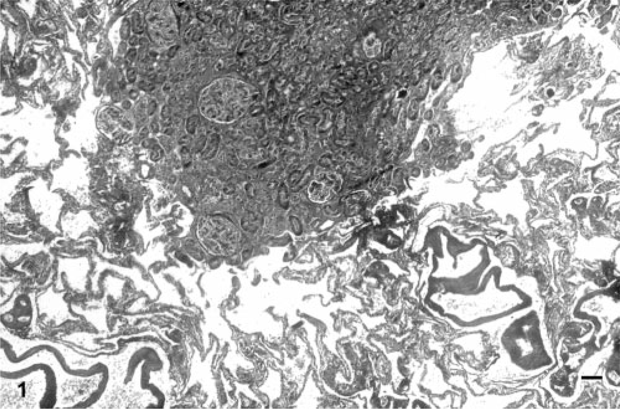
Lung; common dolphin (Delphinus delphis). HKT. The concerned foreign tissue island harbors several regularly shaped, apparently mature, renal glomeruli and tubules, that are not encapsulated and poorly demarcated from the surrounding lung parenchyma. Masson's trichrome. Bar = 120 μm.
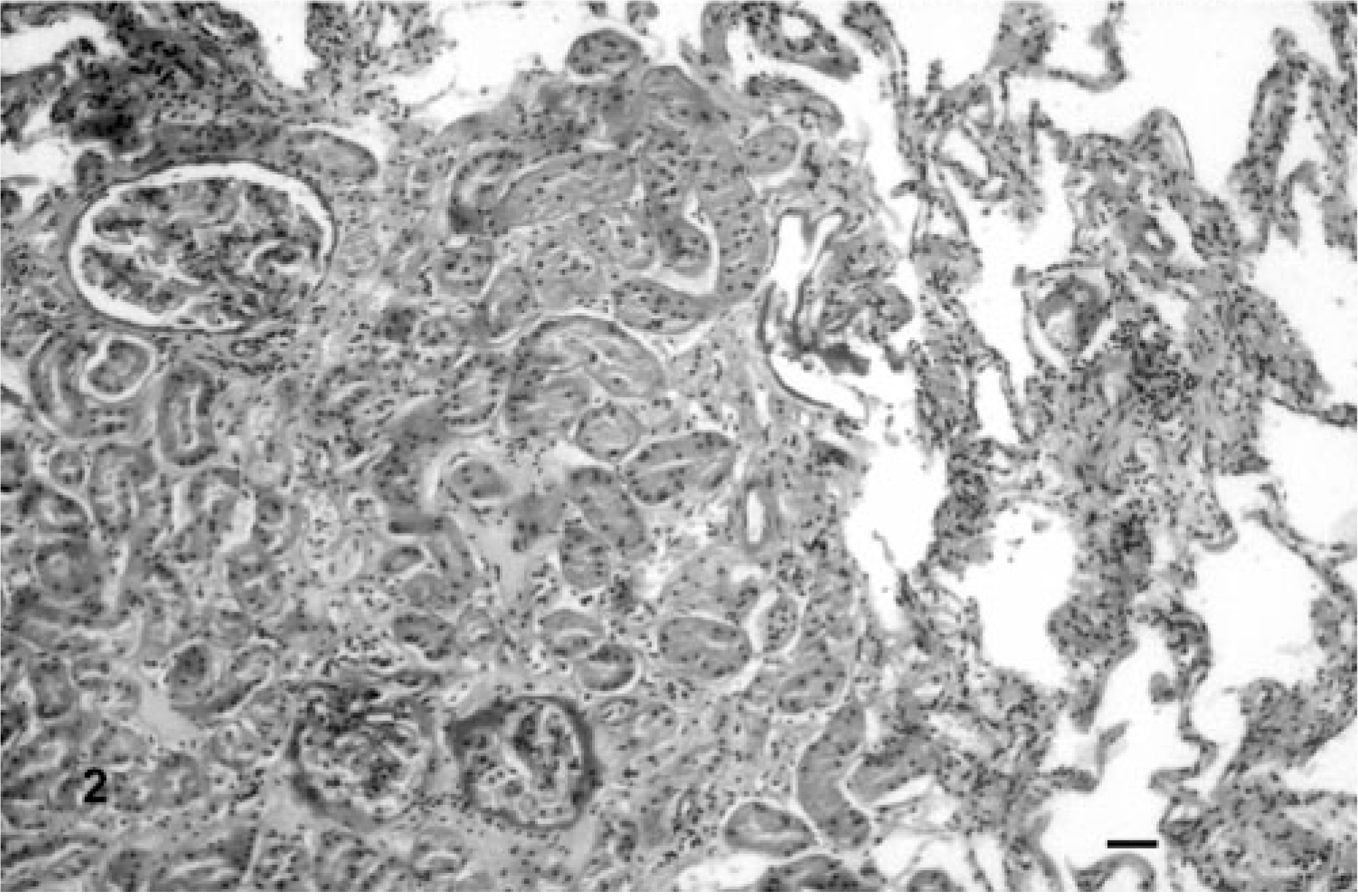
Lung; common dolphin (Delphinus delphis). HKT. Higher magnification appearance of the lesion consisting of well-developed and mature renal tissue. Some glomeruli show variably thickened Bowman's capsules and basement membranes. No tissue reaction secondary to this heterotopia is observed. Periodic acid-Schiff. Bar = 60 μm.
Microscopic examination of the other major organs confirmed all the aforementioned gross pathology findings. Furthermore, single hyaline, weakly PAS-positive intracytoplasmic inclusions similar to those reported in striped dolphins (Stenella coeruleoalba), for the first time, by Domingo et al. 3 were detected in some hepatocytes.
No additional laboratory examinations were carried out.
On the basis of the above results, associated with the presence of two normally located and shaped kidneys, a diagnosis of intrapulmonary HKT was made.
More in detail, the intrapulmonary HKT case in the dolphin under study showed morphologic features largely justifying its classification into the category of “renal tissue imperceptibly mixed with normal tissue,” with the other two categories being represented by “grossly visible nodules of often disorganized renal tissue” and by “teratomas with a HKT component,” respectively. 4 Renal tissue imperceptibly mixed with normal tissue, as in the case reported here, should be regarded as an exceptional and often incidental finding, which is commonly detected in humans associated with other congenital anomalies. 4 In this case, no other developmental defects were observed in the cetacean under study.
Interestingly enough, the intrapulmonary HKT described in this dolphin was not encapsulated and poorly demarcated from the surrounding lung parenchyma, showing lack of any inflammatory reaction. This would suggest, similarly to that reported in a previous study, 6 that any glomerular filtrate formed was resorbed, thus preventing its leakage into the neighboring pulmonary tissue.
This “serendipitous” and absolutely exceptional pathologic finding apparently had no influence in causing the cetacean's stranding and subsequent death, which was most likely caused by respiratory failure because of chronic progressive pneumopathy. However, we would still like to emphasize its potential value and use in comparative pathology studies. This is especially true as far as both HKT etiology and pathogenesis are concerned, provided that human HKT has been also hypothesized to undergo neoplastic transformation. 4
As a concluding remark, in open contrast with their denomination, common dolphins have experienced a dramatic numerical reduction in the Mediterranean Sea during the past three or four decades. 2 The reasons for such population's decrease are poorly understood. Nevertheless, the likely reduction of genetic variability secondary to the above phenomenon should be adequately taken into account, among others, as a possible explanation for the occurrence, in the concerned cetacean species, of developmental defects such as the one reported here.
Footnotes
Acknowledgements
The outstanding technical assistance of Prof. Leonardo Della Salda, DVM, Dipl. ECVP, is greatly appreciated.