
Abstract
This article describes the gross, histopathologic, and ultrastructural findings of the livers of cetaceans stranded on the coast of the Canary Islands between 1992 and 2000. A total of 135 cetaceans were included in the study, among which 25 were common dolphins (Delphinus delphis), 23 Atlantic spotted dolphins (Stenella frontalis), 19 striped dolphins (Stenella coeruleoalba), and 15 other species of dolphins and whales. The most common lesion observed in these animals was a nonspecific chronic reactive hepatitis (47/135), followed by hyaline intracytoplasmic inclusions in hepatocytes (33/135). Parasitic cholangitis was detected in 8/135 animals, whereas hepatic lipidosis was presented in 7/135 animals. The ultrastructure of hyaline hepatocytic cytoplasmic inclusions is described, and possible causes of these inclusions are discussed.
Information on cetaceans found stranded on the coast of the Canary Islands has been collected by the Histology and Pathology Unit of Las Palmas of Gran Canaria University during an 8-year period (1992–2000). One hundred and thirty-five cetacean strandings occurred on the shores of these islands, involving both single and multiple strandings of adults and juvenile cetaceans, but to date there have been no surveys of the causes of illness in the wild, free-living dolphin and whales found dead in the Canary Islands.
In the study of the stranded cetaceans, the liver, as in other studies, 2,12,13 was extensively analyzed and a variety of lesions that merited further investigation were found. Chronic liver disease of unknown cause is a common ailment in dolphins and is clinically characterized by wasting but is rarely characterized by jaundice. Hepatic lesions consist of hepatocyte degeneration, fatty change, and fibrosis. 33 Chronic liver disease is generally regarded as being nutritional in origin, but there is no definite evidence to support this. 19,33 Different types of acute hepatitis associated with nutritional or environmental toxins and biologic agents, such as viruses, bacteria, fungi, and parasites, have also been reported in stranded dolphins. 30
Parasitic infections have been recognized amongst the pathologic changes found in marine mammals. 34 Campula spp. is a common trematode that primarily inhabits the bile and pancreatic ducts, stomach, and intestine of cetaceans. 36 Affected ducts are dilated, fibrosed, and infiltrated with mononuclear cells, and the epithelium is hyperplastic. 8,27
Dolphins, as other cetaceans, are at the top of the marine food chain, and they accumulate pollutants in their tissues during their life. Therefore, this species is an excellent bioindicator to evaluate contamination of the marine environment. Numerous studies have been carried out to analyze concentrations of pollutants in dolphins from different areas of the world. 1,4–6,16 Despite some studies that have described hepatic lesions in stranded dolphins, 2,9,12,22,29 histopathologic studies of the liver in a large series of animals have not been conducted.
The aim of this article is to describe the gross, histopathologic, and ultrastructural features of the livers of cetaceans (dolphins and whales) found stranded along the coasts of Canary Islands between 1992 and 2000.
Materials and Methods
A total of 135 stranded cetaceans were included in the study, 25 of which were common dolphins (Delphinus delphis), 23 were Atlantic spotted dolphins (Stenella frontalis), 19 were striped dolphins (Stenella coeruleoalba), and the remaining 68 animals represented 15 different cetacean species (Table 1). Necropsies were carried out according to standard protocol for cetaceans. 24 A range of tissues were fixed in neutral-buffered formalin. For this study, three samples from each liver were routinely processed for histologic examination. Selected sections were stained for the presence of morbillivirus antigen. 21 For ultrastructural study, liver tissue initially fixed in formalin was postfixed in osmium tetroxide (pH 7.4) and embedded in epoxy resin. Semithin sections were cut and stained with toluidine blue for light microscopic examination. Ultrathin sections were cut from selected blocks, stained with lead citrate and uranyl acetate, and examined using a Philips CM-10 transmission electron microscope. In cases where liver parasites were found, they were processed, gold sputtered, and examined using a Jeol JSM 6300 scanning electron microscope.
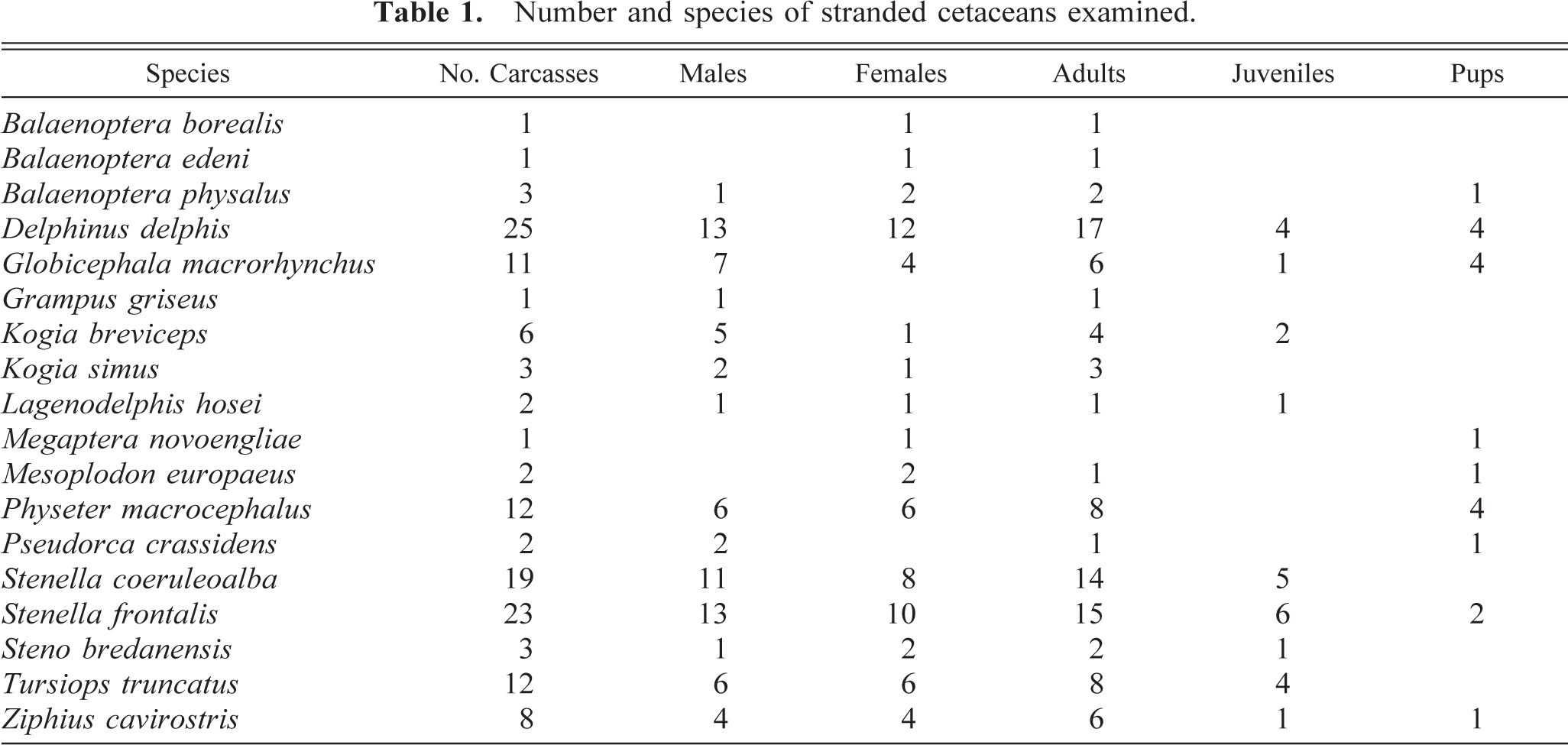
Number and species of stranded cetaceans examined.
Results
Pathologic findings are summarized in Table 2. The most common hepatic lesion was a nonspecific chronic reactive hepatitis, which was diagnosed in 47 animals, 17 of which were common dolphins, 11 Atlantic spotted dolphins, 10 striped dolphins, 7 bottle-nosed dolphins, 1 rough-toothed dolphin, and 1 false killer whale. The only macroscopic evidence of this disease was a mild enlargement of the liver. The histologic appearance of this lesion was a diffuse inflammatory infiltrate in portal areas, around the hepatic veins, and within hepatic sinusoids. This inflammatory infiltrate varied from mild to severe in the different cases, and it was composed of lymphocytes and plasma cells (Fig. 1).
Hepatic lesions recorded in cetaceans.
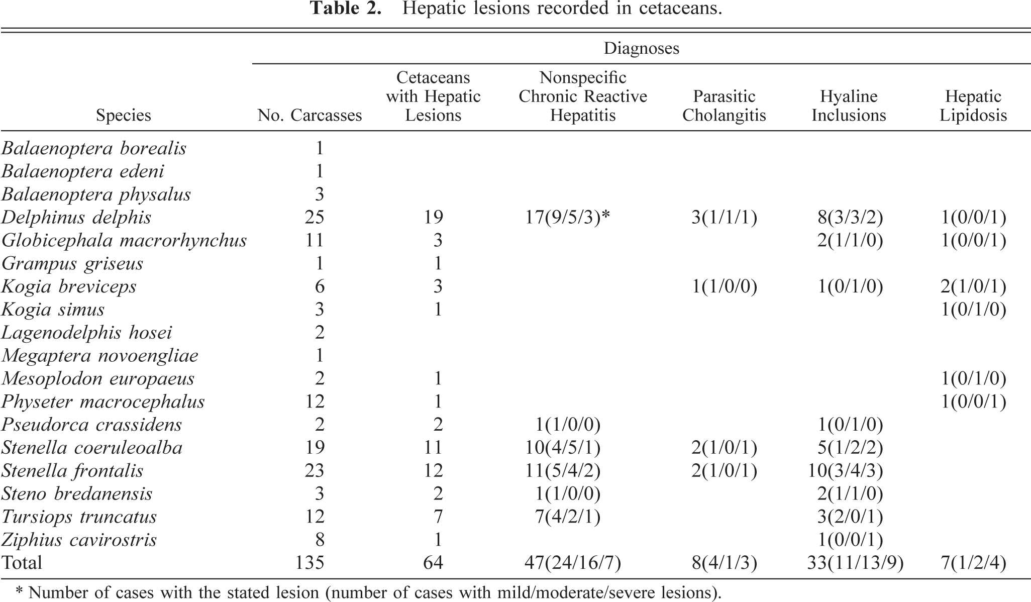
∗ Number of cases with the stated lesion (number of cases with mild/moderate/severe lesions).
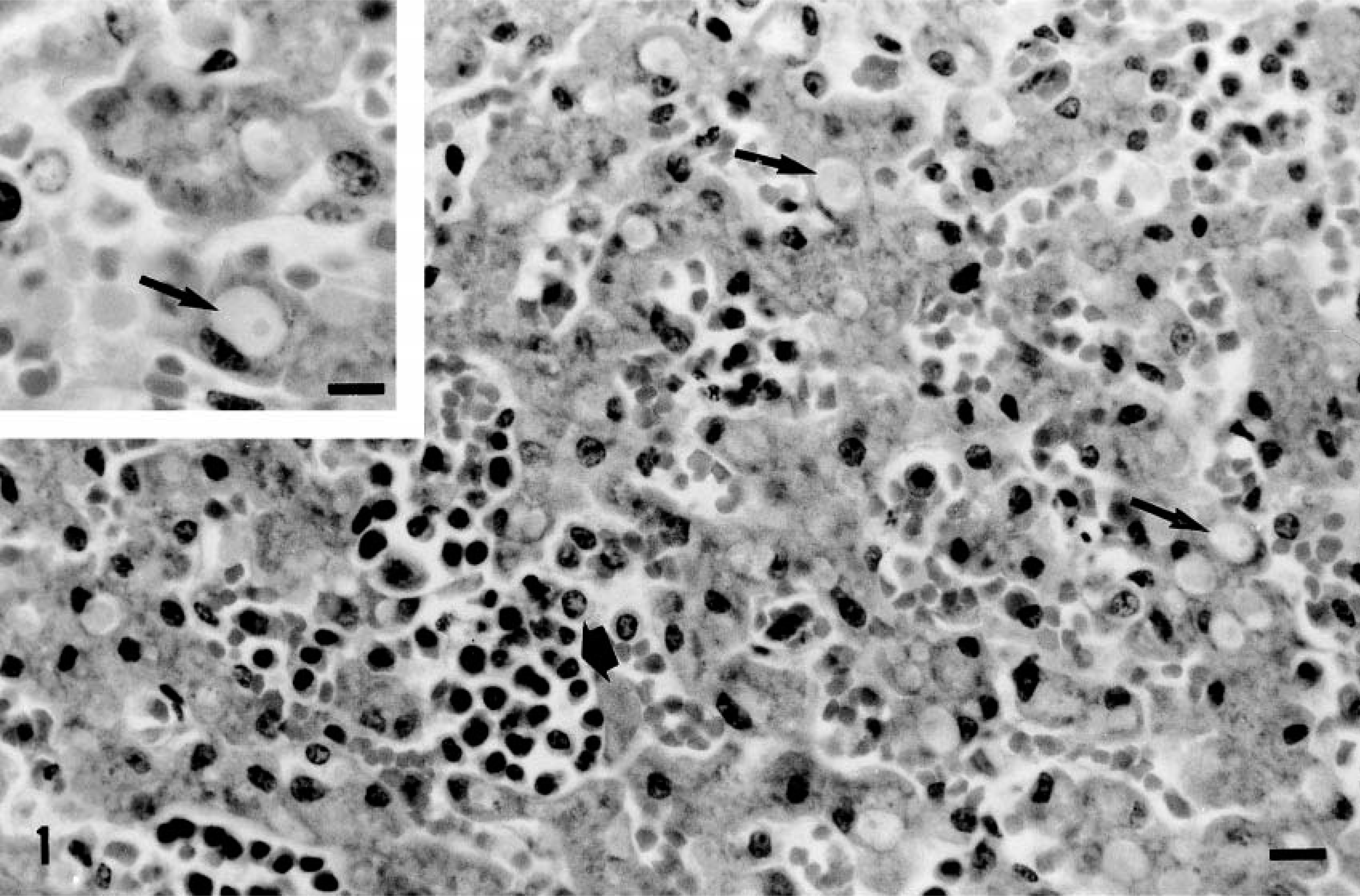
Steno bredanensis. Nonspecific chronic reactive hepatitis showing lymphoplasmacytic infiltrate within hepatic sinusoids (thick arrow). Numerous hyaline globules are found in hepatocytes (arrows). HE. Bar = 22 µm. Inset: detail of hyaline globules (arrow) displacing the nucleus to the periphery. HE. Bar = 15 µm.
Trematodes identified as Campula spp. were found in bile ducts of three common dolphins, two striped dolphins, two Atlantic spotted dolphins, and one pygmy sperm whale. These trematodes produced severe damage to the bile ducts, eliciting focal suppurative cholangitis, severe necrotizing cholangitis, or chronic granulomatous cholangitis.
Focal suppurative cholangitis was observed in one Atlantic spotted dolphin. This focus contained parasite ova and was surrounded by fibrotic tissue with a mixed inflammatory infiltrate composed of numerous neutrophils and plasma cells more peripherally. This inflammation extended to the adjacent parenchyma along portal spaces.
One common dolphin had a severe multifocal chronic necrotizing cholangitis of parasitic cause (Fig. 2). Grossly, the visceral surface of this liver showed enlarged bile ducts containing numerous trematodes identified as Campula spp., several whitish foci, and a 4 × 7 cm in diameter cyst also containing numerous trematodes. Histopathologic analysis revealed multiple necrotic foci in the bile ducts, with surrounding abundant fibrosis and inflammatory cells, predominantly eosinophils, lymphocytes, and plasma cells. The bile ducts contained numerous parasite eggs and some trematodes (Fig. 2).
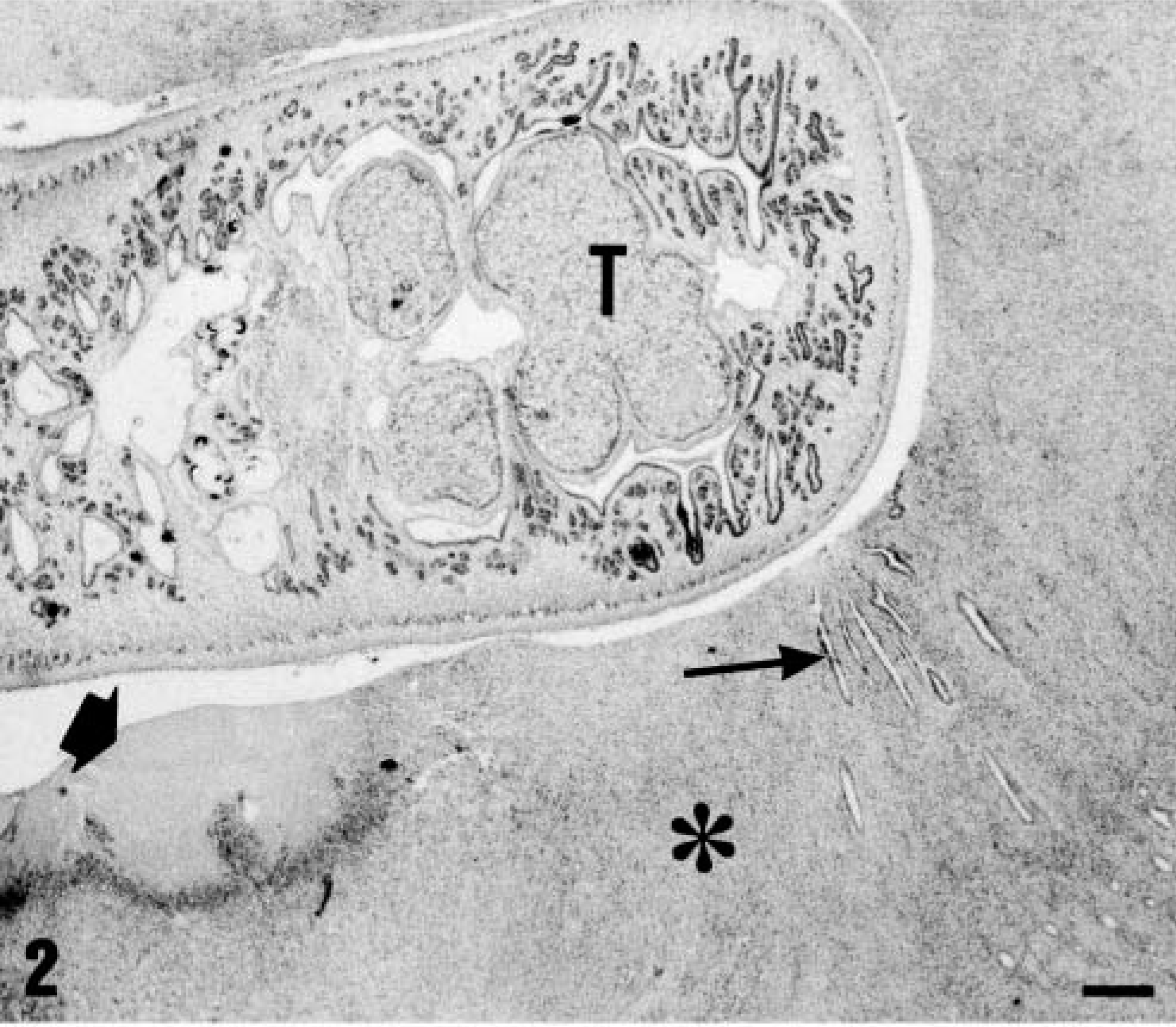
Stenella coeruleoalba. Severe necrotizing cholangitis showing a trematode (T) adjacent to a necrotic band (thick arrow) and severe inflammatory infiltration (asterisk) among proliferating bile ducts (thin arrow). HE. Bar = 190 µm.
Parasitic chronic granulomatous cholangitits was detected in two striped dolphins, two common dolphins, one Atlantic spotted dolphin, and one pygmy sperm whale. Grossly, these livers showed enlarged bile ducts with multiple whitish solid foci ranging from 1 to 4 cm in diameter. Histopathologically, the lesions were characterized by the presence of numerous parasitic eggs surrounded by macrophages, multinucleated giant cells, lymphocytes, and plasma cells in ruptured bile ducts and portal stroma (Fig. 3). Portal bridging fibrosis was also observed (Fig. 4). In these areas, fibrosis was accompanied by bile duct proliferation and marked infiltrates of plasma cells and lymphocytes, admixed with multifocal to coalescing lymphoid follicle formation with germinal centers (Fig. 5).
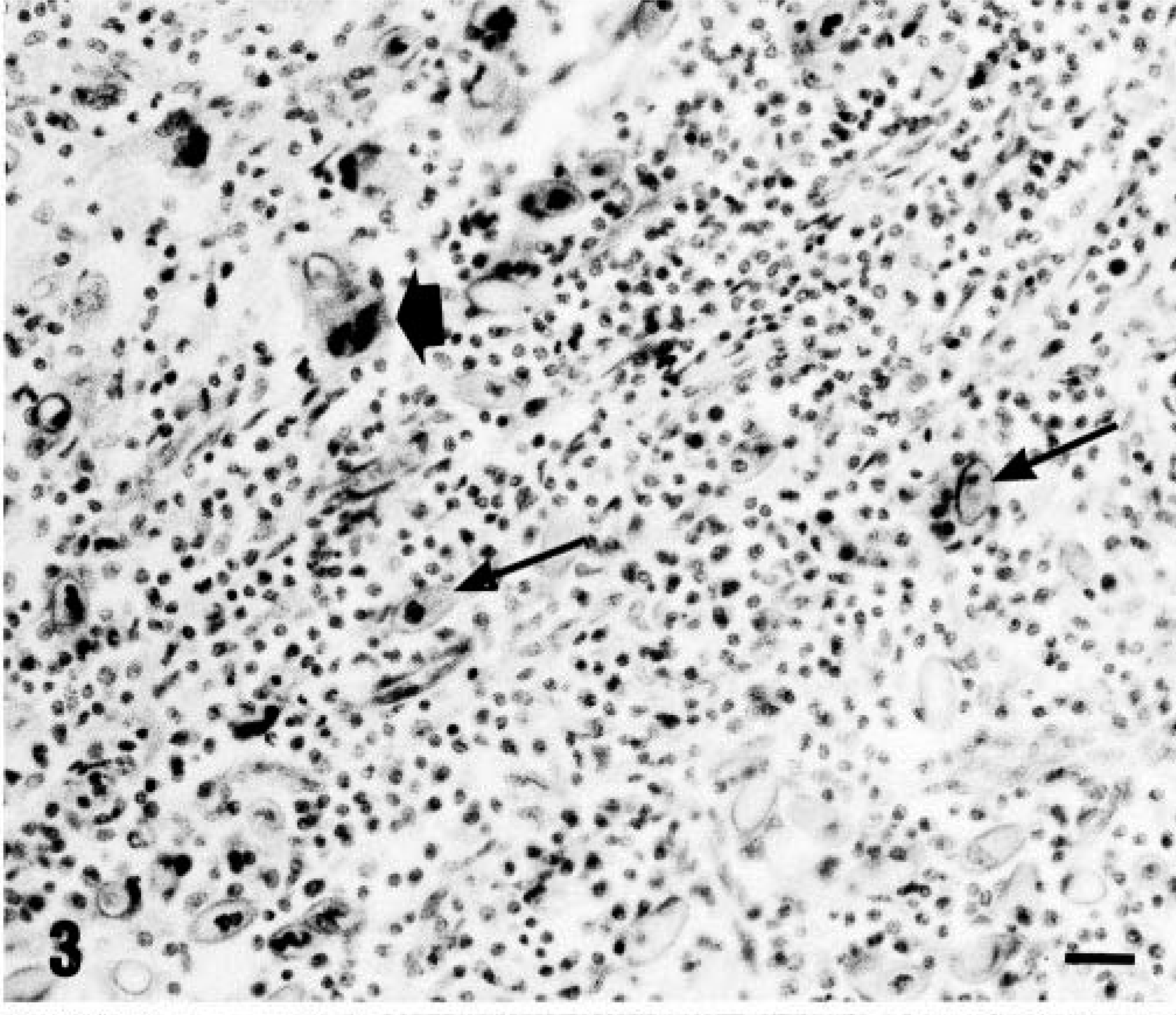
Stenella coeruleoalba. Parasitic cholangitis with numerous parasite eggs (arrows), with occasional multinucleate giant cells, some of which contained egg debris (thick arrow) and severe lymphoplasmacytic infiltrate. HE. Bar = 36 µm.
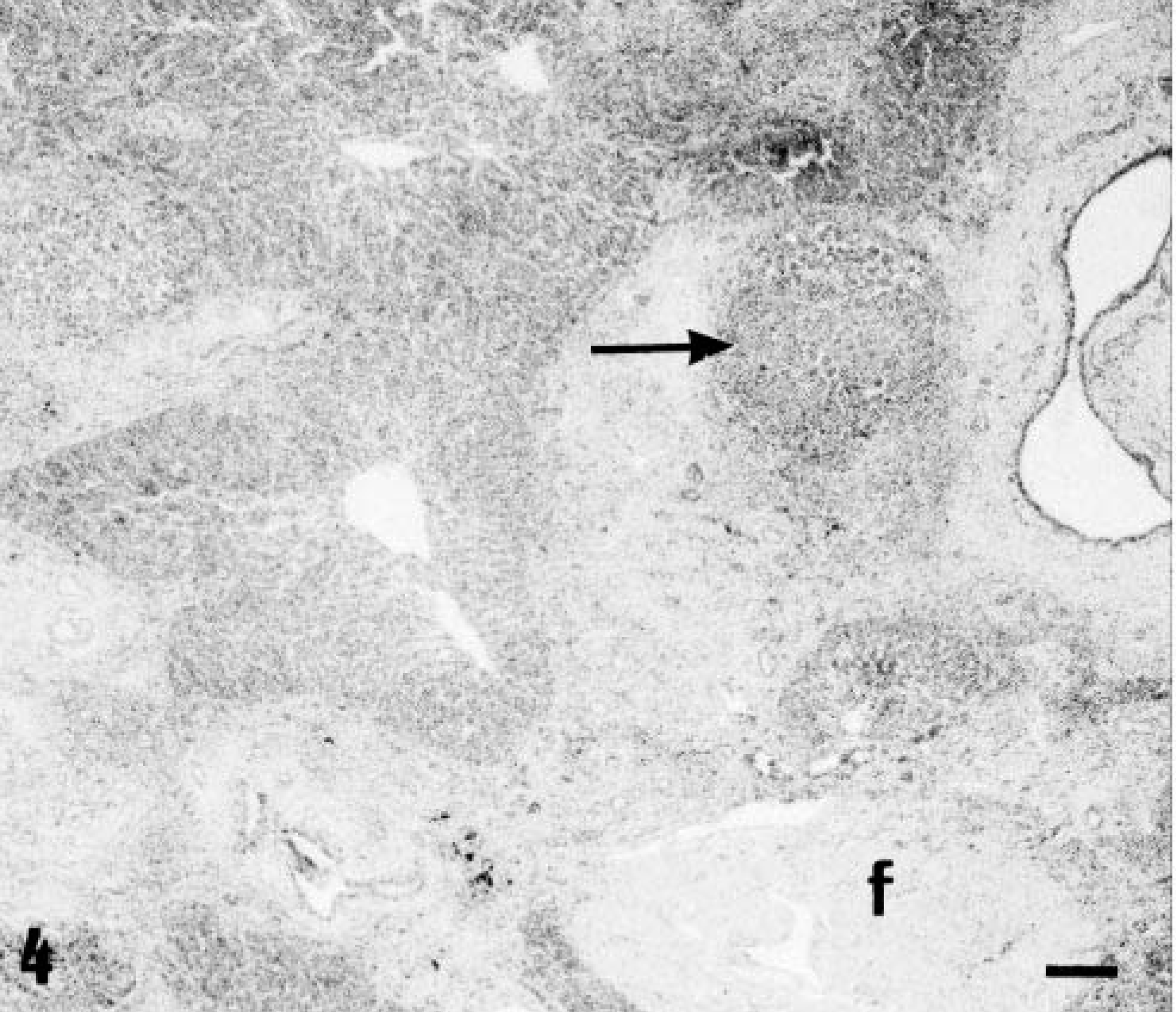
Stenella coeruleoalba. Severe chronic parasitic cholangitis with portal fibrosis (f) and formation of isolated lymphoid follicles (arrows). HE. Bar = 75 µm.
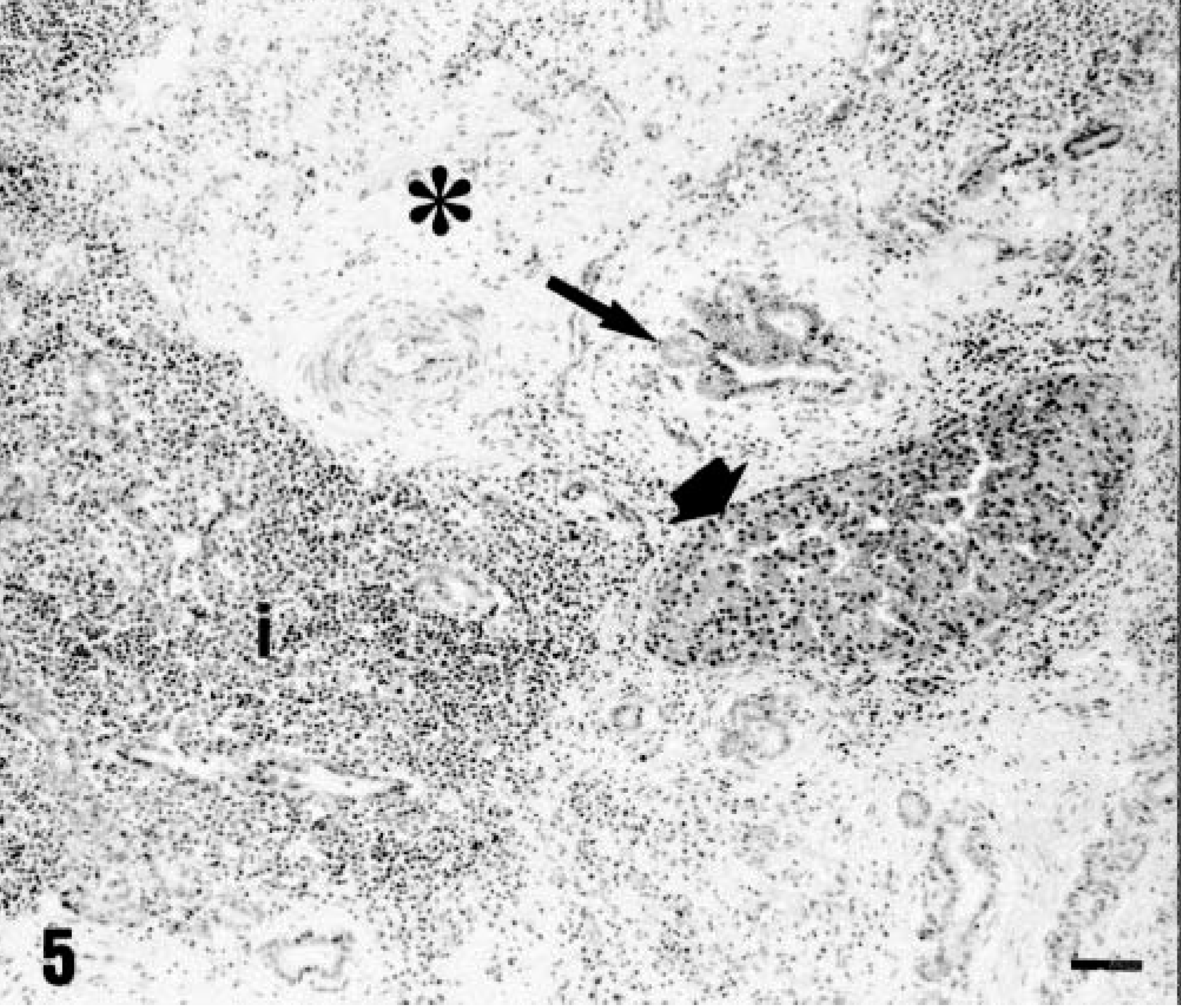
Stenella coeruleoalba. Chronic parasitic cholangitis with portal fibrosis (asterisk), bile duct proliferation (arrow), diffuse inflammation (i) and distortion of hepatic lobules (thick arrow). HE. Bar = 45 µm.
Hepatocytes containing cytoplasmic hyaline inclusions were observed in 33 animals: 10 Atlantic spotted dolphins, 8 common dolphins, 5 striped dolphins, 3 bottle-nosed dolphins, 2 rough-toothed dolphins, 2 short-finned pilot whales, 1 pygmy sperm whale, 1 false killer whale, and 1 Cuvier's beaked whale. Cytoplasmic inclusions were round to oval, moderately eosinophilic periodic acid–Schiff positive, and ranged from 4 to 20 µm in diameter, often displacing the nucleus of the hepatocyte to the periphery of the cell (Fig. 1). Inclusions were randomly scattered through the hepatic parenchyma in varying numbers, usually as single, but occasionally multiple inclusions. A central or eccentric densely eosinophilic staining core was frequently apparent in some cases. By transmission electron microscopy, these inclusions were usually diffusely moderately electron dense but occasionally had a central or eccentric core of highly electron-dense material (Fig. 6).
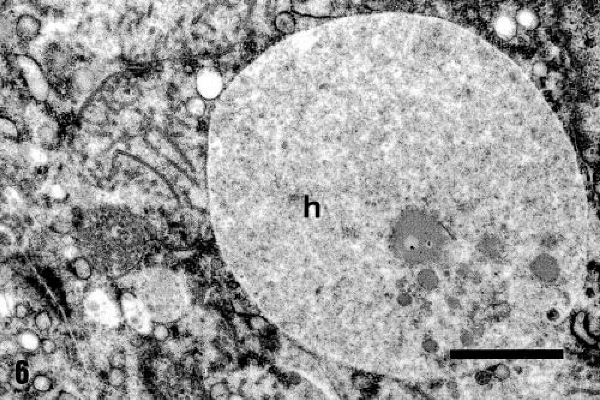
Delphinus delphis. Detail of a hepatocyte showing a intracytoplasmic hyaline inclusion (h). Transmission electron microscopy. Bar = 1.8 µm.
Moderate to severe hepatic lipidosis was observed in seven animals: two pigmy sperm whales, one sperm whale, one common dolphin, one short-finned pilot whale, one dwarf sperm whale, and one Gervais' beaked whale. These livers were diffusely pale. Diffuse hepatic lipidosis was observed in the majority of younger animals examined, but occasionally a periacinar distribution was observed. Neoplastic lesions were not seen in any of the livers examined. All tissue samples tested were negative for morbillivirus.
Discussion
The most common lesion in the cetaceans in this study was a nonspecific chronic reactive hepatitis, which was characterized by infiltration of lymphocytes and plasma cells thorough the liver parenchyma and in the portal or perivenular stroma with or without minimal hepatocytic necrosis. Nonspecific reactive hepatitis, which may be acute or chronic, has been reported as a type of hepatitis different from chronic active hepatitis in human beings 25,26,32 and dogs. 15,31 In both species, histopathologic features of nonspecific chronic reactive hepatitis were quite similar to that observed in the cetaceans of the present study. Several types of hepatitis, particularly nonpurulent, interstitial hepatitis and subacute to chronic pericholangitis, have been commonly found in wild, free-living, and captive dolphins. 2,9,12,30,32 However, nonspecific reactive hepatitis has not been reported in dolphins before this study. In human beings, nonspecific reactive hepatitis is a morphologic entity widespread within the liver, representing either the residuum of previous inflammatory intrahepatic disease or a response to a variety of extrahepatic disease processes, especially febrile illnesses and inflammation somewhere in the splanchnic bed. 25,26,32 In domestic animals, it is almost always related to systemic disease or diseases in the splanchnic bed. 15,31 In the cetaceans of the present study, this lesion may be related with previous inflammatory processes of parasitic, viral, or bacterial origin.
Chronic cholangitis was associated with parasites or parasite eggs within bile ducts or in the stroma of portal spaces. Morphologic features of parasites and eggs were consistent with Campula spp. 10,36 This trematode is commonly found in the liver and pancreas of cetaceans. 7,8,27,35 Mild infestation may be asymptomatic, but heavy infestations may produce biliary hyperplasia, portal fibrosis (or periductular fibrosis), and an infiltrate of lymphocytes, plasma cells, and eosinophils. 25 These changes were observed in the present study, although the most severe lesions were found when parasite eggs were found in the stroma of portal spaces causing severe granulomatous hepatitis. Perivascular clusters of lymphocytes and plasma cells present were most likely the result of persistent local or systemic presence of parasitic antigens.
Cytoplasmic hyaline inclusions in hepatocytes have been described in anesthetic accidents, internal hemorrhage, and right ventricular failure in human beings, 28 endotoxemia in dogs, 17 pyrrolizidine alkaloid toxicity in sheep and horses, 3 and hepatic necrosis in rabbits. 23 In cetaceans, hyaline cytoplasmic inclusions in hepatocytes were previously described in 33–82% of dolphins affected by morbillivirus in the Mediterranean Sea. 11,13,14 However, in accord with previous studies 20 morbillivirus antigen or viral particles were not detected in any of these inclusions from stranded cetaceans examined in our study; therefore, a relation between this virus and the cytoplasmic inclusions is unlikely.
In this study, most of the animals with inclusions suffered from an active stranding. During stranding, thoracic and abdominal vasculature compression compromises blood flow leading to acute to subacute liver congestion and subsequent hepatocellular hypoxia. The hyaline inclusions bear close similarity to those seen in human beings with congestive heart failure. 28 This suggests that the morphologic change is reversible and is not a direct effect of toxic pollutants, as previously postulated. 1,4–6,16
Hepatic lipidosis is a relatively common finding in stranded cetaceans with metabolic disorders produced by toxic injuries or nutritional deficiencies. 18 Stranded animals with severe starvation, or breast-feeding animals that have a dietary intake high in carbohydrates, may accumulate excessive triglycerides within hepatocytes. 18 The hepatic lipidosis observed in this study occurred in very young animals and, therefore, may be considered a physiologic change in these species. However, some of the animals showed zonal lipidosis, which could be related for example, to poor nutrition or a toxic insult, thus making it very difficult to establish a common etiology for the lesions in these animals.
Nonspecific reactive hepatitis, hepatic parasitism lesions, and hyaline intracytoplasmic inclusions were the main histologic findings in the livers of cetaceans analyzed in this study. Additional ultrastructural and toxicologic studies are under way to compare the presence of pollutants with hepatic lesions.
Footnotes
Acknowledgements
This work was supported by Ministerio de Ciencia y Tech-nología (REN 2002-04162-C02-01/MAR), Consejería de Educación, Cultura y Deportes del Gobierno de Canarias, and by Junta de Andalucía (AGR 137). The authors are also grateful to Dr. Jorge González and Dr. Eduardo Degollada for their assistance in collecting and providing some of the material included in this study.