
Abstract
Seminoma with metastasis was diagnosed in a spotted dolphin (Stenella frontalis) and an Atlantic bottlenose dolphin (Tursiops truncatus). Sertoli cell tumor and pheochromocytoma were also diagnosed in the spotted dolphin. The spotted and bottlenose dolphins were adult males that stranded and died on the coasts of northwest Florida and southeast North carolina, respectively. Neoplasia is infrequently reported in cetaceans. This is the first report of seminoma, Sertoli cell tumor, and pheochromocytoma in a dolphin, the first report of three distinct neoplasms in a dolphin, and one of the few reports of malignant neoplasia in dolphins.
Keywords
There are few reports of neoplasia in cetaceans. Two reviews 1, 3 enumerate only 80 confirmed neoplasms in cetaceans. Fifteen of these were considered malignant. Twenty-eight of the 80 tumors (35%) were found in beluga whales (Delphinapterus leucas) from the St. Lawrence estuary. 7 This report describes malignant seminoma with metastasis, Sertoli cell tumor, and pheochromocytoma in a spotted dolphin and malignant seminoma with metastasis in an Atlantic bottlenose dolphin.
A spotted dolphin stranded alive on the southeastern shore of Choctawhatchee Bay, Florida, in July 1993. Clinical signs included dyspnea, back arching, and profuse diarrhea. The dolphin died shortly after arrival at the oceanarium, and the necropsy was performed within an hour of its death. The dolphin was 210 cm long and the teeth were worn, suggesting that the animal was a mature adult. At necropsy, the left testis was markedly enlarged (58.0 cm in length, 8.4 kg) (Fig. 1) and displacing the intestines. The cut surface had multiple, firm gray-white nodules, measuring up to 8 cm, with interspersed areas of hemorrhage. The right testis was small (34.5 cm in length, 1.0 kg) and had a smooth gray exterior and uniform tan parenchyma. Numerous gray-white nodular masses (resembling those in the left testis) were present in the retroperitoneal area extending into the adjacent muscle. Also, both adrenal glands were enlarged. Specimens of heart, lung, stomach, liver, spleen, adrenal gland, right kidney, left and right testis, retroperitoneal lymph node, and brain were preserved in formalin, embedded in paraffin, sectioned at 5 μm, and stained with hematoxylin and eosin.
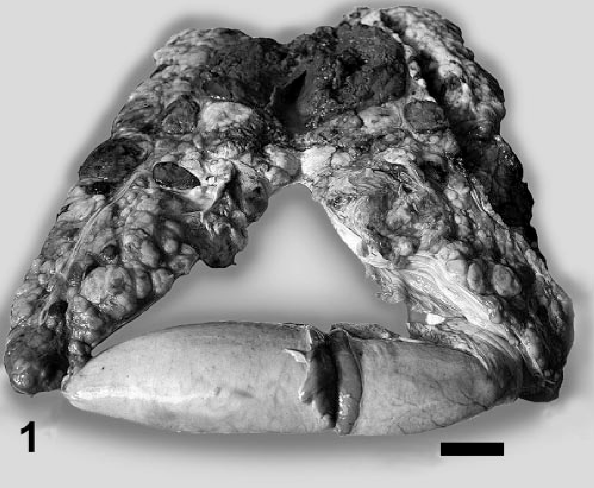
Testes; spotted dolphin No. 1. Sectioned left testis (above) is enlarged and nodular. Compare with normal right testis (below). Bar = 7 cm.
Histologic examination revealed that two discrete neoplasms had largely replaced the left testis. The first was unencapsulated and composed of polygonal cells arranged in sheets or ill-defined packets (Fig. 2). The neoplastic cells had variably distinct cell borders, a moderate amount of foamy eosinophilic cytoplasm, round nuclei with finely stippled chromatin, and, occasionally, a single, small, magenta nucleolus. Mitotic figures averaged one per high-power field and ranged up to three per high-power field. The microscopic characteristics were consistent with a seminoma. Neoplastic cells identical to those of the seminoma had effaced most of a retroperitoneal lymph node.
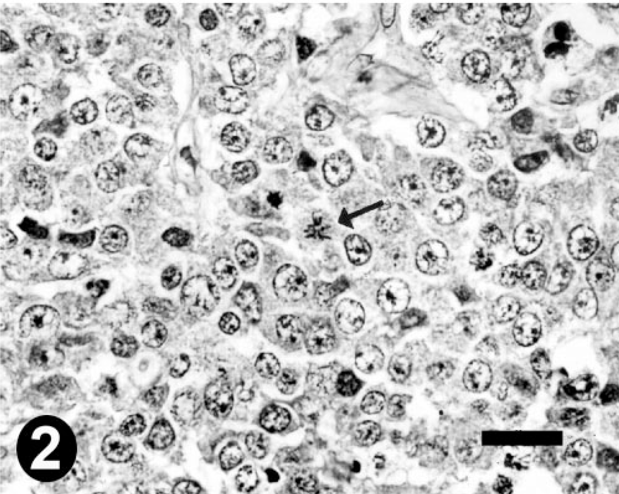
Seminoma; spotted dolphin No. 1. Neoplastic polygonal cells arranged in packets. Note abnormal mitotic figure (arrow). HE. Bar = 20 μm.
The second testicular tumor was unencapsulated and composed of packets of elongate to spindle-shaped cells that sometimes palisaded along a fine fibrovascular stroma (Fig. 3). Neoplastic cells had small to moderate amounts of wispy eosinophilic cytoplasm. Nuclei were round to oval with finely stippled chromatin and had small magenta nucleoli. Mitoses averaged one per two high-power fields. This second tumor was consistent with a Sertoli cell tumor. Hemorrhage and necrosis were present multifocally in both testicular tumors.
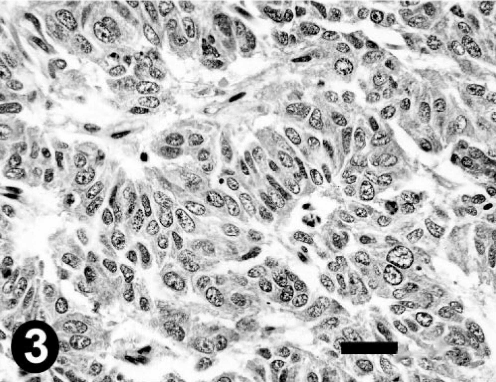
Sertoli cell tumor; spotted dolphin No. 1. Elongated neoplastic cells in palisading pattern. HE. Bar = 25 μm.
The medulla of one adrenal gland was markedly expanded by an unencapsulated neoplasm composed of packets and cords of polygonal cells with distinct cell borders and abundant gray granular cytoplasm (Fig. 4). Nuclei were irregularly round and vesicular with indistinct nucleoli. Mitotic figures were rare. Multifocally, cytoplasmic granules were argyrophilic by the Churukian-Schenk method. These findings were consistent with pheochromocytoma.
Pheochromocytoma; spotted dolphin No. 1. Packets and cords of neoplastic cells with granular cytoplasm. HE. Bar = 40 μm.
An Atlantic bottlenose dolphin stranded and died on a beach at Topsail Island, North Carolina, in November 1997. The animal (DAP 027) was 283 cm long and teeth were worn and chipped, consistent with a mature adult. The dolphin was extremely emaciated with protruding ribs and scapulae. At necropsy, a large neoplasm that originated in the right testicle was fused with organs of the caudal peritoneal cavity (Fig. 5), including intestine, mesenteric lymph node, kidney, pancreas, liver, and spleen. There were multiple pedunculated and infiltrative masses attached to the mesentery, and the abdominal cavity was filled with abundant red-brown fluid.
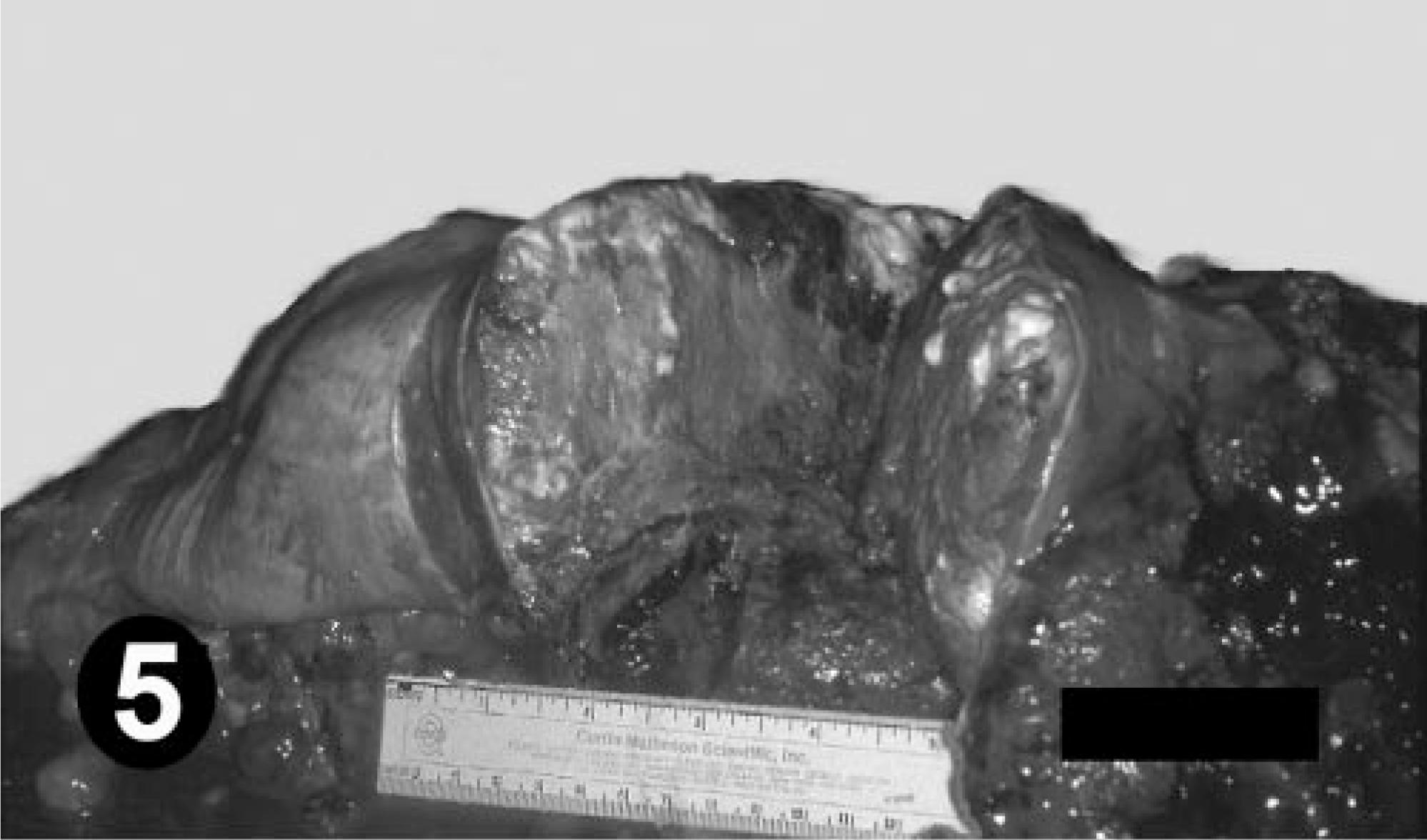
Right testis; bottlenose dolphin No. 2. Sectioned right testis, attached intestine, and mesenteric nodules. Bar = 6 cm.
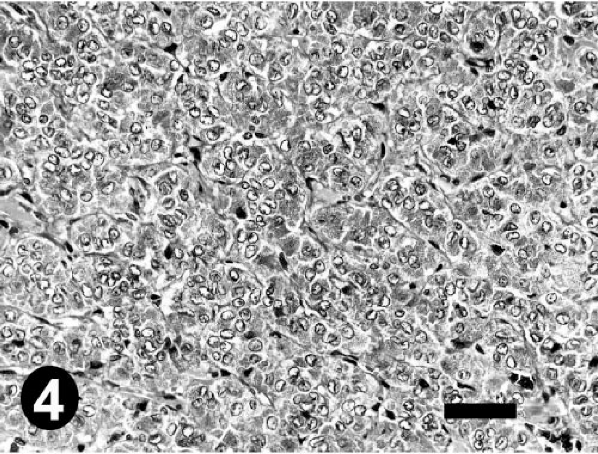
Histologically, the neoplasm in the testis and mesentery was composed of variably sized nests and packets of round cells, separated and supported by thick bands of fibrous connective tissue. Neoplastic cells varied in size and had distinct cell borders and granular eosinophilic cytoplasm. There was moderate anisokaryosis. Nuclei were vesiculate with clumped chromatin and 1–4 variably sized and shaped magenta nucleoli. Mitotic figures averaged one per high-power field and ranged up to three per high-power field (Fig. 6). The findings were consistent with a testicular seminoma with metastasis to the mesentery.
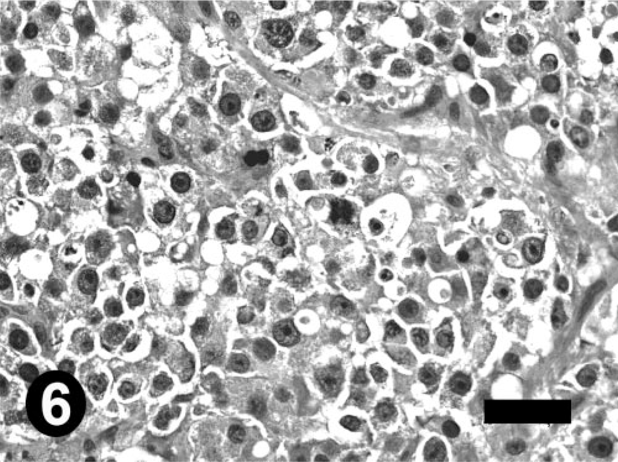
Seminoma; bottlenose dolphin No. 2. Neoplastic polygonal cells; note anisokaryosis, mitotic figure, and binucleate cell. Bar = 20 μm.
There have been very few reports of testicular neoplasia in dolphins or other marine mammals. Cowan et al. (1986) described a Leydig cell tumor in a common dolphin (Delphinus delphis). 2 An unspecified testicular tumor was noted by Mawdesley-Thomas (1974) in a bottlenose dolphin (T. truncatus). 8 Malignant seminoma with metastasis was reported in a sea otter (Enhydra lutris) (Reimer and Lipscomb, 1998). 10
Similarly, few adrenal neoplasms have been reported in cetaceans. De Guise et al. (1994) reported a pheochromocytoma in a beluga whale (D. leucas). 3 Geraci et al. (1987) described adrenal adenomas in two Atlantic white-sided dolphins (Lagenorhynchus acutus). 5 Mawdesley-Thomas (1974) noted an unspecified adrenal cortical tumor in a bottlenose dolphin. 8
There has been only one reported neoplasm in a spotted dolphin, an immunoblastic malignant lymphoma reported by Bossart et al. (1997). 1 Other neoplasms reported in bottle-nose dolphins include: pulmonary squamous cell carcinoma, 4 uterine adenocarcinoma, 12 immunoblastic malignant lymphoma, 1 splenic lymphoma, 5 hepatic adenoma, 5 pancreatic carcinoma, 5 lung and liver reticuloendotheliosis, 5 renal adenoma, 9 thyroid adenoma, 6 and sublingual squamous cell carcinoma. 11
This is the first report of seminoma, Sertoli cell tumor, and pheochromocytoma in a dolphin, the first report of three neoplasms in a dolphin, and one of the very few reports of metastatic neoplasia in cetaceans. Our findings indicate that seminomas have metastatic potential in dolphins.
Footnotes
Acknowledgements
We thank the participants in the Marine Mammal Stranding Network, the Gulfarium (Fort Walton Beach, FL), and the Florida Marine Patrol for their participation in the spotted dolphin's rescue and examination; the UNCW Marine Stranding Program and the VABLAB for response to the bottlenose dolphin stranding; Drs. F. Mostofi, C. Davis, and I. Sesterhenn from the Department of Genitourinary Pathology, Armed Forces Institute of Pathology (Washington, DC) for review of histologic sections of the testicular tumors; and R. A. Ferris and A. Moraytaya, Armed Forces Institute of Pathology for photographic assistance. Formalin-fixed tissues and paraffin-embedded blocks for this dolphin have been archived at The Armed Forces Institute of Pathology (AFIP accession number 2428264 [dolphin 1] and 2660824 [dolphin 2]). The opinions and assertions contained in this study are those of the authors and are not to be construed as official or representing those of the Department of Defense. J. S. Estep, R. E. Baumgartner, and D. G. Dunn are members of the US Army.