
Abstract
Hepatic biliary cystadenoma is a well-delineated neoplasm in some domestic animals, especially in cats, but it has not been reported in equines. We report on a case of hepatic biliary tumor, incidentally found in a 10-year-old horse, with gross and microscopic features similar to those observed in biliary adenofibroma of humans. The tumor presented as a solid mass measuring 16 cm in diameter and histologically was composed of complex tubulocystic biliary components embedded in an abundant fibrotic stroma. We regarded this tumor as a morphological variant of biliary cystadenoma of domestic animals. Differential diagnoses from other hepatic biliary tumor-like and tumor lesions are provided.
Case Report
Hepatic biliary cystadenoma, also known as cholangioma, 6 is a relatively uncommon benign tumor reported in some domestic animals, including sheep, 6 pigs, 6 dogs, 6 and cats. 1,4,6 The clinicopathologic features of such tumors are well delineated especially in cats, being reported in two large series in the literature, 1,4 whereas to the best of our knowledge, no case has been recorded in equines to date. We report on the first case of a hepatic benign biliary tumor showing gross and microscopic features similar to those reported in the so-called biliary adenofibroma in humans. 5,9 In the authors' opinion, the present case is a morphological variant of the biliary cystadenoma of domestic animals.
A 10-year-old horse was normally slaughtered and a mass protruding from the superior surface of the left lobe of the liver was found during the routine meat-inspection procedures. The cut surface revealed a well-circumscribed, solid, oval-shaped mass, measuring 16 cm in greatest diameter, with multiple round, small-sized cysts (2 mm in greatest diameter) filled with clear, viscous fluid. Necrosis was absent. Periportal lymph nodes were unremarkable. The specimen was fixed in 10% neutral-buffered formalin, embedded in paraffin, processed routinely, and stained with hematoxylin and eosin (HE), periodic acid-Schiff (PAS) reaction with and without diastase digestion and elastic Van Gieson for reticulin. Immunohistochemical studies were performed by the standard labeled streptavidin–biotin technique, using commercially available reagents (LSAB kit, Dako, Glostrup, Denmark). In brief, sections were dewaxed in xylene for 15 minutes, rehydrated, and treated with 3% H2O2 for 10 minutes to block endogenous peroxidase activity, and then rinsed in distilled water and a 15 minutes wash in 0.01 M phosphate-buffered saline (PBS), pH 7.4. Incubation with primary antibodies (pancytokeratins, prediluted, Dako, Glostrup, Denmark; cytokeratins 7, 8, 18, 19, 20, all from Novocastra Laboratories (Newcastle upon Tyne, UK), dilution 1:50; vimentin, prediluted, Immunotech, Marseille, France; α-smooth muscle actin, dilution 1:200, Dako, Glostrup, Denmark) was performed overnight at 4 C, and then incubated with the linking antibody (biotinylated anti-mouse immunoglobulins) and with the peroxidase-conjugated streptavidin for 20 minutes at room temperature. Peroxidase activity was developed in 3,3′-diaminobenzidine (Sigma Chemical Co., St. Louis, MO) substrate with 0.01% H2O2 for 5 minutes. Slides were counterstained with hematoxylin, dehydrated, and mounted. Negative control sections were incubated in PBS in place of the primary antibodies.
Histologic examination revealed an unencapsulated tumor with compressing borders composed of epithelial structures such as tubules, acini, and cysts embedded in an abundant amount of fibrotic stroma (Figs. 1, 2). Tubules were frequently dilated, branched, and focally exhibited papillary projections into lumens; cysts were variable in size and shape, ranging from round to oval to tortuous configurations. The epithelium lining tubules and acini was predominantly composed of a monolayer of low columnar-type cells sometimes showing apocrine-like changes (Fig. 3), whereas cysts were lined by cuboidal to flattened cells. Proteinaceous fluid within cysts was stained with PAS stain. Immunohistochemical analysis, showing that all the epithelial cells were immunoreactive to cytokeratins 7 and 19 (Fig. 4), confirmed their biliary nature because this profile is typical of developing and mature normal biliary ducts. 7 Cellular debris were found within some tubules and cysts. Cellular atypia, mitoses, necrosis, inflammatory infiltrates, stromal, vascular, or lymphatic invasion were not observed. The stroma surrounding the epithelial structures was fibrotic in nature and scantly to moderately cellular (Figs. 1, 2). The cellular component was represented by spindle-shaped fibroblasts, haphazardly arranged and positive only to vimentin. Vascularization was poor. The liver parenchyma outside the tumor was morphologically unremarkable. The diagnosis of “biliary adenofibroma” was made on the basis of the gross, histological, and immunohistochemical features identical to the counterpart described in humans. 5,9
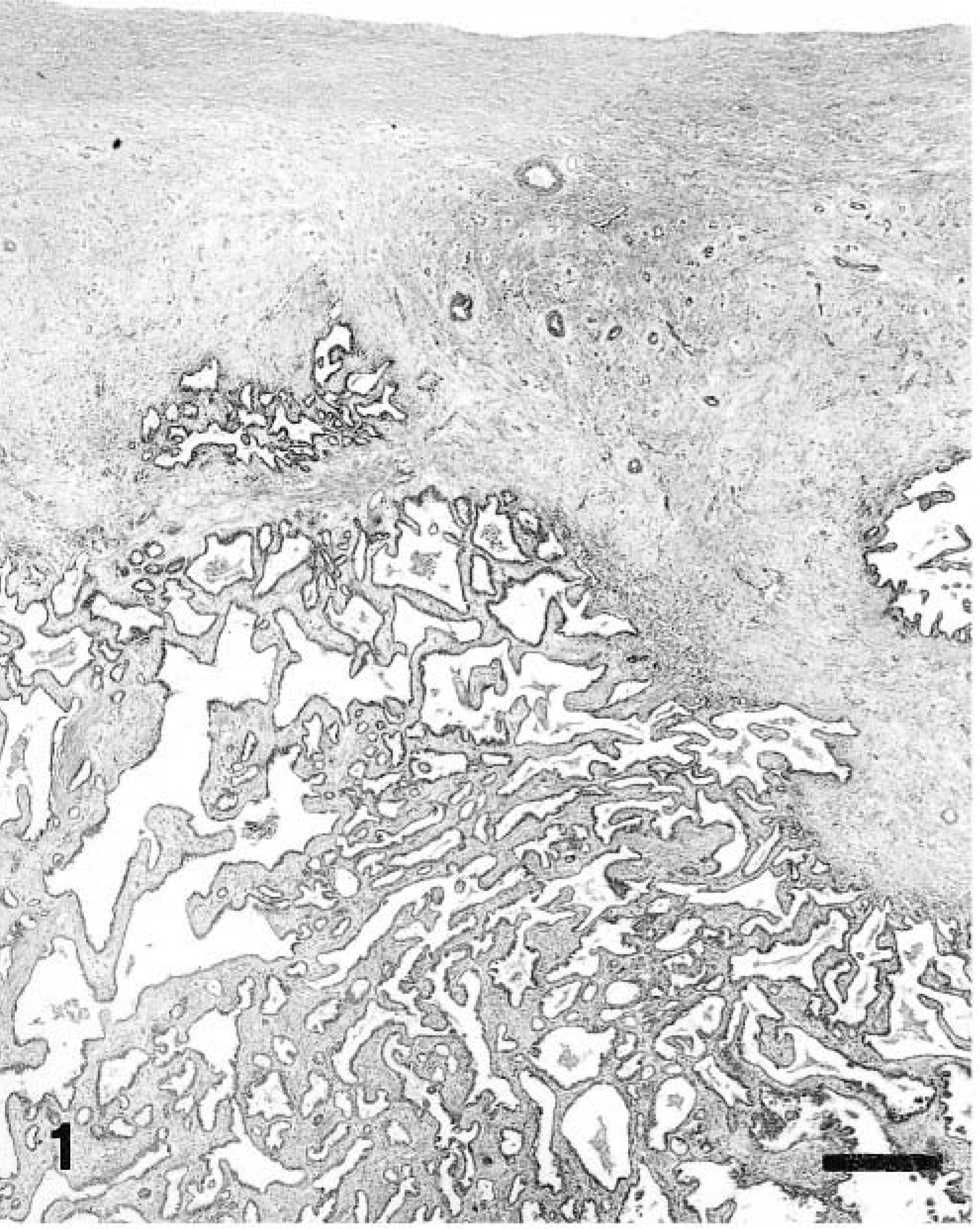
Liver; equine. Subcapsular tumor composed of branching tubules, some of them cystically dilated, embedded in abundant fibrous stroma. HE. Bar = 80 μm.
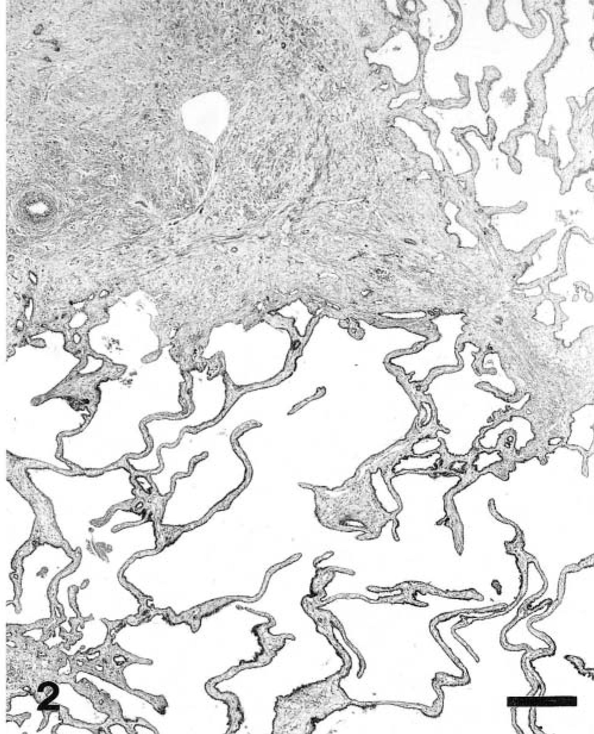
Liver; equine. Variable-sized cysts set in fibrous stroma. HE. Bar = 80 μm.
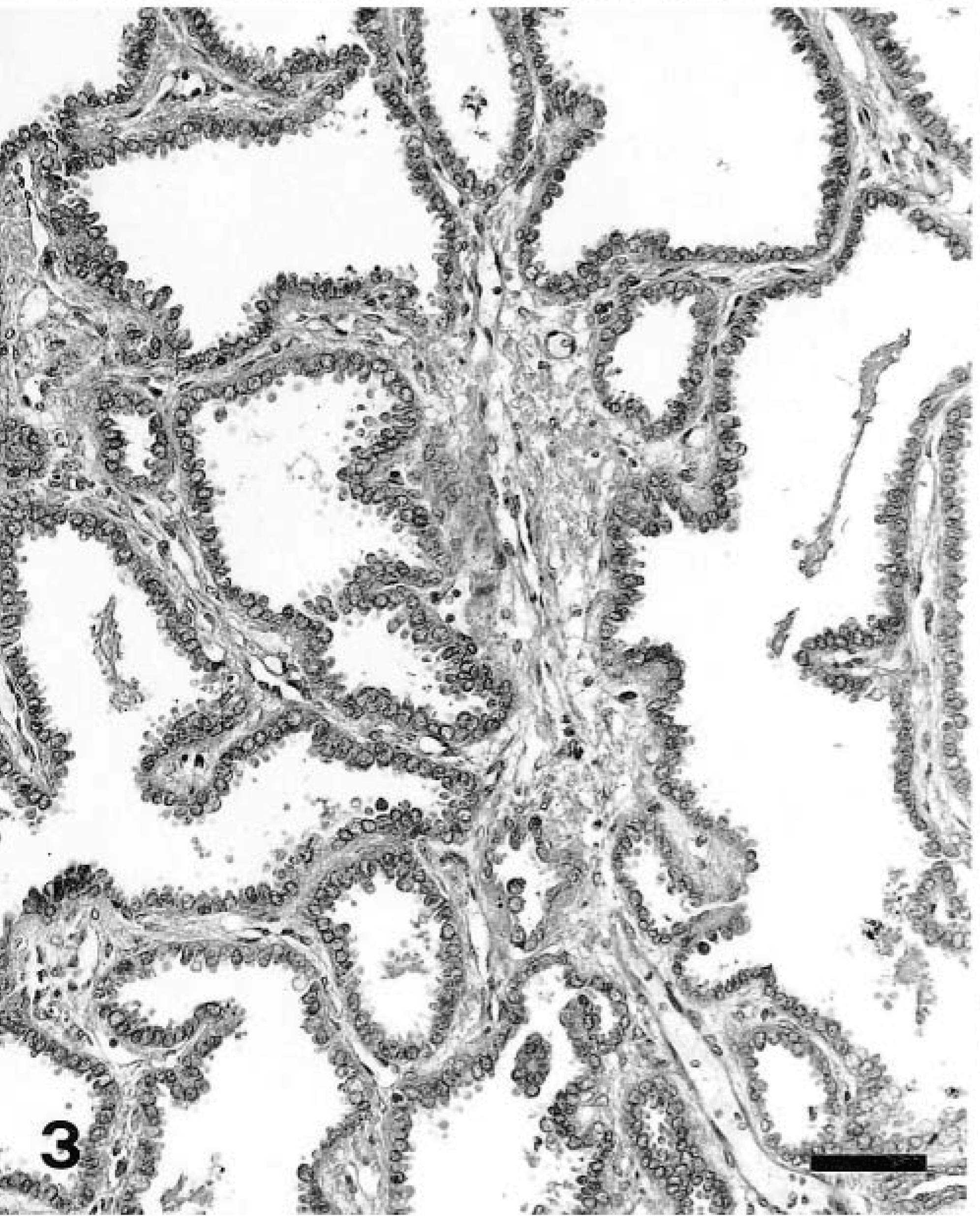
Liver; equine. High-power view showing epithelial structures lined by an apocrine columnar type epithelium. HE. Bar = 25 μm.
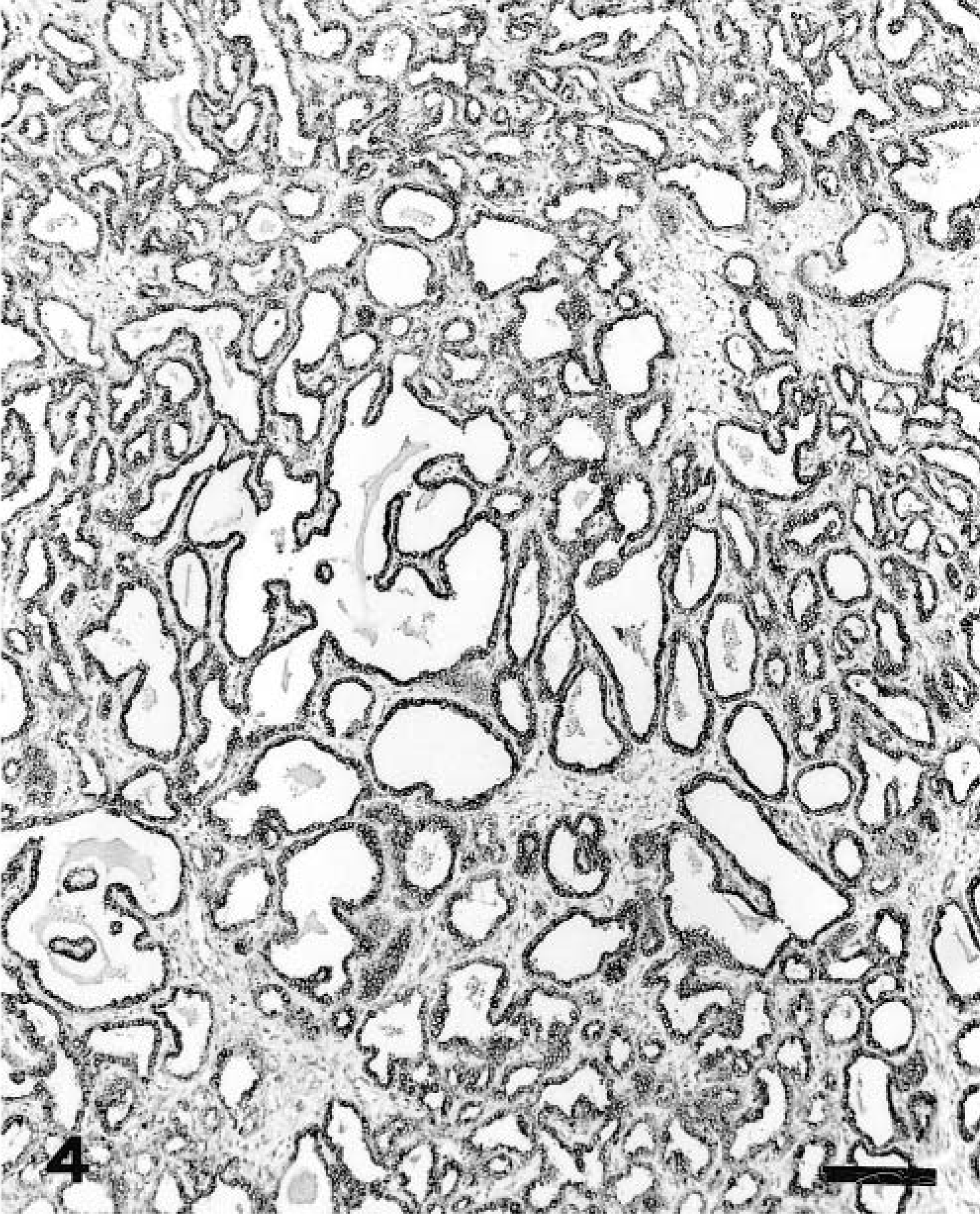
Liver; equine. Epithelium is positive for cytokeratin 19. Streptavidin-biotin–peroxidase complex method, Mayer's hematoxylin counterstain. Bar = 31.25 μm.
Although this tumor is histologically similar to biliary cystadenoma of domestic animals, we prefer to maintain the term “adenofibroma” because it is basically a solid lesion with microcysts not exceeding 2 mm and containing abundant fibrotic stroma. On the contrary, the typical biliary cystadenoma of domestic animals presents grossly as a multilocular cystic mass, with the single cysts ranging is size up to 15 mm, 1 and usually the stromal component is less represented. But we are aware that we are dealing with a variant of the biliary cystadenoma, likely representing its early phase before acquiring a multilocular cystic appearance.
Because both biliary cystadenoma and its variant “adenofibroma” share a basic common morphological theme—numerous biliary ducts, variably dilated and embedded in a fibrous stroma—especially with congenital hepatic fibrosis and liver polycystic disease, biliary cystadenoma and its variant should be distinguished from these two entities. In humans, both these conditions are part of the so-called fibropolycystic disease, a spectrum of congenital abnormalities involving bile ducts, mainly because of an abnormal remodeling of the developing bile-duct plate. 7 Both congenital hepatic fibrosis 3 and polycystic liver disease 2,8 are reported in equines. Although the former may exhibit small and dilated biliary ducts, its hallmark is the diffuse presence of porto-portal bridging fibrotic septa surrounding islands of normal liver parenchyma. 3 This finding is lacking in our case. Unlike congenital liver polycystic disease, a genetic disorder characterized by multiple lesions that may be associated with cystic lesions even in the kidney, our case presented as a single liver mass without any evidence of other hepatic or kidney abnormalities. Another biliary lesion that could be histologically confused with biliary adenofibroma is bile-duct adenoma. 7 The latter differs in that it presents as a small solid neoplasm, ranging in size from a few millimeters to 2 cm, composed of small, usually not dilated biliary ducts and not containing bile. Malignant tumors such as cystadenocarcinoma and cholangiocellular carcinoma were easily ruled out because of the absence of cytological atypia, mitoses, and necrosis.
The present case deals with a hepatic tumor basically showing the morphological features of the biliary cystadenoma of domestic animals. 1,4 A comparative pathological analysis between biliary cystadenoma in these animals and humans reveals a significant difference in the composition of the epithelial lining, being represented by mucin-secreting cells in the latter and normal-appearing biliary epithelium in the former. 7 Because the tumor with gross and microscopic features identical to our case is the so-called biliary adenofibroma, a very rare neoplasm with only two cases reported in humans to date, 5,9 we prefer to maintain such a designation over that of cystadenoma. It is the authors' intention to highlight that biliary cystadenoma/adenofibroma should be included in the differential diagnosis when a solitary, huge, solid mass with microcysts is discovered at necropsy or incidentally found both at slaughter or ultrasonographic examination of a hepatic mass in the equine.