
Abstract
An ovarian mucinous cystadenoma is described in a gray brocket deer (Mazama gouazoupira). The tumor was histologically characterized by the presence of cysts and proliferation of papillae, both lined by single- or multi-layered pleomorphic epithelial cells that contained alcian blue–positive mucins.
Ovarian epithelial neoplasms are uncommon tumors in domestic and nondomestic animals, 24 but have been reported in the cow, pig, 18 small ruminants, 25 dogs, 20 cat, 6 mare, 8 deer, 10,27 and jaguar. 2 Except in the dog, 14 epithelial ovarian neoplasms occur less frequently than those of mesenchymal origin. As in other species, tumors of the female genital tract are rarely reported in wild ungulates, with few described cases of ovarian tumor in cervids. 10,27 Additionally, there are no documented cases of female reproductive tract tumors in gray brocket deer. The present report documents a case of an ovarian mucinous cystadenoma in a gray brocket deer (Mazama gouazoupira).
A free-ranging female gray brocket deer, estimated to be 7–8 years old, was found alive but recumbent after colliding with a vehicle in May 2010 (autumn in Brazil). The animal was initially stabilized but died during surgery to correct femoral and pelvic fractures caused by the car accident. The doe was subsequently presented for postmortem examination to the Veterinary Pathology Service of São Paulo State University (Botucatu, São Paulo, Brazil). At necropsy, a firm, slightly raised mass measuring 8 cm × 10 cm × 5 cm was identified on the right ovary; the contralateral ovary was not enlarged and measured 2 cm × 1 cm × 1 cm. On cut surface, the right ovary contained multiple cysts, predominantly in the ovarian cortex, that were surrounded and separated by gray to tan thickened foci with scattered to coalescing hemorrhage and necrosis (Fig. 1). The vulvar region of the animal was tumescent, and mammary gland examination revealed enlargement with milk production. All other organs, including the remainder of the reproductive tract, were within normal limits.
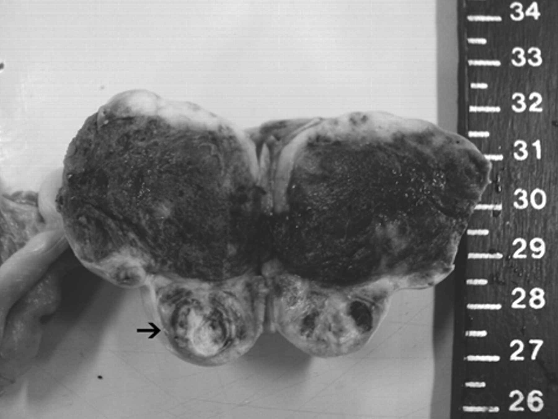
Ovarian papillary cystadenoma; multiple cysts, extensive hemorrhage, and thickened areas are present in the ovarian cortex (arrow).
Microscopically, the left ovary had follicles in different stages of development but no corpus luteum. On the other hand, the ovarian tumor was composed of numerous prominent cysts ranging from approximately 50 to 400 µm. The cysts were lined by single- to multi-layered cuboidal to columnar, pleomorphic, epithelial cells that occasionally formed arboriform papillae (Fig. 2). No neoplastic stromal invasion was observed. Alcian blue staining (pH 2.5) was performed to evaluate mucin production by neoplastic cells, and positive intracellular and intracystic staining was observed (Fig.3). Based on gross and histologic features, the tumor was considered to be an ovarian mucinous cystadenoma.
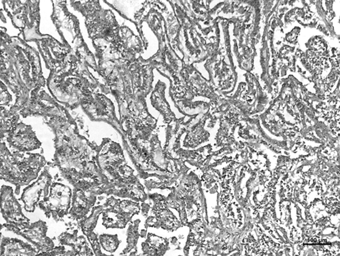
Ovarian papillary cystadenoma; arboriform papillae composed of atypical epithelial cells are present. Hematoxylin and eosin. Bar = 100 µm.
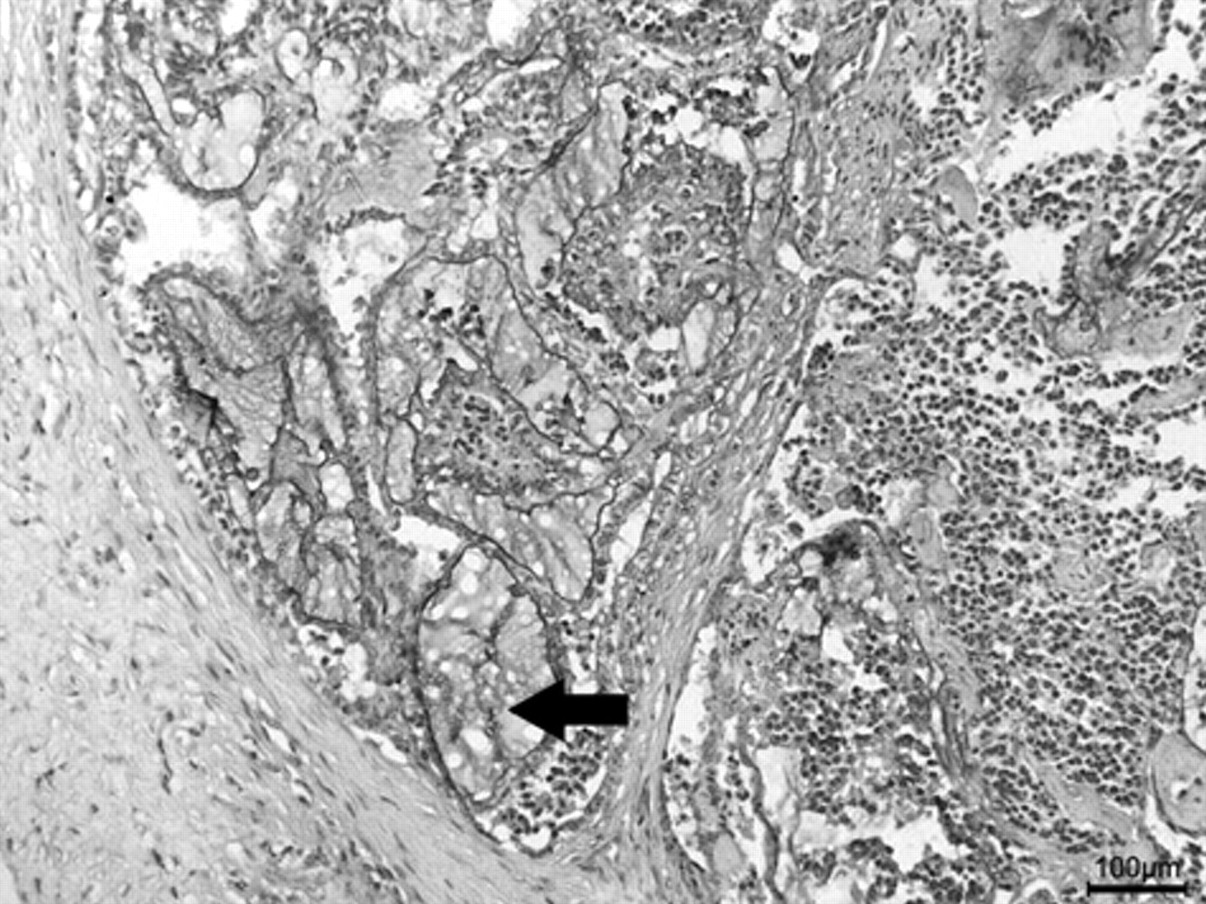
Ovarian papillary cystadenoma; intratumoral mucin production is present (arrow). Alcian blue stain, pH 2.5. Bar = 100 µm.
The gray brocket deer is distributed throughout Central and South America, occupying nearly all biotopes from rainforests to savannahs, and is one of 8 species of Cervidae living in Brazil. 3 The International Union for Conservation of Nature status of the gray brocket deer is “least concern,” which means the species is not threatened, near threatened, nor depends on conservation. However, according to some authors, 3,7 the deer has an endangered status due to hunting, competition with livestock, and the continuous destruction of its habitat. Consequently, it is important to document cases of morbidity and mortality in gray brocket deer as a part of a database for improved management of this species.
Various spontaneous neoplasms have been reported in wild deer species, but the majority of these neoplasms have been cutaneous fibromas, fibropapillomas, and squamous cell carcinomas. 1,17,25 In addition, there are reports of various noncutaneous neoplasms in the white-tailed deer (Odocoileus virginianus), such as ependymoma, 19 astrocytoma, 11 hepatocellular carcinoma, 21 and lymphoma. 15 A uterine adenocarcinoma in a sika deer (Cervus nippon) 22 was also documented. Additionally, fibrosarcoma, 12 mammary adenocarcinoma, 26 concurrent multicentric hemangiosarcoma, and ovarian teratoma 27 have been described in the Pere David’s deer (Elaphurus davidianus). However, ovarian tumors of epithelial origin have not yet been reported in deer, and such tumors are rare in both ruminants and other species. 14
Epithelial tumors of the ovary usually develop from the surface epithelium, which is coelomic mesothelium; however, they can also arise from the rete ovarii. 14,24 Those that arise from the surface epithelium initially are confined to the cortex of the ovary, whereas those that arise from the rete ovarii are initially confined to the medulla adjacent to the hilus of the ovary. 14 In the present case, since neoplastic areas were predominantly adjacent to the ovarian cortex, the tumor was considered to have originated from the surface epithelium.
In domestic animals, both benign and malignant ovarian epithelial tumors predominantly have a serous character and are similar in location and morphology. Similarly to those that occur in human beings, benign and malignant ovarian tumors of animals are sometimes difficult to differentiate when there is absence of metastasis or obvious vascular invasion. 13 In such cases, the distinction between adenoma and adenocarcinoma is based on the size of the tumor, cellular atypia, tendency for the neoplastic cells to pile up, and the presence of necrotic and hemorrhagic foci. The mitotic index and evidence of stromal invasion are also used to differentiate benign and malignant neoplasms and are said to be of great importance in achieving an accurate diagnosis. 14 However, there is no available standardized threshold for these features in the veterinary literature to date. In the present case, since there were no piled-up cells and stromal or vascular invasion was not observed, the tumor was considered to be histologically benign.
Ovarian neoplasms can occasionally be associated with clinical hormone production; however, this feature is more common in sex-cord stromal tumors. 14 In dogs and human beings, ovarian neoplasms of epithelial origin have been associated with steroidal hormone production, causing signs of hyperestrogenism such as enlargement of the mammary gland and vulva. 23,24 Additionally, stromal components of mucinous ovarian tumors in human beings may produce estrogens, androgens, or rarely, progestagens, or any combination of these hormones. 23 This hormonal imbalance can lead to different clinical signs such as milk production, a feature that can be derived from higher estrogen levels due to increase in prolactin production. However, such signs are also noted in recent parturition, making the differentiation between the 2 causes very important.
In the present case, milk production was observed in conjunction with mammary gland and vulvar enlargement. These findings, in the absence of uterine implantation sites or alteration in the size of the uterus, raised a suspicion of tumor-associated steroidal hormone production. Additionally, pseudopregnancy, a syndrome described in some ruminant species that is associated with the persistence of corpus luteum, 9 was considered in the differential diagnosis. However, pseudopregnancy was not thought to be related to the alterations observed in this animal because the ovaries did not contain luteal tissue. In order to confirm hyperestrogenism, measurement of blood estrogen concentrations could have been performed to confirm patient values that were higher than corresponding reference intervals. Unfortunately, this evaluation was not performed in the present case because reference intervals for blood estrogen concentration has yet to be determined experimentally for M. gouazoupira, as has been done for other species of deer. The season of the year also tends to exclude the association of milk production with recent parturition. However, since tropical climate deer species (especially the ones of Mazama genus) do not experience seasonal influence on their reproduction, 4 this feature could not be used in the present case.
Ovarian epithelial tumors are uncommon neoplasms of ruminants. In a study of 1,489 ovarian neoplasms in cows, 5 only 4 tumors of epithelial origin (all of them cystadenomas) were diagnosed. Of these 4 cystadenomas, only 1 had mucin production similar to that observed in the present case.
Other authors 16 have described a mucinous adenocarcinoma of the ovary associated with ascites in a goat. This neoplasm presented as an indistinct, firm, slightly raised, gray to tan tumor of the left ovary. The gross appearance of the tumor and the mucin production observed in histologic sections were very similar to what was observed in the current case. However, ascites, a characteristic thought to be related to lymphatic obstruction by neoplastic emboli, 14 was not observed, probably because the tumor in the present case was benign. In addition, the usual proliferative cauliflower-like growths that project from the surface of the ovary, 14,24 particularly in papillary adenomas and adenocarcinomas of coelomic surface-origin, were not observed. Instead, the major histologic characteristic of the tumor was its prominent solid to cystic nature that was consistent with a cystadenoma. Ovarian adenomas are uncommon tumors of domestic ruminants, and they are rare in cervids.
Footnotes
The authors declared that they had no conflicts of interest with respect to their authorship or the publication of this article.
The authors declared that they received no financial support for their research and/or authorship of this article.