
Abstract
The role of tumor suppressor genes in the pathogenesis of canine melanoma is incompletely understood. The genes encoding the tumor suppressors p53, Rb, p21 (waf-1), p16 (ink-4a), and PTEN have been postulated to contribute to the pathogenesis of melanoma in humans and experimental animal models. To assess whether inactivation of these genes similarly contributes to the origin and progression of canine melanoma, we examined their expression in seven distinct canine melanoma cell lines and in 31 retrospective samples (representing 29 dogs) of spontaneous canine melanoma. Various patterns suggestive of loss of tumor suppressor function emerged in these cell lines. The most frequently observed abnormality was loss or significant reduction of p16 expression in six of seven cell lines and in 21 of 26 tumor samples. Loss or significant reduction of PTEN expression was seen in four of seven cell lines and in 13 of 27 tumor samples. Although p53 was detectable in all the cell lines and in 24 of 30 tumors, exclusion of p53 from the nuclear compartment was observed in each of the cell lines and in 18 of 25 tumor samples. These results indicate that loss of function of these tumor suppressor proteins is a common occurrence that may contribute to the origin of canine melanoma. In our sample population, abnormalities in the expression or localization of one or more tumor suppressor proteins occurred with similar frequency in malignant and benign tumors; thus, additional work is necessary to determine how these proteins may impact disease progression and response to therapy.
Melanoma is a common tumor in dogs that arises from melanocytes or melanoblasts. It is the most common malignancy found in the oral cavity and on the digits in the dog.21,65 Dermal melanomas are usually benign, but uveal, digital, and oral melanomas generally are malignant and respond poorly to standard therapies.46 There are, however, exceptions to these generalities; a few dogs with oral melanoma are cured of the disease, whereas others with dermal melanoma die from metastasis. Thus, shared genetic characteristics may be as or more important than location.
The natural history and pathogenesis of canine melanoma are incompletely understood, but recent work has begun to elucidate the role of growth regulation genes in the development of this disease.46 Cellular growth and division is a complex pathway governed by competing growth inhibitors and promoters. Proto-oncogenes encode proteins that promote cell cycle progression, proliferation, and survival in normal cells; tumor suppressor genes do the opposite, encoding proteins that inhibit cellular proliferation and promote cell death.68 For cells that have undergone malignant transformation, the balance between proliferation and inhibition is upset.
For this study, we used reverse transcription (RT) polymerase chain reaction (PCR) assays, immunoblotting, and immunocytochemistry to examine gene expression and protein accumulation of the p53, p21 (waf-1), p16 (ink-4a), Rb, and PTEN tumor suppressor genes in seven independent cell lines and immunohistochemistry to assess the presence and subcellular location of these proteins in 31 retrospective tumor samples. Our hypotheses were that abnormalities leading to loss of function of these pathways contribute to the pathogenesis of canine melanoma and that defects in multiple tumor suppressor genes would predict a worse outcome. These genes and proteins are members of three interrelated negative growth regulatory pathways. The p53 gene encodes a nuclear protein (p53 or TP53) that is integral to maintenance of DNA integrity and is a cornerstone of the DNA repair machinery.36 Induction of p53 can result in apoptosis or growth arrest in cells with irreparable DNA damage.36 The retinoblastoma susceptibility gene RB-1 is a tumor suppressor gene that encodes a protein (Rb) that regulates the transition from the G1 phase to the S phase of the cell cycle.35,69 Rb is initially made in the cytoplasm and is then transported into the nucleus, where it exerts its effects. The cyclin-dependent kinase inhibitors (CDKI) p21 (the product of the waf-1 gene) and p16 (the product of the ink-4a gene) prevent cell cycle progression and promote growth arrest or apoptosis.25,62,63 PTEN is a gene that encodes a bifunctional phospholipid and tyrosine phosphatase, which antagonizes growth and survival signals transmitted through the phosphoinositide-3 kinase pathways4,40,43,66 and may play a role in angiogenesis.5,29
Reduced or absent expression of p16 and PTEN and accumulation of p53 in an inappropriate subcellular location were the most frequent abnormalities identified in this study. Loss of these tumor suppressor pathways seems to be a common occurrence that may contribute to the pathogenesis of canine melanoma. Further studies are necessary to confirm the prognostic significance of these biomarkers in canine melanoma.
Materials and Methods
Cell lines and normal tissues
The melanoma cell lines utilized for this study were derived from primary tumors of dogs with oral melanoma (CML-2, CML-13, JENNY, SCOOTER, TLM-1), a lymph node metastasis of cutaneous melanoma (CML-6MC2), and a lung metastasis of oral melanoma (SHADOW) and have been described previously.12,33,56,73 The Cf2Th line, originating from normal canine thymic epithelial cells, was obtained from the American Type Culture Collection (Rockville, MD). Normal tissue samples obtained from healthy dogs at the time of euthanasia were generously provided by Dr. John Bauer. The procedures for handling and euthanasia of the dogs and for the transfer of samples to this study were approved by the Institutional Animal Care and Use Committee of Texas A&M University.
Clinical cases
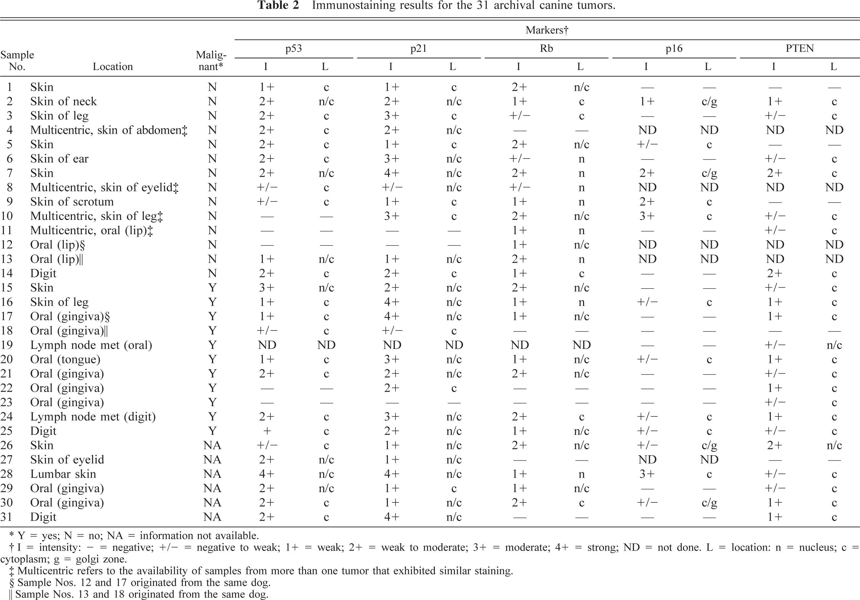
Clinical cases were retrieved from the medical records database of the Texas Veterinary Medical Center. Every case that had a histologic diagnosis of melanoma between 1994 and 1997 was reviewed for eligibility; 31 tumor samples with formalin-fixed archival material that were suitable for immunostaining were utilized. Samples were excluded when 1) blocks did not contain sufficient material for serial sectioning, 2) tumor material was not evident on routine hematoxylin and eosin–stained recuts, 3) blocks had evidence of improper or incomplete fixation, such as loss of cellular detail along plasma membrane and nuclear membrane or decreased staining intensity, 4) blocks had evidence of improper or incomplete infiltration of paraffin (based on their gross appearance), and 5) tissues were negative for vimentin control stains, where normal stromal tissues in the block were evaluated for vimentin expression by immunostaining. The features of some of these cases were reported previously.33 Some tissue blocks did not yield sufficient tissue to analyze all of the markers included in this study. The eligible cases included 11 primary oral tumors (one tongue, three lip, seven gingiva), 18 dermal melanomas (three digit, three limb, two eyelid, three trunk, one ear, one scrotum, five unspecified), and two lymph node metastasis (one from an oral tumor and one from a digital tumor). Twenty-one tumors were pigmented, and ten were amelanotic. The tumors were categorized as malignant or benign based on pleomorphism, degree of proliferation, local invasion, and documentation of metastasis. Pleomorphism and proliferation were not by themselves considered adequate indicators of malignancy in the absence of known recurrence, invasiveness, or documentation of metastasis. Using these criteria, 14 tumors were benign, 11 were identified as malignant, and the behavior of six was not evident from the histology or history. The samples represented 29 dogs and 17 different breeds: four Gordon Setters, four Doberman Pinschers, three Labrador Retrievers, two Miniature Schnauzers, two Boxers, one each of Giant Schnauzer, Brittany Spaniel, Bichon Frise, Cairn Terrier, Bull Terrier, Weimaraner, Bull Mastiff, Dachshund, Shetland Sheepdog, Golden Retriever, English Pointer, and Greyhound, and two mixed-breed dogs. The dogs ranged in age from 2 to 14 years, with an average age of 11 years and a median age of 12 years. The age of one dog was unknown. Thirteen of the dogs were neutered males, three were intact males, 12 were spayed females, and one was an intact female.
Cell culture
Tissue culture materials were obtained from Nalge Nunc (Naperville, IL). Cells were cultured in Dulbecco's modified Eagle medium (DMEM, Gibco BRL, Grand Island, NY) containing 10% heat-inactivated fetal calf serum (FCS, Hyclone, Logan, UT) in a humidified atmosphere containing 5% CO2 at 37 C. All the cell lines grew as monolayer cultures and were maintained by passage when they reached >90% confluence.
Identification of a partial cDNA for canine ink-4a
The sequence for canine ink-4a was unknown. To obtain a partial sequence of canine ink-4a, we used a set of commercially available PCR primers (Research Genetics, Huntsville, AL) designed specifically to examine deletions of ink-4a in human tumors. The commercial primers consist of a conserved sequence within the first intron of ink-4a (sense) and within exon 2 (antisense). Genomic DNA was isolated from normal canine liver using a commercially available kit (Gentra Systems, Minneapolis, MN) as per the manufacturer's instructions. PCR was performed using the primers and conditions specified (Research Genetics). The PCR using the Research Genetics primers and genomic DNA resulted in amplification of a 393-base pair (bp) product. To confirm the identity of the putative ink-4a DNA, we isolated a cosmid clone from a library (provided by Dr. E. Ostrander, Fred Hutchinson Cancer Research Center, Seattle, WA) using a human ink-4a cDNA (provided by Dr. D. Beach, Cold Spring Harbor Laboratories, Cold Spring Harbor, NY) as a screening tool. One positive clone was selected from a tertiary screen. PCR amplification using the same primers and the cosmid as a template produced an identical sequence to that obtained by RT-PCR from canine liver RNA. Figure 1 shows the arrangement of the partial canine ink-4a DNA (GenBank accession No. AF234176), including 150 bp of sequence within intron 1 and 243 bp within the coding domain of exon 2 (highlighted). The 243 bp of canine ink-4a exon 2 encompass approximately 56% of the highly conserved sequence that encodes the ankyrin repeats based on the human homologue. Figure 2 shows that the translated partial protein product consists of 81 amino acids (of a predicted 145, based on the human sequence), with 78/81 identities, one conserved substitution, and two nonconserved substitutions. The canine ink-4a fragment also has up to 88% identity at the nucleotide level with porcine, murine, rat, and equine ink-4a (also known as CDKN2A) and with the closely related genes ink-4b, ink-4c, and ink-4d that encode the p15, p18, and p19 proteins, respectively. The partial canine p16 protein also shares 76–88% identity at the protein level with the porcine, murine, rat, equine, and opossum p16 proteins and 55% identity with the platyfish (Xiphophorus maculatus) p16 protein. Predictably, this amino acid sequence is also highly homologous to human and rodent p15; the ankyrin repeats in all the INK-4 proteins are highly conserved.
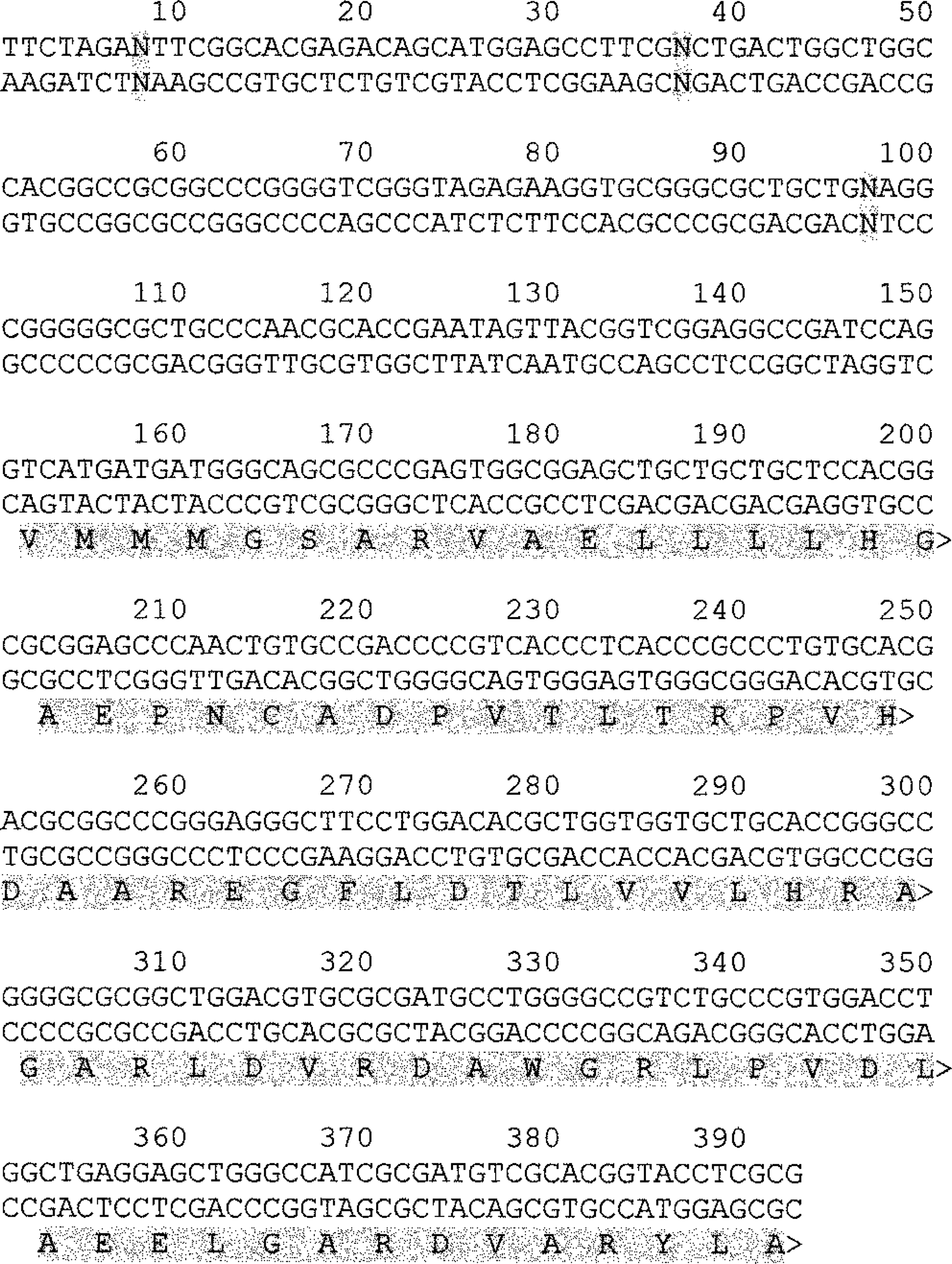
Double-stranded nucleotide sequence of a partial canine ink-4a DNA region encompassing 393 bp including 150 nucleotides of intron 1 sequence and 243 nucleotides of exon 2 sequence. The coding domain for exon 2 is represented by the partial amino acid sequence shown below the nucleotide sequence (highlighted). Nucleotides within the intron represented by an “N” and highlighted in gray represent bases where the precise sequence is uncertain.
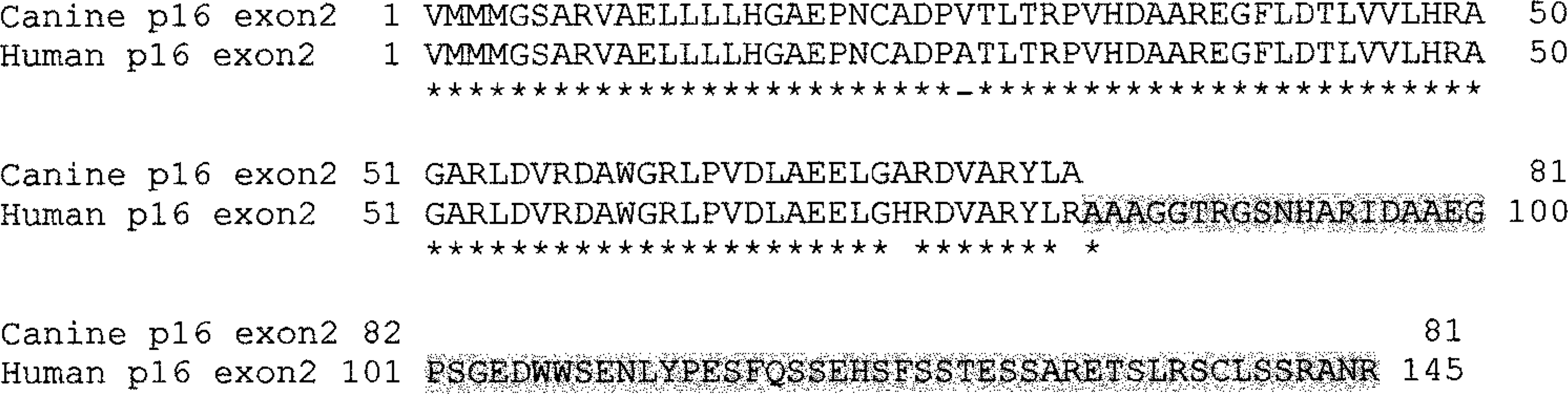
Aligned canine and human amino acid sequences for p16 exon 2. Asterisks represent sequence identity. Conserved substitutions are represented by a dash under the sequence. A blank space under the sequence represents nonconserved substitutions. The regions of the human protein for which the matching canine sequence is unknown are highlighted in gray.
Gene expression
Gene expression was analyzed by RT-PCR as previously described.55 RNA was isolated using the RNAWiz kit (Ambion, Austin, TX) as recommended by the manufacturer. Complementary DNA synthesis was accomplished using a kit (Roche Diagnostics, Chicago, IL) followed by PCR amplification with dog-specific primers. The sequence of the complete coding domains for the p53 gene is available in GenBank (accession No. AB020761). Amplification of p53 mRNA was done using a primer pair (sense 5′-CTGGCTAGACGAAGACTCAG-3′, antisense 5′-AGGCAGTGCTCGCTTGGTAC-3′) under conditions described previously38 to produce a 738-bp amplification product representing amino acids 52–296. The amplification products were gel purified and submitted for sequencing to the sequencing core facility of the University of Colorado Cancer Center. Reproducible sequences representing amino acids 72–285 were obtained for all cell lines. We previously reported a partial sequence for canine waf-1 (GenBank accession No. AF076469).55 For this study, we extended the sequence by an additional 25 bases in the 5′ (N-terminal) direction and identified 32 potential polymorphisms, resulting in seven partially conserved nucleotide substitutions and six conserved nucleotide substitutions. Two primer pairs were used for waf-1 amplification (pair 1: sense 5′-GATGGAACTTGGACTTGGGC-3′, antisense 5′-GAGTGGTAGAAATCTTTAAGGCTGG-3′; pair 2: sense 5′-GACTGTGATGCGCTAATGGC-3′, antisense 5′-GGGTACAAGACAGTGACAGG-3′) to generate products of 314 and 259 bases, respectively. For ink-4a, a primer pair (sense 5′-AGCTGCTGCTGCTCCACGG-3′, antisense 5′-ACCAGCGTGTCCAGGAAGCC-3′) was designed to amplify 103 bp within the canine sequence for exon 2. The canine homologue of PTEN has been cloned (GenBank accession No. U92435), and it is 95% identical to the human sequence. Two sets of primer pairs were designed from the published canine sequence (GenBank accession No. U92435). One primer pair (sense 5′-ATGACAGCCATCATCAAGGAG-3′; antisense 5′-CACACACAAATCACAAAAGTC-3′) was designed to amplify the complete PTEN coding domains (1,208 bp), and the other (sense 5′-GCTATGGGGTTTCCTG-3′, antisense 5′-TAAGGACCAGAGACAAAAAG-3′) amplified an internal product of 393 bp that included the catalytic domains of the phosphatase. β-Actin expression was used to control for integrity of the RNA. The oligonucleotide primers used for amplification of β-actin were 5′-ATGTTCGAGACGTTCAACACCCC-3′' (sense) and 5′-GCCATCTCCTGCTCGAAGTCCAG-3′ (antisense), based on the GenBank sequence for Canis familiaris β-actin (accession No. AF021873) to produce an amplification product of 318 bp. Normalization of the levels of expression for each tumor suppressor gene to the levels of ß-actin allowed for semiquantitative assessments of mRNA expression.
Immunocytochemistry and immunohistochemistry
For cell lines, at the end of the culture period plastic chambers were removed, and slides were rinsed in phosphate buffered saline (pH 7.4), fixed in acetone at 4 C for 5 minutes, and air dried. For archival tissues, 5-μm serial sections from paraffin-embedded blocks were mounted onto positively charged slides (Probe-on, Fisher Scientific, Pittsburgh, PA). Immunostaining was performed using a modified streptavidin–biotin complex method previously described.55,56 For paraffin-embedded sections, antigen retrieval procedures included microwave heating for 6 minutes in a buffer of 0.1 M sodium citrate (pH 6) for p21, p16, Rb, and PTEN staining and microwave heating for 6 minutes in Retrieve All (Signet Pathology Systems, Dedham, MA) for p53. Antigen retrieval was not used for staining of the cell lines, except in the case of p21, where microwave heating for 6 minutes in sodium citrate buffer was used to unmask nucleolar staining. Slides were incubated for 1 hour at room temperature with primary antibodies against p53 (antibody CM-1, diluted 20-fold, Signet Pathology Systems), p21 (antibody C-20, 5 µg/ml, Santa Cruz Biotechnology, Santa Cruz, CA), Rb (mouse monoclonal antibody G3–245, 5 µg/ml, Pharmingen, San Diego, CA), p16 (antibody H-156, 0.5 µg/ml, Santa Cruz Biotechnology), or PTEN (antibody A2B1, diluted 100-fold, Santa Cruz Biotechnology). Secondary antibodies consisted of goat anti-rabbit IgG or goat anti-mouse IgG conjugated to biotin (Kirkegaard & Perry Laboratories, Gaithersburg, MD). The presence of the relevant antigens was detected using streptavidin conjugated to alkaline phosphatase. Following a rinse in Tris buffer, the color reaction was accomplished using the Histomark red kit (Kirkegaard & Perry Laboratories). Negative controls were prepared by using irrelevant isotype matched antibodies in place of the primary antibodies. Sections obtained from canine tissues (liver, kidney, brain, spleen, gut, skin) served as positive controls for the immunostains. To our knowledge, there are no previous reports of immunohistochemical staining for p16 and PTEN in fixed and embedded canine tissues. The conditions and specificity for staining were optimized in one of our laboratories (J. Wojcieszyn) based on those reported by Levine.37 Expression of p16 in the normal tissues examined was most intense in hepatocytes, with localization to the cytoplasm and prominent accumulation in Golgi areas. Moderately intense staining also was seen in cell bodies of the gray matter in the brain. Mononuclear leukocytes showed faint cytoplasmic staining. Expression of PTEN in the normal tissues examined was prominent in vascular structures, with the most intense staining in renal glomeruli.
Cell cultures and cell lines were graded for intensity and percentage of positive cells. Samples were considered negative when no staining was seen above that seen in negative controls. Staining that was fine, diffuse, and not always visible at low magnification (40–100×) was considered weak. Staining that was punctate or regional but prominent enough to be seen at low magnification was considered moderate. Staining that was diffuse, prominent, and easily visualized at low magnification was considered strong. In negative to weak (− to −/+) samples, weak staining was visible in <10% of the cells in the tumor. Samples were only considered positive when expression of these proteins was clearly detectable in melanocytic tumor cells (versus supporting stroma or inflammatory cells) based on the expression of S100a protein, Melan A, or neuron-specific enolase in serial sections from the tumors.
Immunoblotting
Whole cell lysates were made by disrupting cells in a high-salt buffer (300 mM sodium chloride, 50 mM Tris, pH 7.6, 0.5% Triton X-100, 1 mM N-ethylmaleimide, 2 µg/ml aprotinin, and 1 µg/ml leupeptin). Insoluble material was removed by centrifugation, and protein concentrations of the cell lysates were determined with a protein assay kit (BioRad, Hercules, CA). Cellular proteins (20 µg) were separated by sodium dodecyl sulfate polyacrylamide gel electrophoresis and transferred to nitrocellulose membranes (Hybond, Amersham, Arlington Heights, IL) as previously described.56 The same antibodies against p16 and PTEN used for immunocytochemistry were used for immunoblotting at a 1:1,000 dilution. The anti-β-actin antibody (mouse monoclonal, Sigma Chemical Co., St. Louis, MO) was used at a 1:5,000 dilution. Membranes were incubated with primary antibodies for 1 hour at room temperature, followed by secondary anti-rabbit antibody conjugated to horseradish peroxidase (Amersham). Detection was performed using the enhanced chemiluminescence system (ECL, Amersham) according to the manufacturer's instructions.
Statistics
Statistical analyses were performed at the Biostatistics Core facility of the AMC Cancer Research Center and the University of Colorado Cancer Center. Data were analyzed using the SAS statistical package. Correlation among the different variables was determined using Spearman rank correlation analysis.
Results
Expression of p53
Alterations in p53 appear to be a late event in the progression of melanoma. Such alterations can include deletion of the gene or mutations that silence the gene, disrupt the DNA binding domains, or alter the stability or subcellular localization of the protein.26,36 However, many other proteins that interact with p53 affect its stability and subcellular localization,26,36 thus, overexpression of p53 protein in tumor cells does not necessarily imply mutation or loss of the p53 gene.10,26 To determine whether the melanoma cells harbored mutant p53 genes, we amplified p53 mRNA by RT-PCR. Each of the cell lines produced amplification products of the predicted size (Fig. 3), although the expression of the p53 gene appeared to be reduced in JENNY cells. The sequences encompassing amino acids 77–285 for each of these products were identical to the published wild type sequence for the canine p53 gene. The expression of p53 mRNA suggested that p53 protein also would be present in these cells. We showed previously that immunoreactive p53 protein was undetectable in cells from each pigmented lesion analyzed in a dog with multiple melanocytic tumors, but p53 was present in normal cells in the tissue sections.56 To continue this line of investigation, we used immunocytochemistry to examine accumulation of p53 protein in the canine melanoma cell lines (Figs. 4, 5). The CM-1 antibody recognizes the wild type as well as various mutant forms of p53 protein, and we previously verified the specificity of this antibody against canine p53 by immunoblotting.56 Each of the melanoma cell lines showed cytoplasmic accumulation of p53, but none of the cell lines had p53 in the nucleus (Table 1, Fig. 4b). Staining in CML-2, CML-13, and SCOOTER cells was faint, diffuse, and uniform. In CML-6MC2 cells, the staining was perinuclear, in JENNY cells it was perinuclear with a polar to bipolar distribution, in SHADOW cells it was polar to diffuse, and in TLM-1 cells it was faint, perinuclear, and polar.
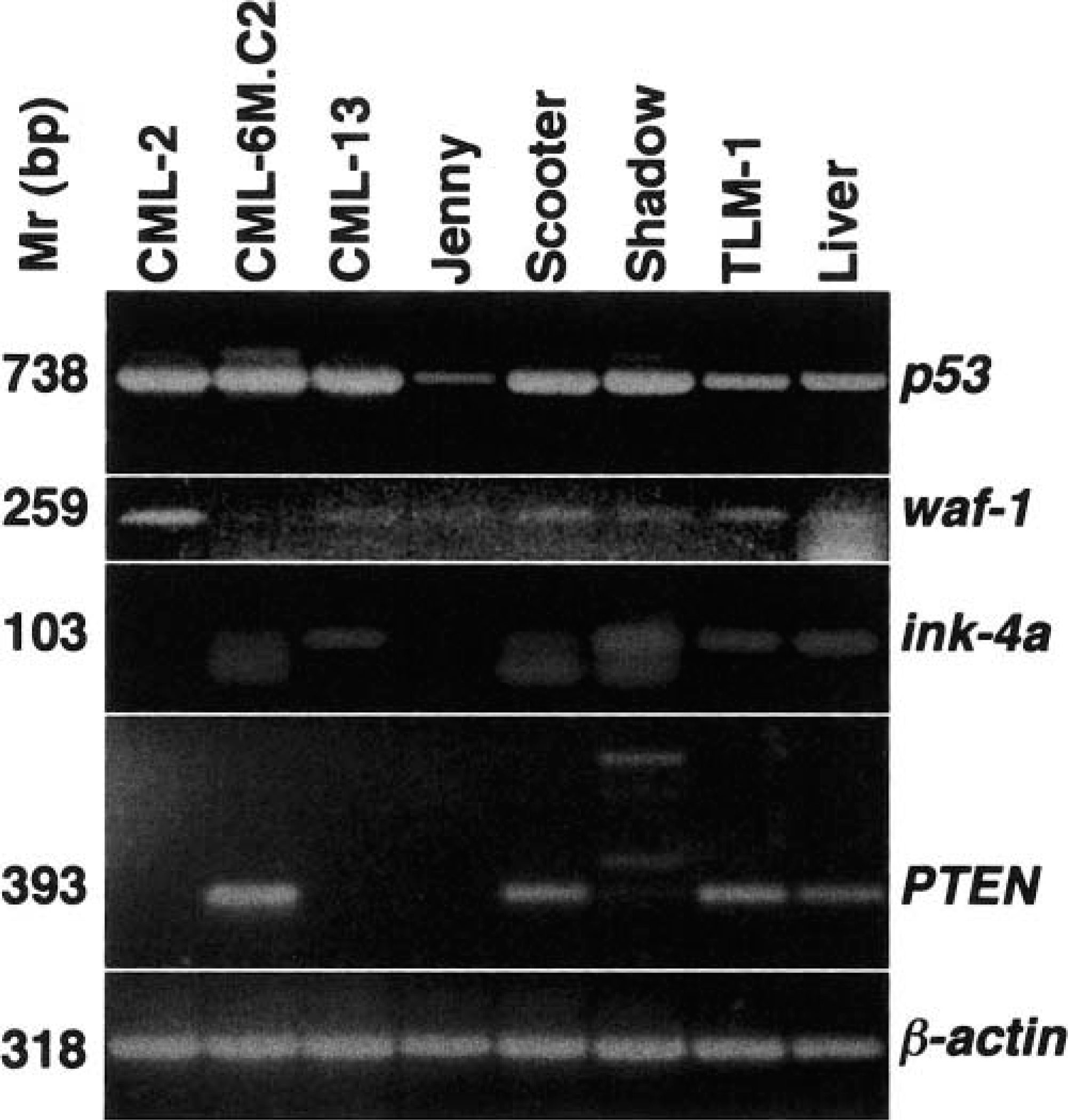
Expression of mRNA for each tumor suppressor gene (p53, waf-1, ink-4a, and PTEN) was examined by RT-PCR in the canine melanoma cell lines CML-2, CML-6MC2, CML-13, JENNY, SCOOTER, SHADOW, and TLM-1 using canine-specific primers. The expression of ß-actin was used as a loading control for the reactions and to ensure integrity of the RNA. Normal canine liver was used to evaluate the expression of these genes in a nonneoplastic tissue sample. The predicted sizes for the amplification products were 738 bp for p53, 259 bp for waf-1, 103 bp for ink-4a, and 393 bp for PTEN.
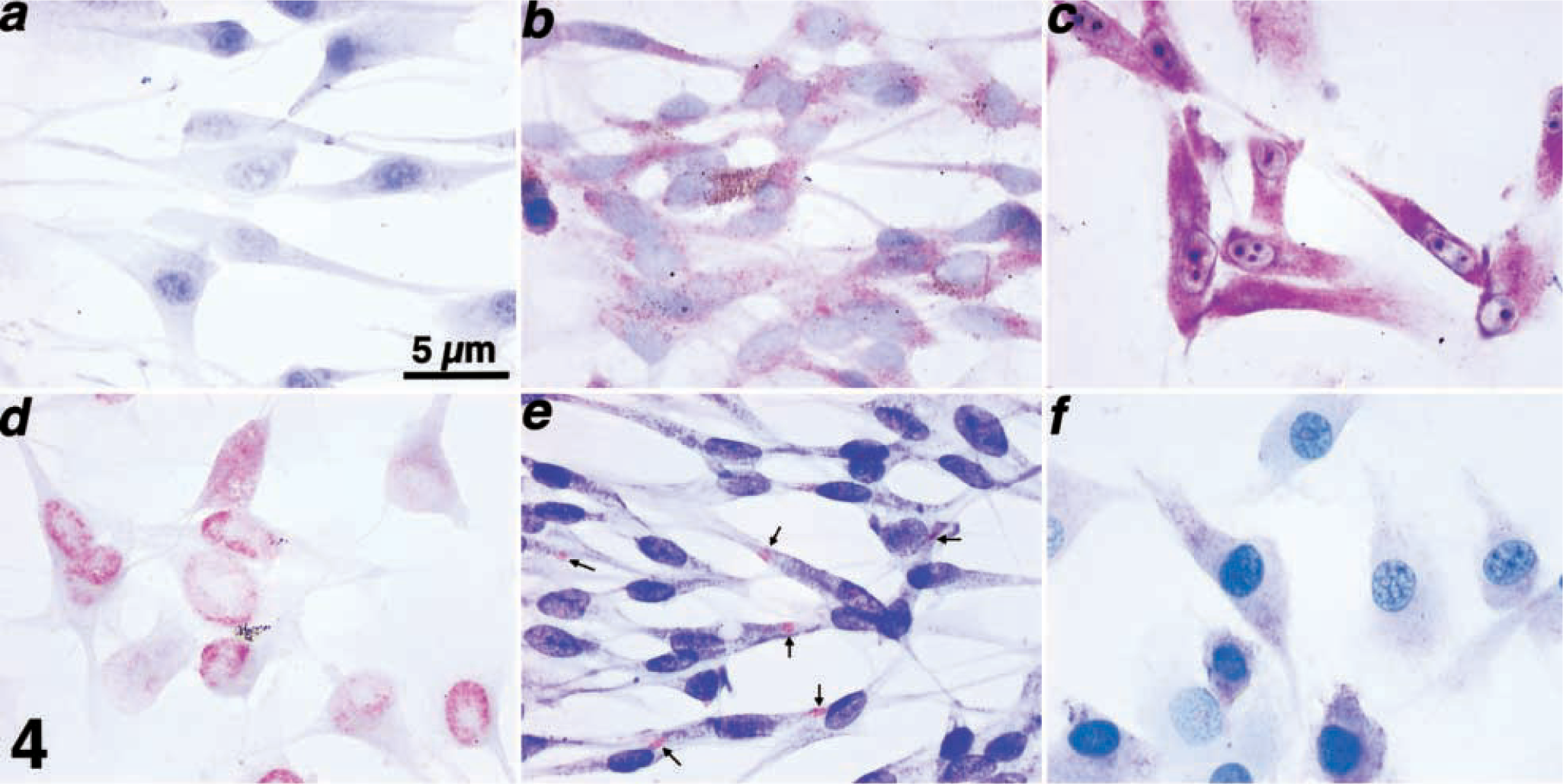
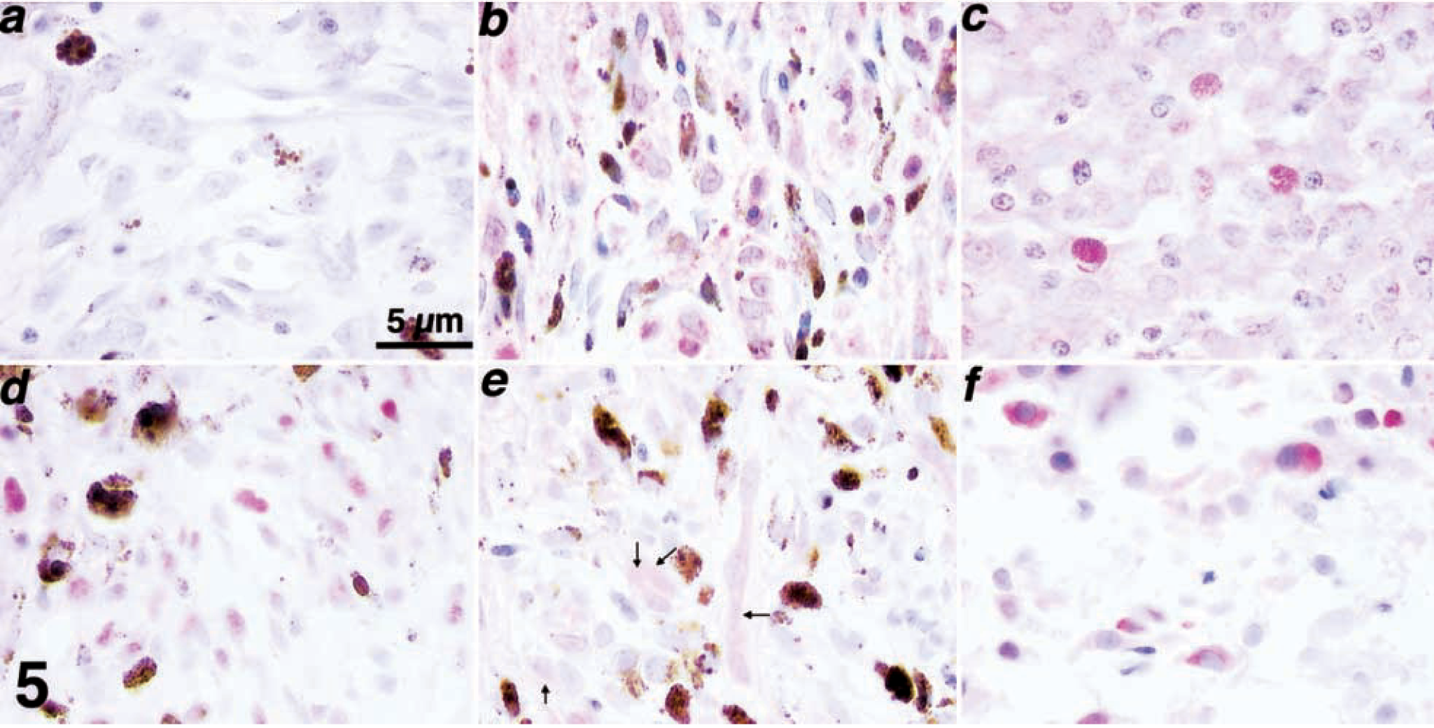
Immunocytochemical assessment of the expression and accumulation patterns for p53, p21, Rb, p16, and PTEN in canine melanoma cell lines.
Immunohistochemical assessment of the expression and accumulation patterns for p53, p21, Rb, p16, and PTEN in samples from spontaneous cases of canine melanoma.
Immunostaining results for the seven canine melanoma cell lines.
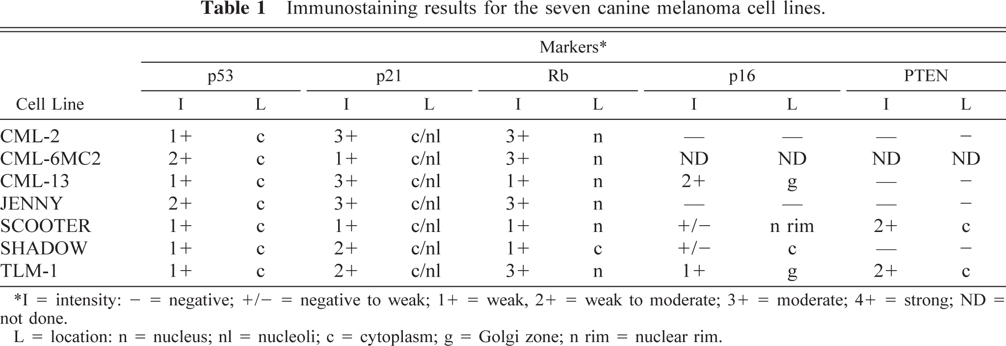
I = intensity: - = negative; +/− = negative to weak; 1+ = weak, 2+ = weak to moderate; 3+ = moderate; 4+ = strong; ND = not done.
L = Location: n = nucleus; nl = nucleoli; c = cytoplasm; g = Golgi zone; n rim = nuclear rim.
Loss or dramatic reduction of p53 expression (graded as negative or negative to weak) was seen in nine of 30 samples (five benign, three malignant, one of indeterminate behavior, Table 2). Three of these samples (Nos. 8, 10, 11) were from dogs with multiple, cutaneous, benign melanocytic tumors, with loss or reduction of p53 in every independent tumor examined. Exclusion of p53 from the nuclear compartment occurred in 18 of 25 samples with detectable p53 staining (eight benign, seven malignant, three of indeterminate behavior; Table 2). Figure 5b shows the p53 staining for sample No. 2, where the protein was shown to localize to the cytoplasmic and nuclear compartment. The two samples (Nos. 4, 8) from dogs with multiple benign melanocytic tumors with detectable p53 expression that were part of this latter group similarly showed localization of p53 exclusively in the cytoplasm in every independent tumor examined.
Immunostaining results for the 31 archival canine tumors.
Y = yes; N = no; NA = information not available.
I = intensity: + = negative; +/− = negative to weak; 1+ = weak; 2+ = weak to moderate; 3+ = moderate; 4+ = strong; ND = not done. L = location: n = nucleus; c = cytoplasm; g = golgi zone.
Multicentric refers to the availability of samples from more than one tumor that exhibited similar staining.
Sample Nos. 12 and 17 originated from the same dog.
Sample Nos. 13 and 18 originated from the same dog.
Expression of p21
The waf-1 gene plays an important role in melanocyte growth and differentiation.30,55 We previously documented the loss of p21 expression in a dog with multiple dermal melanocytic tumors56 and the requirement for localization of p21 protein to the nuclear compartment for contact-induced growth arrest (and possibly apoptosis).55 Our previous work suggests that waf-1 expression is not highly regulated in cultured melanoma cells; thus, qualitative changes of waf-1 expression would likely be significant. We used RT-PCR with two distinct sets of primer pairs to amplify products encompassing approximately 70% of the coding domains of waf-1, including the cyclin and CDK-interacting domains.55 Each of the seven cell lines generated amplification products of the predicted size and sequence using the C-terminal primer pair, with decreased expression apparent in JENNY, SCOOTER, and SHADOW cells (data not shown). Amplification products obtained using the N-terminal primer pair showed similarly decreased levels of waf-1 expression in the CML-6MC2, CML-13, JENNY, and SHADOW cell lines (Fig. 3). Sequencing of the waf-1 amplification products from CML-2, SHADOW, and TLM-1 cell lines revealed no mutations distinct from the polymorphisms identified for the wild-type canine gene. The presence of waf-1 mRNA in each cell line suggested these cells would show accumulation of p21 protein. We used immunocytochemistry to examine accumulation of p21 protein in the canine melanoma cell lines. We previously verified the specificity of the C-20 antibody against canine p21 by immunoblotting.55,56 Each of the seven cell lines examined showed accumulation of p21 that was predominantly cytoplasmic and nucleolar (Table 1, Fig. 4c). The staining was intense and uniform in the CML-2, CML-13, and JENNY cell lines. The SHADOW and TLM-1 cell lines showed granular staining with occasional cells having focal staining in the perinuclear or Golgi regions. The CML-6MC2 and SCOOTER cell lines stained faintly for cytoplasmic p21 but still had prominent nucleolar staining. The absence of nuclear p21 was predictable in subconfluent cells in the logarithmic growth phase.55
Loss or dramatic reduction of p21 expression (graded as negative or negative to weak) was seen in six of 30 samples (three benign, two malignant, one of indeterminate behavior; Table 2). Two of these samples (Nos. 8, 11) were from dogs with multiple, cutaneous, benign melanocytic tumors, with loss of p21 in every independent tumor examined. Exclusion of p21 from the nuclear compartment occurred in nine of the 27 samples with detectable p21 staining (six benign, two malignant, one indeterminate, Table 2). One of the three samples from dogs with benign multiple melanocytic tumors with detectable p21 expression (sample No. 10) that was part of this latter group similarly showed localization of p21 exclusively in the cytoplasm in every independent tumor examined. Figure 5c shows the p21 staining for sample No. 21, where the protein was localized in the cytoplasmic and nuclear compartments.
Expression of Rb
Mutations of Rb do not appear to be prevalent in human melanoma, possibly because mutations of other genes that participate in the same growth control pathway occur so often as to render Rb expression inconsequential.8,51,54,59,60,75 The expression of Rb in canine melanoma has not been evaluated extensively. We used immunocytochemistry to evaluate the expression and subcellular localization of Rb in the canine melanoma cell lines. We previously verified the specificity of the G3–245 antibody against canine Rb by immunoblotting.47,55 The results show nuclear accumulation of Rb in six of the seven cell lines (Table 1, Fig. 4d). Not all the cells from the CML-13 and SCOOTER lines showed Rb staining, although this inconsistency could be attributable to the fact that these cells were growing asynchronously. Alternatively, loss or reduction of ink-4a in SCOOTER cells may have eliminated the constraints of Rb expression and localization. We showed previously that the predicted phosphorylation state of Rb in TLM-1 cells corresponded with quiescence (hypophosphorylated) or proliferation (hyperphosphorylated), and overexpression of cyclin-dependent kinase inhibitors p21 or p16 in TLM-1 cells and CML-2 cells resulted in hypophosphorylation of Rb with subsequent growth arrest and apoptosis.47,55 Thus, nuclear Rb in these melanoma cell lines probably represented the product of a functional wild-type gene. Rb staining in the SHADOW cell line was restricted to faint accumulation in the cytoplasmic and perinuclear compartment of few cells, suggesting that this cell line might harbor a mutant Rb gene product (Table 1).
Loss or dramatic reduction of Rb expression (graded as negative or negative to weak) was seen in nine of 30 samples (four benign, three malignant, two of indeterminate behavior; Table 2). Two of these samples (Nos. 4, 8) were from dogs with multiple, cutaneous, benign melanocytic tumors, with loss of Rb in every independent tumor examined. Exclusion of Rb from the nuclear compartment occurred in four of the 24 samples with any detectable Rb staining (three benign, one of indeterminate behavior; Table 2) but was not seen in any of the samples from dogs with multiple benign melanocytic tumors. Figure 5d shows the localization of Rb staining restricted to the nuclear compartment in sample No. 13.
Expression of ink-4a
Mutation of ink-4a is one of the most frequent genetic abnormalities characterized in human tumors59 and is a feature of >50% of all human melanomas.16,27,48,50 The importance of this gene in tumorigenesis was confirmed in mice with a targeted deletion of the INK-4A locus that includes the ink-4a gene, where the addition of a conditionally active ras oncogene leads to the development of spontaneous melanoma.7 Three of the seven cell lines (CML-2, CML-6MC2, and JENNY) lacked ink-4a mRNA, as determined by RT-PCR (Fig. 3). A more quickly migrating (lower) band that corresponded to primer dimers (confirmed by sequencing) was seen in some samples, most often those that had reduced or absent ink-4a expression (see lanes for CML-6MC2, SCOOTER, and SHADOW cells, Fig. 3). The levels of expression of the predicted ink-4a amplification product in SHADOW cells were slightly higher than those in the liver. An amplification product of the predicted size was obtained from the CML-13, SCOOTER, and TLM-1 cell lines (Fig. 3). Immunoblotting showed that a ∼16 kd protein that was recognized by the anti-p16 antibody and was indistinguishable from the protein found in normal Cf2Th cells was abundant in CML13 cells and was expressed at much lower levels in JENNY, SCOOTER, SHADOW, and TLM-1 cells (Fig. 6). No p16 was detectable in CML2 or CML-6MC2 cells. Analysis of p16 protein expression by immunocytochemistry similarly showed moderate expression in CML-13 cells (comparable to that seen in Cf2Th cells) and weak staining in TLM-1 cells (Table 1, Fig. 4e). The staining was predominantly cytoplasmic with prominent accumulation in the Golgi regions. Less than 5% of SCOOTER and SHADOW cells had faint staining localized to the nuclear rim or to cytoplasmic granules, respectively. CML-2 and JENNY cells had no detectable p16. Immunocytochemical staining for p16 protein was not performed in CML-6MC2 cells.
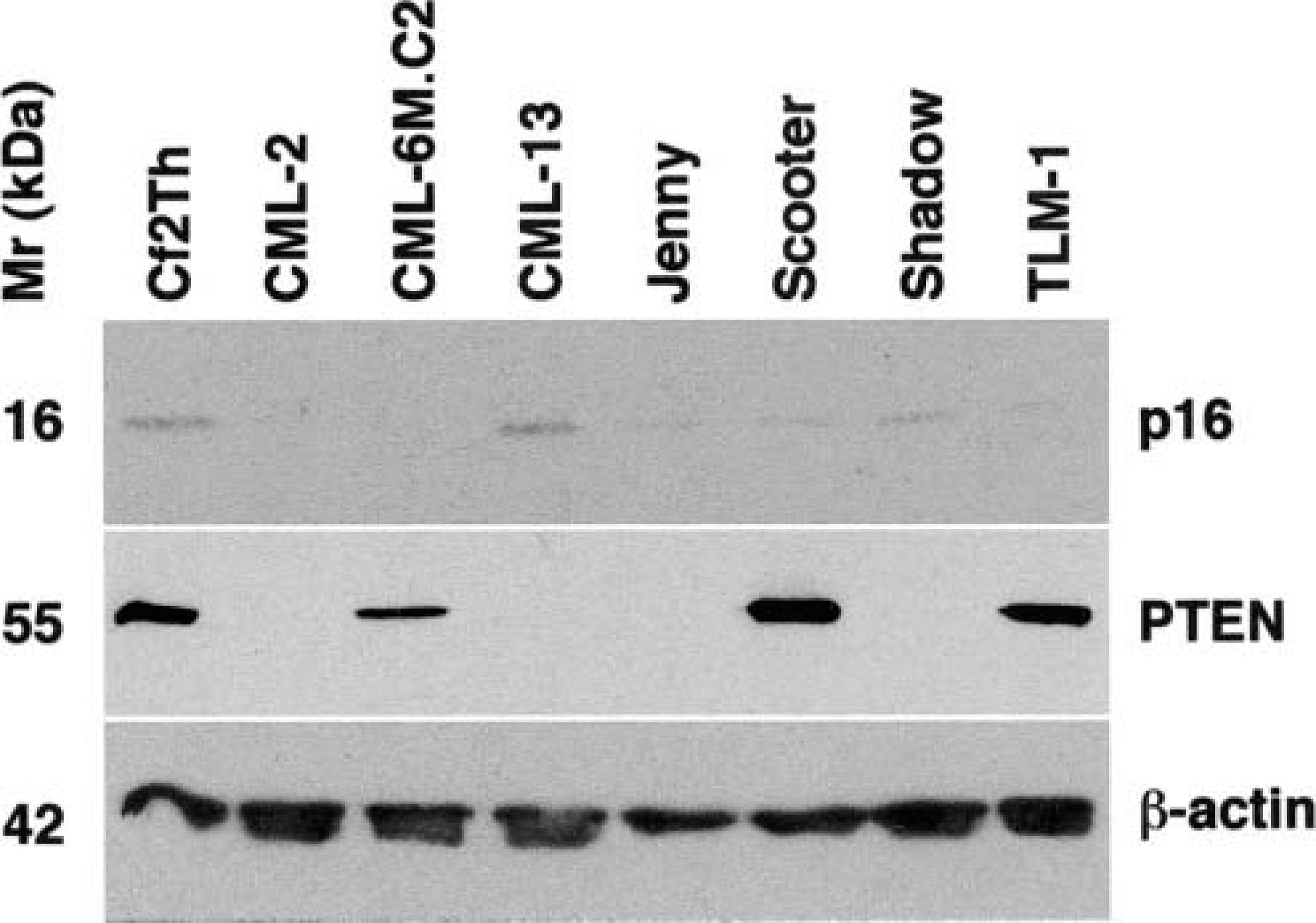
Accumulation of p16 and PTEN tumor suppressor proteins in canine melanoma cell lines CML-2, CML-6MC2, CML-13, JENNY, SCOOTER, SHADOW, and TLM-1 was examined by immunoblotting. Analysis of β-actin was used as a loading control. Normal canine Cf2Th cells were used to evaluate the expression of these proteins in a nonneoplastic tissue sample. The sizes for the proteins are 16 kd for p16, 55 kd for PTEN, and 42 kd for β-actin.
Loss or dramatic reduction of p16 expression (graded as negative or negative to weak) was seen in 21 of 26 samples (six benign, 11 malignant, four of indeterminate behavior; Table 2). When present, staining of p16 was generally confined to the cytoplasm, although intense staining in the Golgi area was seen in four cases (two benign and two indeterminate). The pattern of p16 accumulation is illustrated by the immunostaining for sample No. 7 (Fig. 5e).
Expression of PTEN
The importance of the PTEN gene in the pathogenesis of multiple cancers has recently been a topic of intense interest.3,4,40,43,66 PTEN is an antagonist of the phosphoinositol-3 kinase (PI3K).20 PI3K activation initiates a cascade of events that promote growth factor production, cell division, and survival (resistance to apoptosis).18,19 Thus, loss of PTEN can establish an autocrine growth loop where there is increased production of growth factors and where cells show exaggerated proliferation in response to these growth factors. We examined PTEN gene expression in the canine melanoma cell lines by RT-PCR. Three of the cell lines (CML-2, CML-13, JENNY) lacked PTEN mRNA when the internal primer pair was used for amplification, and cDNA from SHADOW cells generated multiple amplification products that likely represent mutant forms of the protein (Fig. 3), although in some experiments, no amplification products for PTEN mRNA were generated from this cell line. SHADOW cells appear to be hypodiploid based on DNA content analysis (data not shown), suggesting the line may be genetically unstable, and is thus likely to harbor numerous mutations. Each of these cell lines, except JENNY, generated amplification products using the primer pair spanning the complete coding domains (data not shown). The CML-6MC2, SCOOTER, and TLM-1 cell lines produced amplification products of the predicted size with each primer pair, and sequencing revealed no discernible mutations in the 393 bp amplification products generated with the internal primer pair. Consistent with the results observed upon examination of gene expression, immunoblotting showed the presence of a ∼55 kd protein that was recognized by the anti-PTEN antibody in SCOOTER and TLM-1 cells that was indistinguishable from the protein found in normal Cf2Th cells (Fig. 6). A PTEN-immunoreactive protein also was present in CML-6MC2 cells and had a slightly slower electrophoretic mobility than that seen in the other positive cell lines. PTEN protein was not detectable in CML-2, CML-13, JENNY, or SHADOW cells. Analysis of PTEN protein expression by immunocytochemistry showed moderate, diffuse cytoplasmic staining in TLM-1 cells and SCOOTER cells (Table 1, Fig. 4f). No PTEN staining was evident in CML-2, CML13, JENNY, and SHADOW cells. Immunocytochemical staining for PTEN protein was not performed in CML-6MC2 cells. Loss or dramatic reduction of PTEN expression (graded as negative or negative to weak) was seen in 16 of 27 samples (five benign, 11 malignant, five of indeterminate behavior; Table 2). Although PTEN is a cytoplasmic protein, nuclear localization was seen in two samples (one malignant and one of indeterminate behavior; Table 2). Figure 5f shows the staining for PTEN in sample No. 26.
Discussion
Melanomas are malignant tumors of melanocytes or melanoblasts. Cultured cells usually resemble the morphology of the parent tumor from which they were derived;15,33,49,56 however, cultured melanoma cells are not the same as tumors in vivo. Theoretically, cell lines can mutate and may not possess all the same characteristics of the parent tumor. Variation in environmental conditions may also influence growth and differentiation. Nevertheless, cultured melanomas are a useful preliminary model to study the origin and progression of melanoma. Combined with analysis of primary archival sections, this approach provides a robust model to define the frequency with which the expression of particular genes or proteins may be lost (or amplified) in these tumors and to investigate possible biological consequences of these abnormalities.
In this study, we used RT-PCR and immunocytochemistry to examine tumor suppressor gene and protein expression in seven cultured melanoma cell lines and 31 archival samples of melanoma from 29 affected dogs. The gene products we chose to examine, p53, p21, Rb, p16, and PTEN, have each been implicated to some extent in the origin or progression of melanoma in humans or other animal models.1,11,22,28,46,55,56,58,59,70,71 We specifically sought to investigate whether the pathways of growth control, survival, and DNA integrity in which these proteins participate might contribute to the genesis of canine melanoma. Our working hypotheses were 1) mutations in these pathways are common in canine melanoma and 2) mutations in more than one pathway predict a worse outcome. The results presented here are consistent with the first hypothesis. Our results also show that these genetic lesions are common in both the benign and malignant forms of canine melanoma, but additional work is necessary to document their prognostic significance.
We previously proposed a predisposition of individual dogs to develop multiple melanocytic tumors as a result of a mutation in a melanocyte precursor, based on a shared genetic characteristic in the tumors.56 This study includes data from the same dog (sample No. 11) and three additional dogs with multiple, cutaneous, benign melanocytic tumors. Two additional dogs were included that developed two distinct tumors (sample Nos. 12, 17; sample Nos. 13, 18), but in both of these dogs, one of the tumors was benign and one was malignant. Three of these dogs were Gordon Setters, and three of the four Gordon Setters in the study developed more than one melanoma in their lifetime. The results from this study confirm the presence of a shared phenotype in each of the tumors from dogs with multiple, cutaneous, benign melanocytic tumors, supporting the premise that these tumors may have arisen from a common precursor. However, such shared phenotype was not seen in the dogs that developed more than one tumor with different biological behavior (i.e., one benign and one malignant), suggesting that different factors may underlie the pathogenesis of these two apparently related tumors.
The observation that abnormalities indicative of loss of p16/Rb function (including reduced or absent expression of either gene or exclusion of Rb from the nuclear compartment) occurred most frequently in both the cell lines (six of seven) and the clinical samples (22 of 26) suggests that inactivation of this pathway is a critical step in the pathogenesis of benign and malignant melanoma in dogs, as it is in humans and other animals.48,59,61,72 Mutation of ink-4a is a feature of >50% of all human melanomas.16,27,48,50 In the CML-2 and CML-6MC2 cell lines where gene expression and protein accumulation were undetectable, loss of ink-4a expression might have occurred through homozygous deletion of the gene, through mutations of noncoding transcriptional regulatory sequences, or through methylation.14,48,59,72 Our results suggest that JENNY, SCOOTER, and SHADOW cells may harbor mutations in the ink-4a gene that generate unstable proteins. This instability in SCOOTER and SHADOW cells might be related to rapid degradation due to localization of the protein to abnormal subcellular compartments. In JENNY cells, the mutation is likely to involve the ankyrin domains in exon 2; no mRNA was detectable using primers targeting this region. The p16 protein in TLM-1 cells is also expressed at very low levels, raising the possibility of hemizygous deletion of one ink-4a allele or of a mutation that also produces an unstable protein in these cells. The results from our immunoblotting experiments indicate that it is unlikely that the RT-PCR led to amplification of other ink-4 gene family members (i.e., ink-4b, ink-4c, or ink-4d). Although the sequence of p16 exon 2 targeted for amplification is highly homologous to that of p15 (ink-4b) from other species (the sequence for canine ink-4b is unknown) and the anti-p16 antibody used is reported by the manufacturer to have some degree of cross-reactivity with p15, a protein with faster electrophoretic mobility (smaller molecular weight) was not seen in any cell line. The results from the immunoblotting experiments similarly suggest that it is unlikely that the amplification products from the RT-PCR may represent the ARF (alternative reading frame) gene, which is encoded in the same locus as ink-4a. 53
Mutations of Rb do not appear to be prevalent in human melanoma,39 possibly because mutations of ink-4a occur so often as to render Rb expression inconsequential.8,51,54,59,60,75 Normal human melanocytes (and other quiescent cells such as peripheral blood lymphocytes) show little to no detectable Rb by immunostaining,45,58 but Rb was present in all primary and metastatic melanomas in 5–70% of cells and was always localized to the nucleus. Loss or mutational inactivation of both normal Rb alleles leads to loss of Rb expression and can contribute to the development of malignant melanomas.58 Exclusion of Rb from the nuclear compartment such as that seen in the SHADOW cell line and sample Nos. 2, 3, 14, and 30 may represent a novel mechanism of Rb inactivation in melanoma and may reflect a mutation of Rb or possibly other genes that control nuclear translocation of Rb.
Exclusion of p53 from the nuclear compartment occurred almost as frequently as loss of p16/Rb. The cytoplasmic accumulation of p53 in the cell lines and tumor samples was not a feature of the antibody or the conditions used or an artifact of tissue culture; p53 was localized almost exclusively in the nuclear compartment of Cf2Th cells,55 and seven tumor samples contained abundant nuclear p53. In human melanomas, mutations of the p53 gene or abnormalities that result in accumulation of p53 protein that is functionally excluded from the nucleus occur frequently,9,28,31,57,70,71 and melanomas with mutant p53 gene carry a worse prognosis than those without p53 mutations.17,34 However, numerous events may affect the stability and subcellular localization of p53,36 and overexpression of p53 protein in tumor cells does not necessarily imply mutation or loss of function of the p53 gene.10 The sequences for amino acids 77–285 in the p53 mRNAs obtained from the cell lines in this study were indistinguishable from the wild type sequence, suggesting that the p53 proteins in these cells were competent to bind DNA in a sequence-specific manner and to undergo homodimerization. More than 90% of point mutations associated with loss of function of p53 occur within the DNA binding and homodimerization domains between amino acids 102 and 292.36 However, we cannot exclude the possibility that the cell lines had mutations of p53 that affected the mdm-2–interacting domain located between amino acids 13 and 29 or the nuclear localization signal located near amino acids 305 and 306.26,36 These domains are responsible, respectively, for targeting p53 for degradation in the proteasome and for allowing transport of p53 to the nucleus. The inactivation of p16 in many tumors that contained cytoplasmic p53 is somewhat paradoxical because the ARF gene encoded in the same locus stabilizes p53 by targeting mdm-2 for destruction.41,52,78 Thus, p16, but not ARF, had to be selectively inactivated in these tumors (e.g., by methylation), unless mutations of p53 in the mdm-2–binding domain are more common in dogs than they are in humans. The overexpression of cytoplasmic p53 also could reflect a measure of genetic instability (with rapid turnover of the protein in the nuclear compartment) or mutations in other genes that control p53 synthesis and degradation.
Loss or reduction of PTEN expression also was seen frequently in the cell lines but less so in the tumor samples. The cell line that did not exhibit abnormalities of p16 or Rb (CML-13) showed loss of PTEN expression, suggesting that enhanced survival or growth mediated through growth factor pathways that use PI3K may help to overcome the effects of normal p16/Rb pathways in canine melanoma. Three cell lines (CML-2, JENNY, and SHADOW) showed concurrent abnormalities of p16 or Rb and PTEN, suggesting that concurrent loss (or reduction) of both the p16/Rb and PTEN pathways may facilitate progression of the disease. At least two mechanisms may account for loss of PTEN expression. Of the four cell lines that showed mutations of the PTEN gene, three might harbor deletions, insertions, or single nucleotide substitutions that precluded amplification of PTEN mRNA using the internal primer pair, whereas the loss of PTEN gene expression in the other may have been due to aneuploidy or genetic instability. Because amplification products were obtained from each cell line using the primer pair designed to amplify the complete coding domains, it is possible that the internal primer pair may span a “hot spot” and that mutations in this region destabilize the protein and account for the lack of PTEN immunostaining in these cells. However, this is not true for every cell line, and additional experiments by us and others suggest that cultured canine tumor cells can express mutant PTEN genes that can be detected by RT-PCR or immunohistochemistry (E. Dickerson, S. Fosmire, S. Bianco, J. Wojcieszyn, J. Modiano, and S. Helfand, unpublished results). Therefore, because mutations that inactivate PTEN need not necessarily hinder its expression, immunohistochemical analysis of fresh or archival tissues may underestimate the frequency of such mutations in canine melanoma and other tumors.
An important role for p21 in melanocyte growth and differentiation has been documented.30,32,44,47,55 The p21 protein is a CDKI that has nonoverlapping functions with p16, but it also participates in contact-induced growth arrest and controls the function of stress-activated protein kinases, polymerases involved in DNA synthesis, proteins that regulate survival and apoptosis, and possibly cell division (cytokinesis).2,6,23,24,64,67,74 There was no clear correlation between the presence of p21 protein and the morphology or growth properties of the cell lines. One mechanism through which p21 expression can be upregulated is through transcriptional activation mediated by p53,13 and p21 appears to be important in p53-dependent growth arrest in cells that must undergo DNA repair.67 However, p21 expression can also be induced through pathways independent of p53.42,76,77 We saw no apparent correlation between p21 and p53 in the seven cell lines. Loss or reduction of p21 was also an infrequent occurrence in the clinical samples, but exclusion of p21 from the nuclear compartment was disproportionately seen in cutaneous melanomas, suggesting that loss of function of this protein may play a role in this form of the disease.
In this group of samples, anatomic location remained the best predictor for tumor behavior. Multiple abnormalities in these proteins were not significantly more common in malignant lesions than in benign lesions. This lack of significance may be due to the limited statistical power afforded by the relatively small sample size. Alternatively, its could signify that the function of these genes is important to prevent malignant transformation of melanocytes, but once transformation takes place their loss of function does not significantly impact disease progression. Nevertheless, the results showed distinct patterns with shared abnormalities. For example, the absence of functional p53 was significantly correlated with absence of nuclear p21. Similarly, loss or reduction of p16 was correlated with abnormalities of Rb. These results were predictable because p53 is an important regulator of p21 expression and because abnormalities in a single member of the p16/Rb pathway render other members of the pathway inconsequential.
The results from this study show that abnormalities of tumor suppressor genes and proteins are common in canine melanomas. These abnormalities likely reflect mutations that may contribute to the origin of the tumors in vivo. The possibility that malignant tumors accumulate mutations in multiple tumor suppressor gene pathways at an accelerated rate that may influence prognosis merits further investigation in spontaneous canine tumors.
Footnotes
Acknowledgements
We thank Dr. Steve Dow, Dr. Lauren Wolfe, Dr. Elaine Ostrander, and Dr. David Beach for providing cell lines and reagents, Josy Mayor and Dr. Vijayanagaram Venkatraj for assistance cloning canine ink-4a, Dr. Gary Cutter for assistance with statistical analyses, Dr. Roy Levine for information on amplification of p53 by RT-PCR and on the performance of antibodies that recognize canine p16 and PTEN, Dr. Erin Dickerson, Dr. Michelle Ritt, Dr. Kenita Rogers, and Dr. Jennifer Thomas for helpful suggestions, and Dr. Barb Powers for critical review of the manuscript. This work was supported in part by grant 1626 from the AKC Canine Health Foundation and by grants 98PT-16 and 98CA-34 from the Morris Animal Foundation. S. R. Bianco was supported in part by a fellowship from the University of Colorado Cancer Center.