
Abstract
The protein p53 is considered to be one of the most important tumor suppressor factors. Despite this importance, a potential association between TP53 messenger (m)RNA levels and tumor aggressiveness has not been well defined in animal cancer. We assessed and correlated TP53 gene expression in 40 canine mammary carcinomas with histologic grade, tumor size, and aggressiveness. The tumors were subjected to histologic analysis and the TP53 mRNA levels determined by RT-rtPCR. Statistical analysis revealed no correlation between levels of TP53 mRNA and tumor aggressiveness (r = 0.00) or tumor growth (r = 0.06). Histologic grades (r = 0.17) and mitosis count (r = 0.12) showed a weak correlation with TP53 mRNA expression levels. These findings are consistent with molecular studies that revealed heterogeneous expression of TP53 in canine and human mammary tumors. Hence, TP53 gene expression alone cannot be considered a marker for tumor aggressiveness in canine mammary carcinomas.
Keywords
The TP53 gene encodes the tumor suppressor protein p53. This gene initiates protein translation in response to DNA damage or oncogene activation, inducing cell cycle arrest, apoptosis, senescence, DNA repair, or metabolic changes. 1 Most TP53 genetic alterations lead to the synthesis of mutant proteins with longer half-life, 9 and overexpression of the protein p53 observed in human and canine mammary neoplasms has been associated with proliferation increase, differentiation loss, and apoptotic evasion. 8 To the best of our knowledge, only one study has evaluated the transcriptional levels of TP53 in canine mammary tumors, but using a small sample size of malignant tumors. 6 Because of the involvement of protein p53 in tumor development, we investigated the contribution of TP53 gene expression to the process of mammary tumors becoming malignant. Thus, we evaluated whether TP53 gene expression in canine mammary carcinomas could be correlated with histologic grades and aggressiveness rates.
Our study involved 40 canine mammary carcinomas from 25 female patients in stages I–IV, according to the TNM (tumor, lymph nodes, metastasis) system. 2 Tumors were received from the BioBank of Animal Tissue, DNA, and RNA for Pathological and Molecular Diagnostics (UFF, Brazil; approved by Ethics Committee on Animal Use, UFF ref. 326/2013) and histologically classified according to criteria established in the literature. 2 Tumor fragments were stored in liquid nitrogen. RNA preparation was performed (RNeasy mini kit, Qiagen, Germantown, MD) and stored in a freezer at −80°C.
The expression of TP53 was analyzed using a one-step real-time (rt) reverse transcription (RT)-PCR (RT-rtPCR; Power SYBR Green RNA-to-CT 1-step kit, Thermo Fisher Scientific, Waltham, MA) and a platform system from the same manufacturer (Step One real-time PCR system, Thermo Fisher Scientific). The specific primers for TP53 and ATP5B (reference gene) used in the experiments of RT-rtPCR were described previously.3,7 The relative quantity (RQ) of transcript levels was calculated using the comparative ΔΔCT method. In order to compare the levels of TP53 gene expression, the mean RQ values for 5 normal canine mammary tissue samples obtained from 5 dogs was set as 1.0 (calibrator) by statistical normalization. Cutoff values were defined as fold change (FC) ≥2.0 for TP53 overexpression and ≤0.5 for reduced expression.
The neoplasms were analyzed using a proposed aggressiveness index considering the following criteria: tumor size (according to modified TNM system), 2 tubule formation index, nuclear pleomorphism, cellular differentiation, mitotic count, atypical mitosis, invasion of adjacent tissues, tumor capsule, necrosis, neovascularization, tumoral clusters, lymph node metastasis, and histologic grade 4 (Table 1). All tumors were analyzed independently by the same 3 pathologists. The Pearson correlation coefficient (r) was used to determine the relationship between the characteristics of the tumor samples and TP53 expression. Analysis of variance (one-way ANOVA) was used to evaluate TP53 expressions in different carcinoma types found in our study (p < 0.05).
Histologic diagnosis, malignancy factors, and TP53 mRNA expression obtained by RT-rtPCR from 40 canine mammary carcinomas.
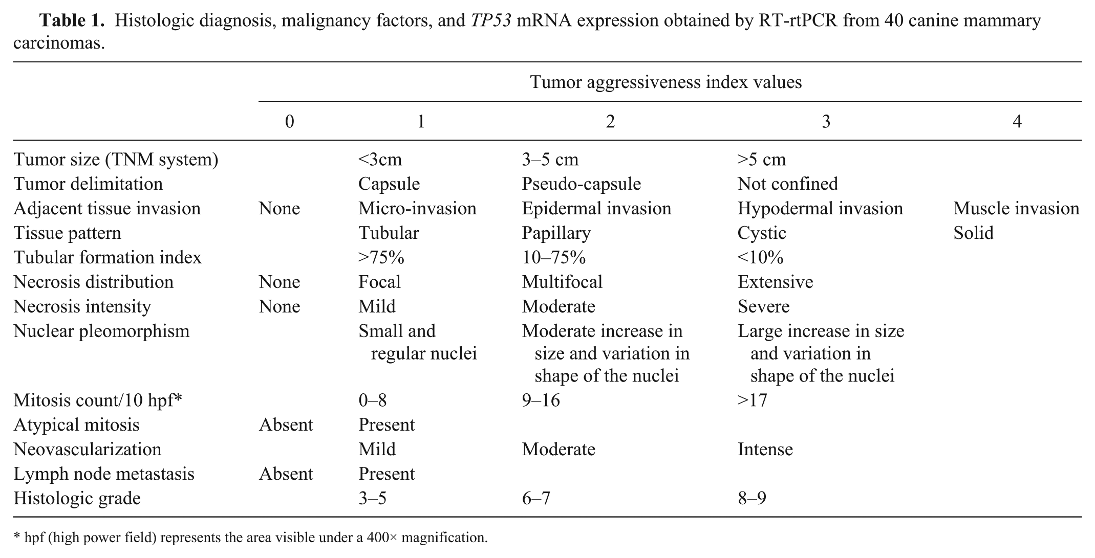
hpf (high power field) represents the area visible under a 400× magnification.
The majority of the 40 mammary tumor samples were histologically classified as carcinoma in a mixed tumor (n = 24), followed by papillary carcinoma (n = 7), complex carcinoma (n = 3), carcinosarcoma (n = 3), solid carcinoma (n = 2), and spindle-cell carcinoma (n = 1; Fig. 1). Tumor size was classified according to the TMN system, 2 and 19 mammary tumors were classified as T1 (≤3.0 cm), 11 as T2 (>3.0; <5.0), and 10 as T3 (≥5.0; Table 2).
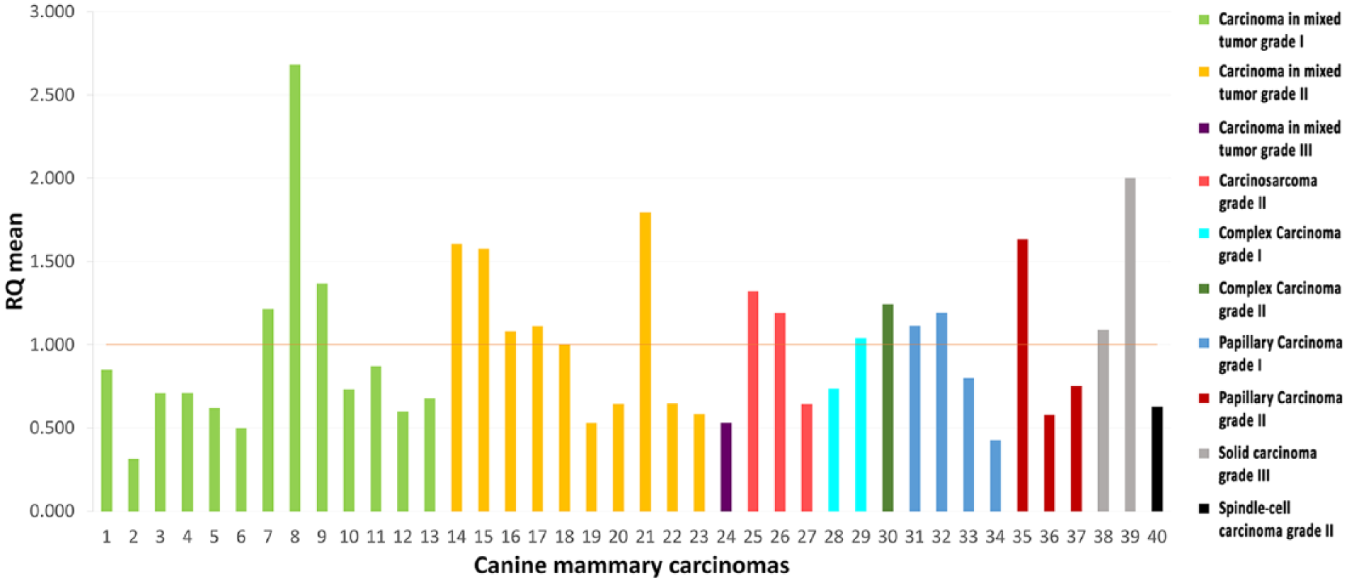
Relative quantity (RQ) of TP53 mRNA levels of canine mammary carcinomas, according to the tumor type and grade, analyzed by RT-rtPCR.
TP53 expression in 40 canine mammary tumors according to histologic type and grade.
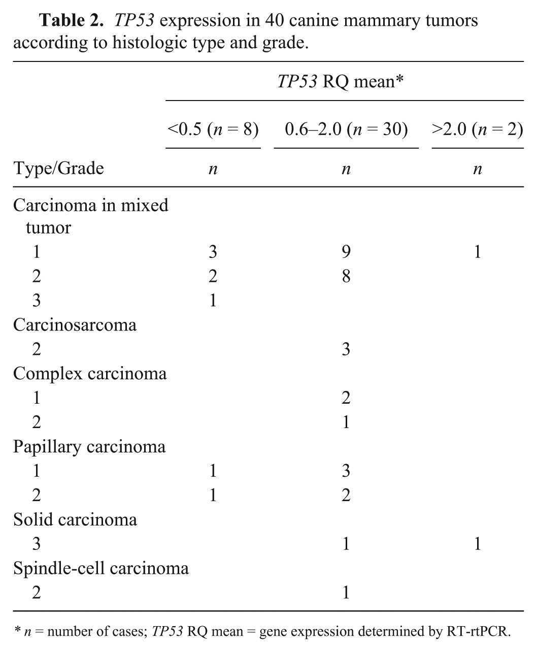
* n = number of cases; TP53 RQ mean = gene expression determined by RT-rtPCR.
TP53 gene expression was compared with the calibrator RNA based on normal mammary tissue. Eight samples had a significant reduction in gene expression (FC ≤ 0.5). Thirty samples had transcription levels similar to that of the calibrator and were considered within normal range; 2 were overexpressed with a significant 2-fold increase (Table 2).
Statistical analysis found no correlation between TP53 gene expression and tumor aggressiveness (r = 0.00; Table 3) or tumor growth (r = 0.06). However, a weak positive correlation was found between the levels of TP53 mRNA and histologic grades (r = 0.17) and mitosis count (r = 0.12). One-way ANOVA showed no significant difference in the TP53 gene expression profile by the different carcinoma types (p > 0.05). Similar results were previously reported revealing heterogeneous expression of TP53 gene in malignant canine mammary tumors. 6 However, those authors considered the number of carcinomas that they studied to be too small to conduct a proper statistical analysis.
TP53 expression in 40 canine mammary tumors according to tumor aggressiveness index.
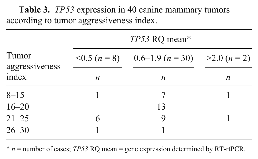
n = number of cases; TP53 RQ mean = gene expression determined by RT-rtPCR.
Our data suggest that variations in TP53 gene expression alone do not seem to be associated with histologic aggressiveness in canine mammary carcinomas. In 2 previous studies of human mammary tumors, when evaluated alone, the TP53 transcriptional profile was not correlated with clinical outcome.5,10 Nevertheless, a significant correlation between TP53 mutation type and gene expression, and patient prognosis, was reported. Accordingly, low levels of TP53 expression were associated with a poor prognosis for patients with a missense mutation. Additionally, increased mRNA levels and TP53 deleterious mutations were indicative of a poor prognosis. 5 A study associating TP53 mRNA expression levels, mutation status, and prognosis has not yet been reported in dogs, to our knowledge.
In spite of the fact that knowledge of the molecular basis of tumor aggressiveness would contribute to a better understanding of the malignant transformation process, and tumor diagnosis and prognosis in canine mammary cancer, such studies are still in the early stages in veterinary medicine. Indeed, advances in molecular veterinary oncology research are essential to specific and individualized cancer treatments.
Footnotes
Acknowledgements
We thank the Surgery Department, Hospital of Veterinary Medicine, Prof. Firmino Mársico Filho, Universidade Federal Fluminense for the animal samples used in this study. Táya F. Oliveira received a fellowship from the Coordenação de Aperfeiçoamento de Pessoal Nível Superior (CAPES).
Declaration of conflicting interests
The authors declared no potential conflicts of interest with respect to the research, authorship, and/or publication of this article.
Funding
The authors received no financial support for the research, authorship, and/or publication of this article.