
Abstract
Early histologic changes in lesions at vaccine sites were compared in cats, mink, and ferrets. Twenty-four 4-month-old cats, 20 4-month-old mink, and 20 12-month-old ferrets were vaccinated with three rabies virus vaccines, two feline leukemia virus vaccines, alum adjuvant, and saline. Injection sites were excised at selected time points up to 21 days postvaccination. Histologic examination of the tissue revealed significant differences among the cats, mink, and ferrets in the local response to the commercial vaccines. When compared with ferrets and mink, cats had more lymphocytes in response to all three rabies vaccines. Production of fibroblasts, collagen, and macrophages differed among the three killed aluminum-adjuvanted vaccines in cats but did not differ significantly in mink or ferrets. Cats produced fewer binucleate cells than did mink or ferrets in response to the two adjuvanted leukemia virus vaccines. Differences seen in early tissue response of cats to commercial vaccines may be related to the increased predisposition of cats to vaccine-associated sarcomas.
Keywords
Vaccine-associated sarcomas (VAS) in cats have been recognized as a potential consequence of vaccination for over a decade.18,21,25,26,33 An estimated ≥0.01% of vaccinated cats in the United States develop a tumor at the site of a killed adjuvanted vaccine.5,29 Several studies have focused on the histology and behavior of the mature neoplasm, which appears to develop 3 months to 3 years after vaccination.31 VAS are usually diagnosed as fibrosarcomas, malignant fibrous histiocytomas, or other mesenchymal tumors.7,11,24,25 Thus far, in only one study have early lesions at vaccine sites been examined histologically.22 Examination of the early stages of the postvaccinal lesions may be valuable for understanding their evolution from inflammation to neoplasia.
Only one report of VAS in a ferret was found.38 No reports of VAS in mink were found, although modified live and killed viral and bacterial vaccines are commonly used in mink.27 Few if any VAS have occurred in domestic canids. Posttraumatic ocular sarcomas have been documented in cats9,10 but not in other domestic species. The occurrence of feline posttraumatic ocular sarcomas and the incidence of VAS in cats suggests that this species is predisposed to these lesions, and there may be one or more common steps in the mechanism of their development. There are multiple endogenous provirus elements (e.g., endogenous feline leukemia virus [FeLV]-related and RD-114 genes) in the cat genome that might be involved in activation of cellular oncogenes under certain circumstances.3 In 1863, Rudolf Virchow first suggested that “wounding and the subsequent proliferative repair processes … can lead to neoplasia.”45 Many studies have provided evidence that factors involved in wound healing or chronic inflammation can serve as tumor promoters.6,8,25,36,37,39,41–44 Feline endogenous retroviruses may provide transcription targets for the growth-promoting effects of wound healing and chronic inflammation.
This study was performed to examine histologic and cellular changes at vaccine sites as a first step toward unraveling the pathogenesis of feline VAS. Twenty-four cats, 20 mink, and 20 ferrets were inoculated at eight sites each with vaccines or relevant controls. The inoculation sites were harvested after 1, 3, 7, 14, and, in cats only, 21 days. Histopathologic changes at the sites were evaluated.
Materials and Methods
Animals
The 24 cats, 20 mink, and 20 ferrets were housed at facilities approved by the American Association for Accreditation of Laboratory Animal Care and by the US Department of Agriculture. The research protocol was approved by the University of Wisconsin–Madison School of Veterinary Medicine Animal Care Committee.
Tissue processing
Twenty 12-month-old neutered male ferrets were anesthetized with ketamine and xylazine given in the semimembranosus muscles (IM). Once immobilized, each animal was shaved along the dorsum from the neck to the sacrum, and 1.0 ml of each of the following was administered subcutaneously: 1) Rabdomun® killed rabies virus vaccine (Schering-Plough), 2) Imrab3® killed rabies vaccine (Merial), 3) Fel-O-Vax Lv-K® killed FeLV vaccine (Fort Dodge), 4) Leukocell2® killed FeLV vaccine (Pfizer Animal Health), 5) aluminum ammonium sulfate (alum) reconstituted to a 10% solution in sterile water and buffered to pH 7.0 with sodium hydroxide (mink and cats received 1.0 ml each, ferrets received 0.7 ml), 6) Purevax® live canarypox-vectored rabies vaccine (Merial), and 7) Purevax® plus 0.001 U neuraminidase type V from Clostridium perfringens (Sigma Chemicals) in sterile water (0.10 ml) for a total volume of 1.1 ml. The same procedure was performed on 20 4-month-old (18 female, two male) mink with the addition of saline as a negative control. Twenty-four 4-month-old cats (13 males, 11 females) were vaccinated similarly. The vaccination sites were marked with one of several permanent markers weekly, or the shaved area was limited to that immediately surrounding the vaccine site.
Animals were euthanatized at 24 hours (day 1) and 3, 7, 14, or (cats only) 21 days postvaccination by random selection of identification numbers. They were anesthetized IM with ketamine-xylazine and euthanatized by intracardiac injection of sodium pentobarbital (Beuthanasia®). The inoculation sites were immediately excised, fixed in 10% formalin, and embedded in paraffin. Four micrometer sections were stained with hematoxylin and eosin (HE).
Lesion scoring
One person (E. Eggers Carroll) scored the lesions at the inoculation site by evaluating 10 parameters: relative numbers of macrophages, fibroblasts, lymphocytes, neutrophils, and eosinophils and relative amounts of granulation tissue, collagen, and adjuvant.7 Each slide was examined at 400× magnification over 10 high-power fields and compared with the normal tissue control slide. Scores of 0, 0.5, 1, 2, or 3 for each parameter represent subjective evaluations: 0 = no increase above background, 0.5 = very few (roughly <10 cells/slide more than the control), 1 = small (roughly 10–25 cells/slide more than the control), 2 = moderate (25–50 cells/slide more than the control), and 3 = >50 cells/slide more than the control). Numbers of bi- or multinucleate cells and mitotic figures per high-power field were counted, and the average of 10 high-power fields (400×) was recorded. The evaluation of fibroblasts, collagen, and granulation tissue was a comparison with amounts observed in the control tissue corresponding to 0 = none, 0.5 = very little, and 1 = small, 2 = moderate, and 3 = large quantities.
Statistical Analysis
Eight of the 10 parameters were based on categorical data and were analyzed using SAS categorical data model (CATMOD) procedures. An analysis of variance (ANOVA) was used to compare three vaccines at a time for the three species, evaluating one parameter at a time. P-values of <0.05 were considered significant. A modified SAS CATMOD program analyzed those parameters that had only two response levels. A fourth SAS program was used to tabulate the raw data into frequency data, which permitted its display with Microsoft Excel 2000® software.
Results
Three species responses to five commercial vaccines
The response to saline injection was negative in all species, with the exception of a very few eosinophils in cats on day 1 and a negligible number of macrophages in cats and mink within the first week (data not shown). In response to aluminum ammonium sulfate, the only differences among the species were fewer fibroblasts in cats than in ferrets and slower collagen production in cats than in mink or ferrets (Fig. 1).
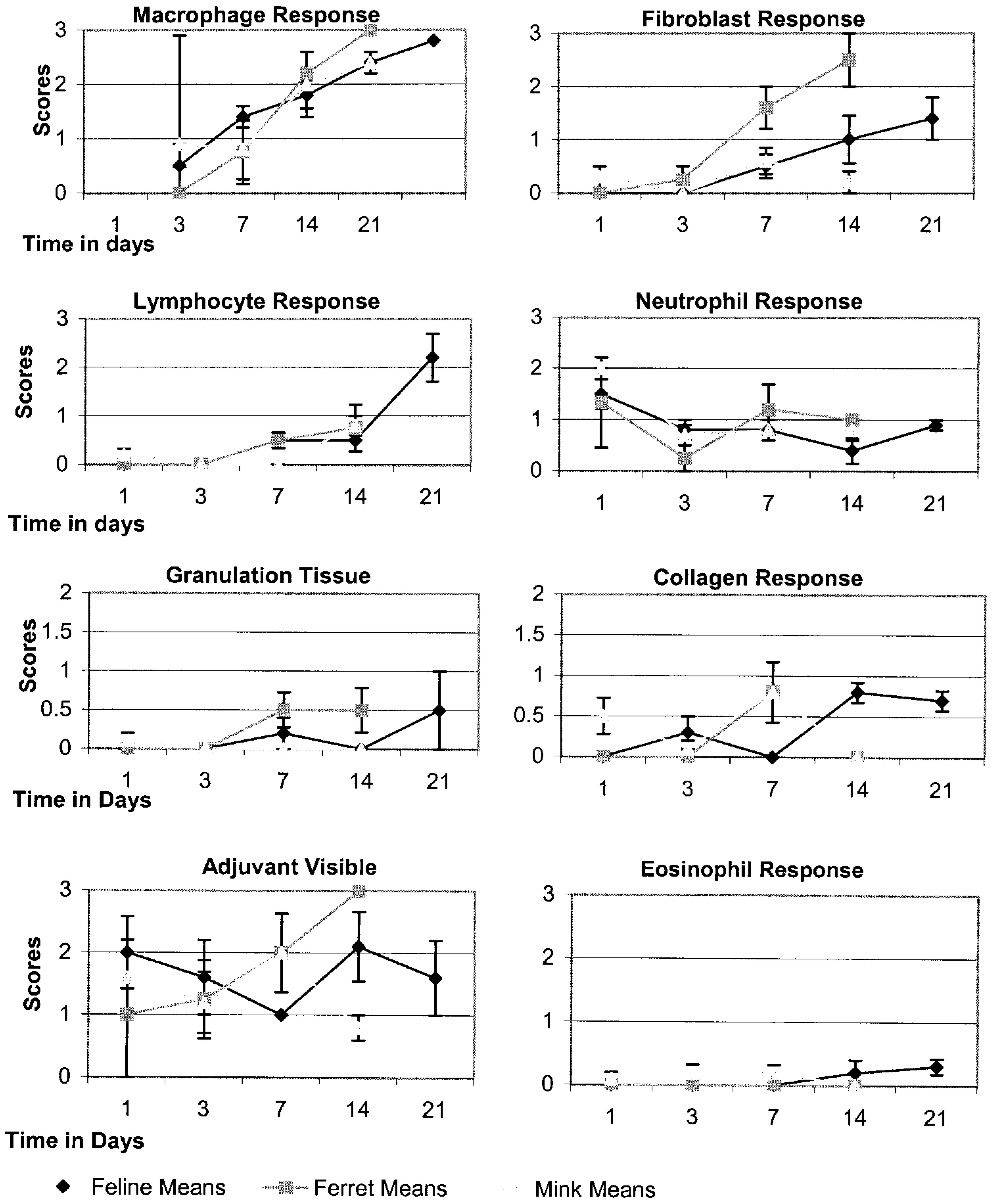
Skin; cat, mink, ferret. Responses to alum inoculation.
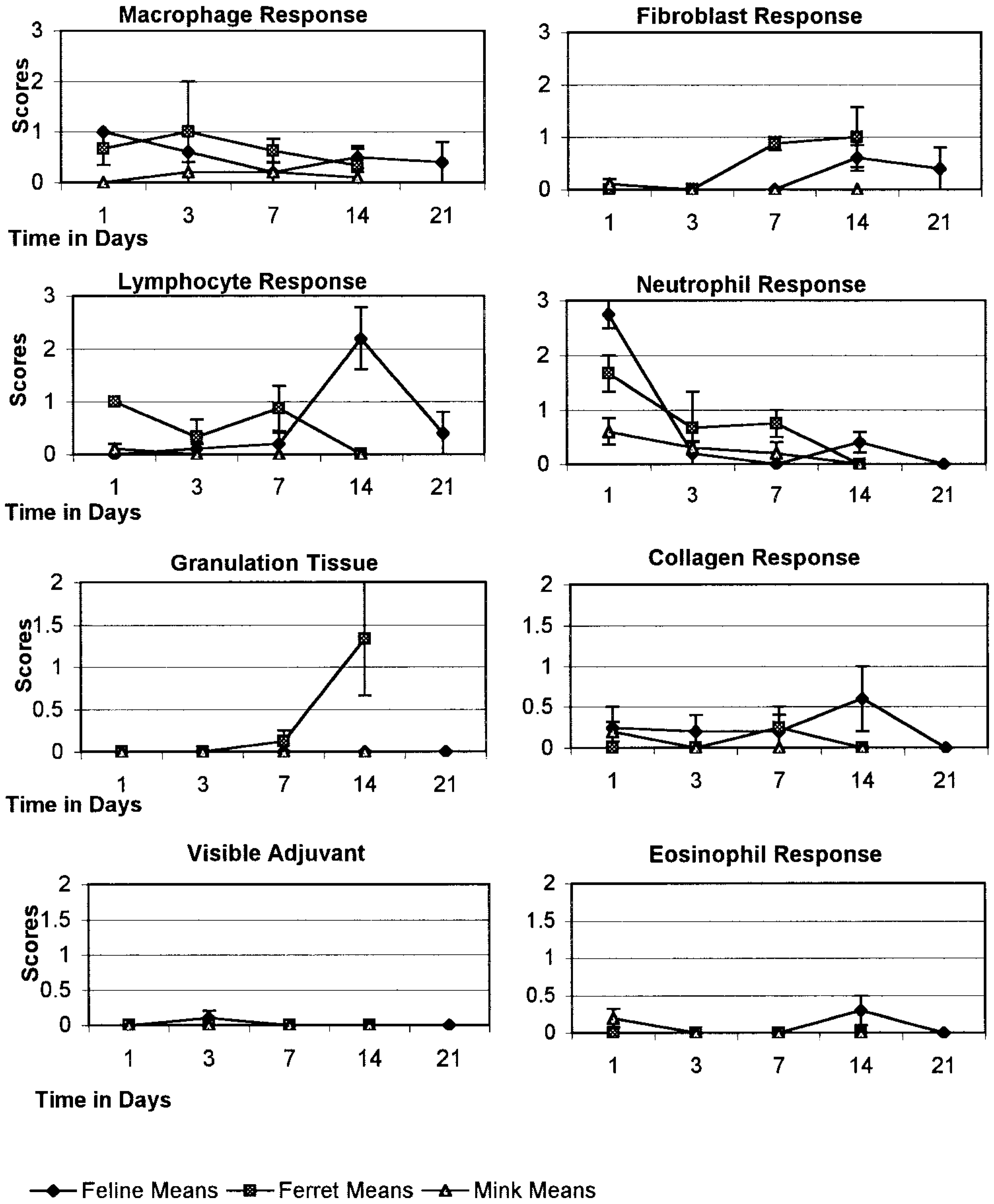
In response to non-aluminum-adjuvanted and aluminum-adjuvanted FeLV vaccines, cats behaved similarly to mink and ferrets (Figs 2, 3). Neutrophils were present in low numbers the first 3 days, then decreased. Macrophages appeared within 24 hours of inoculation, increased during the first 2 weeks, and then decreased in cats. Lymphocytes appeared by day 3, increased slightly by day 14, and then decreased by day 21 in cats. Visible adjuvant decreased over time at the aluminum-adjuvanted leukemia vaccine sites. Adjuvant was barely detectable at any time at sites of the non-aluminum-adjuvanted vaccine. Fibroblasts began proliferating by day 7, increased to moderate levels at the non-aluminum-adjuvanted vaccine site, but remained at low levels at the aluminum-adjuvanted leukemia vaccine sites. Cats differed from mink and ferrets in having fewer binucleate cells in response to the two leukemia vaccines (data not shown).
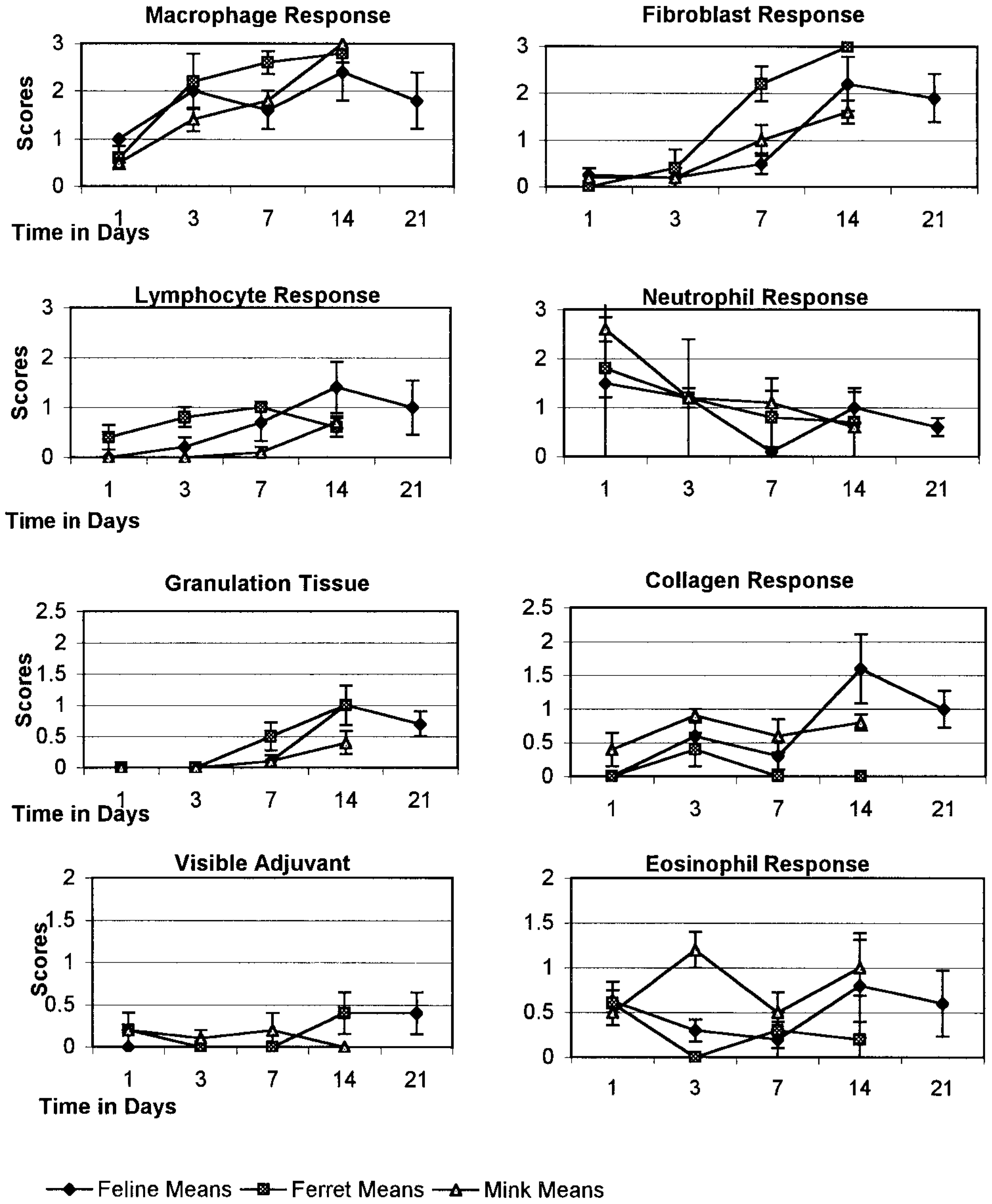
Skin; cat, mink, ferret. Responses to non-aluminum-adjuvanted FeLV vaccination.
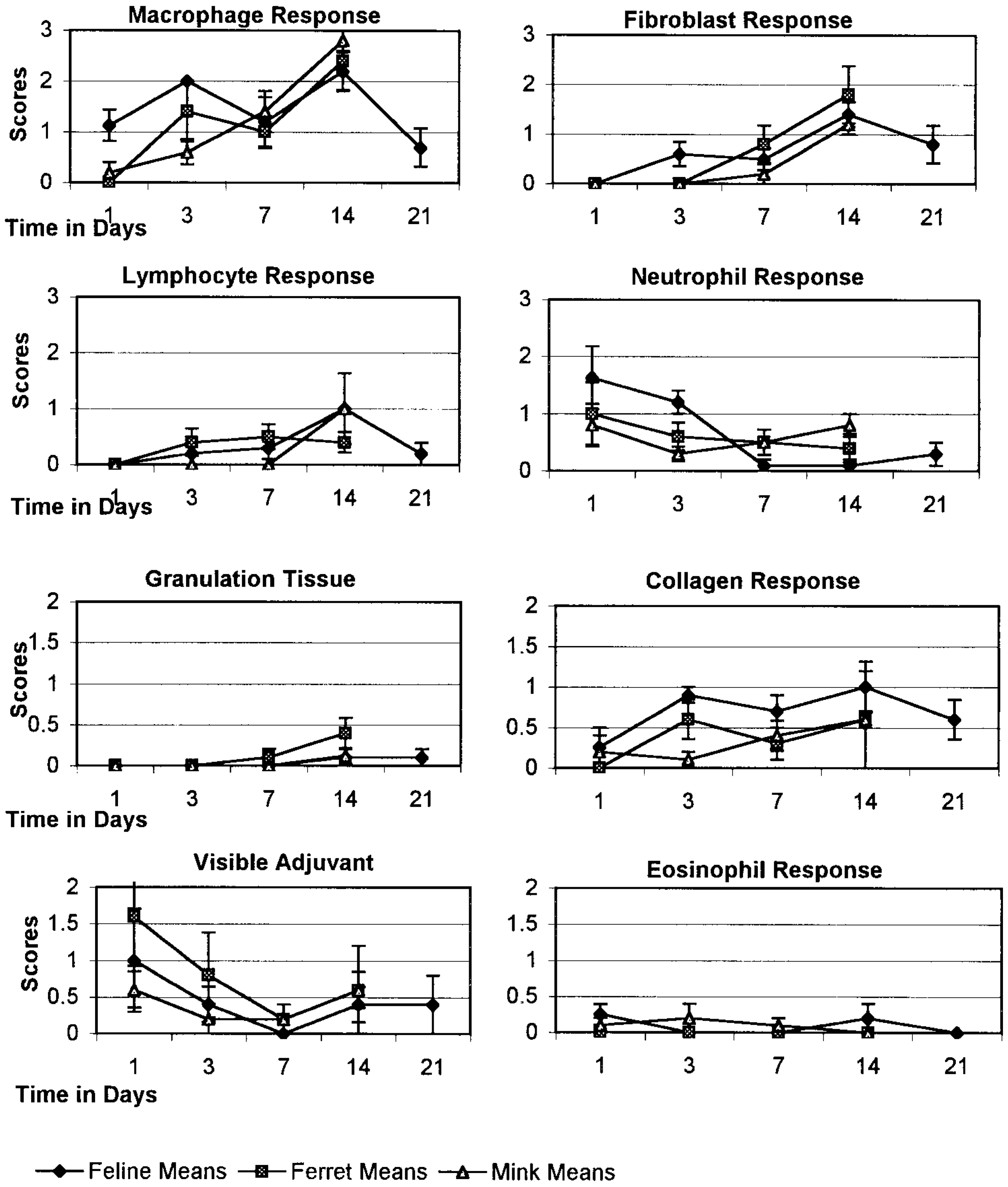
Skin; cat, mink, ferret. Responses to aluminum-adjuvanted FeLV vaccination.
Although the response in mink to the non-aluminum-adjuvanted leukemia vaccine had very few to low numbers of lymphocytes, one mink had well-organized lymphoid aggregates near the vaccine site that was diagnosed by a veterinary pathologist as a reactive deep dermal lymph node. The structure displayed a capsule, cortical germinal centers, attendant vasculature, and reactive histiocytes in the medullary sinuses.
In response to the two killed, aluminum-adjuvanted rabies vaccines, cats had more lymphocytes than either ferrets or mink (Figs. 4, 5).
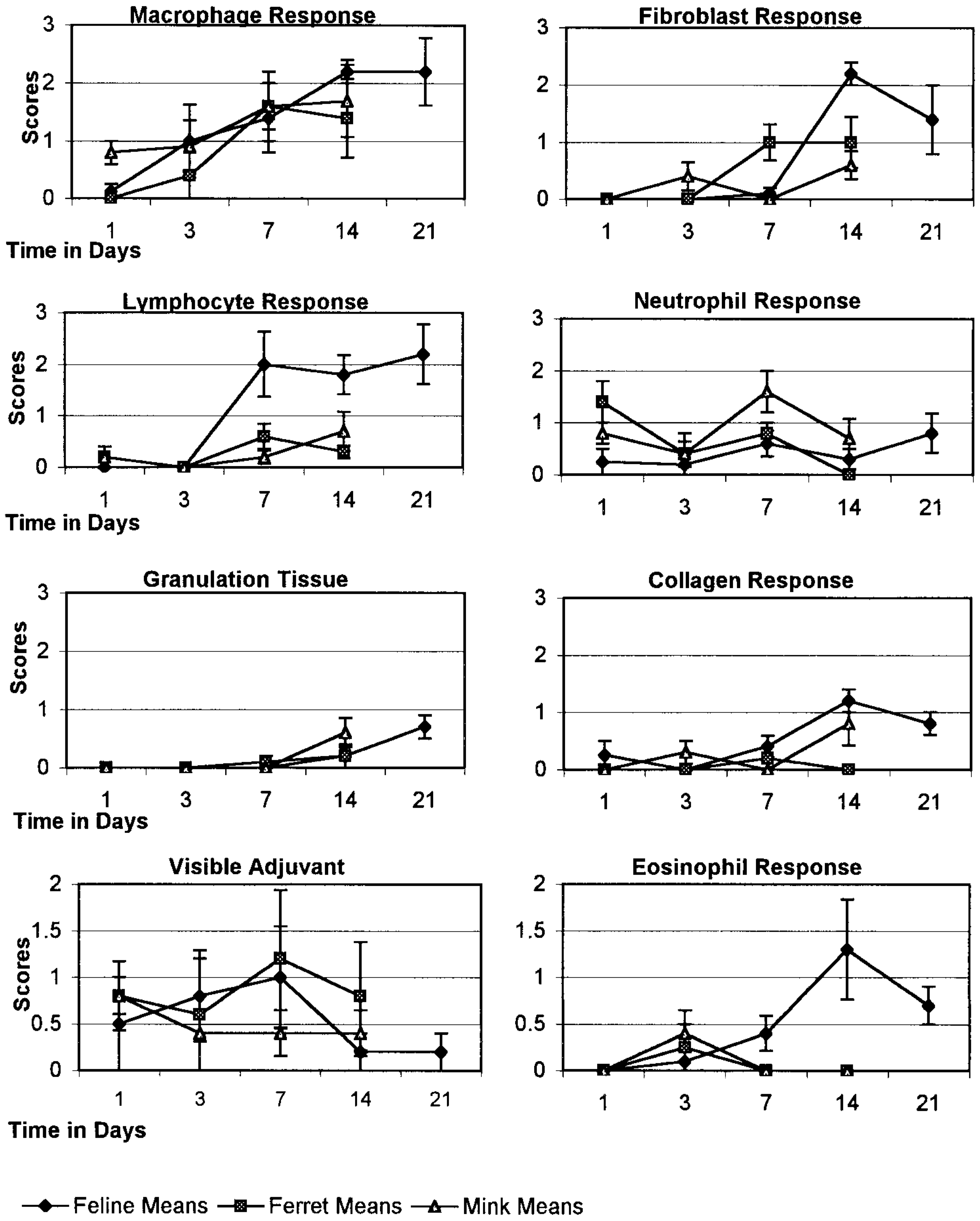
Skin; cat, mink, ferret. Responses to Rabdomun® aluminum-adjuvanted rabies vaccination.
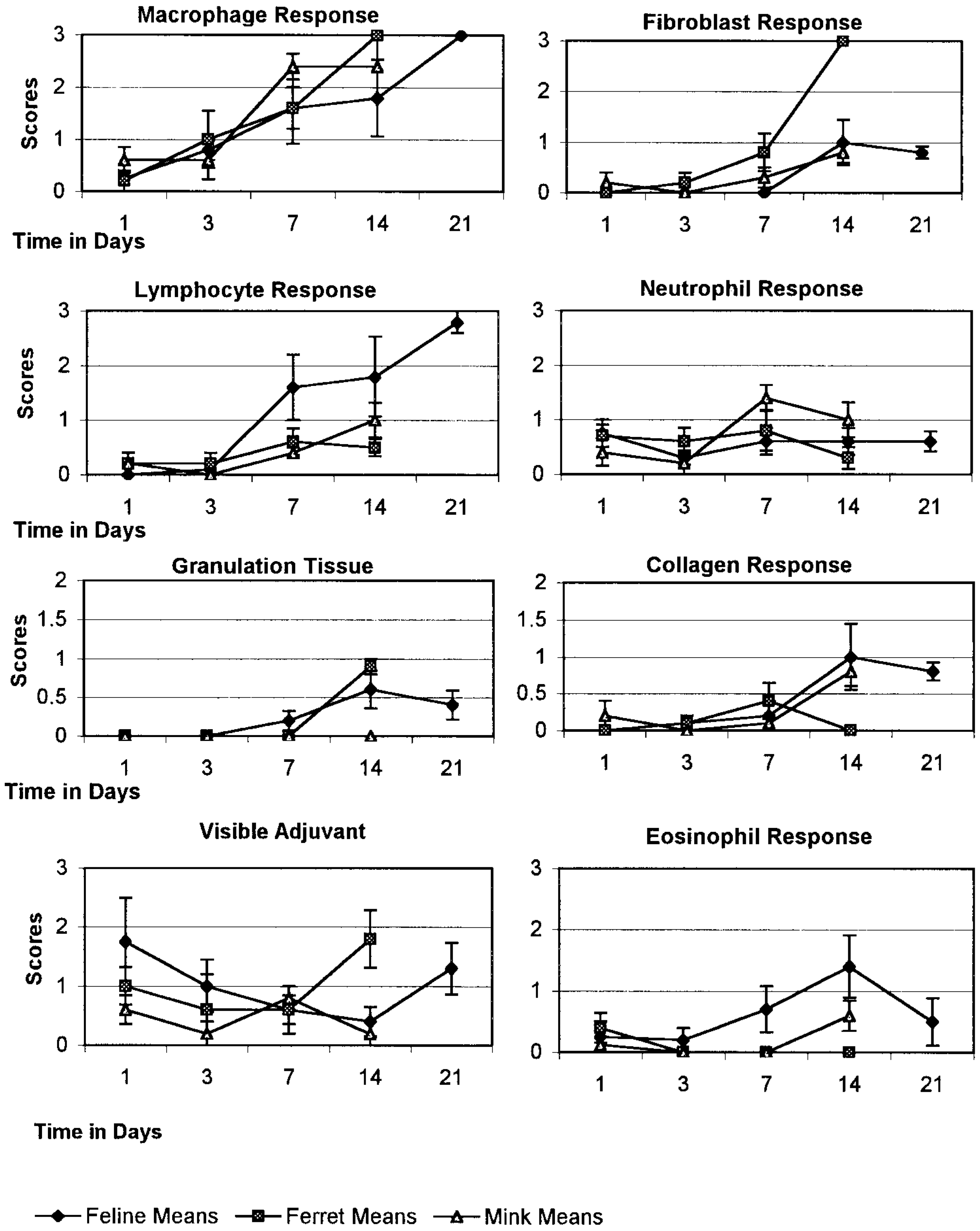
Skin; cat, mink, ferret. Responses to Imrab3® killed aluminum-adjuvanted rabies vaccination.
In response to canarypox-vectored rabies vaccine, cats had a moderate number of lymphocytes on day 14, which was more than in ferrets and mink, but the lymphocytes disappeared by day 21 (Fig. 6). Overall, the cellular response to the canarypox-vectored rabies vaccine was much milder than that to the adjuvanted rabies vaccines in all three species.
Skin; cat, mink, ferret. Response to canarypox-vectored rabies vaccination.
When neuraminidase was added to the canarypox-vectored rabies vaccine, cats did not differ significantly from mink and ferrets in their response nor did they respond to the modified canarypox vaccine differently than they did to the unmodified vaccine.
The mink with a lymph node-like structure near the site of the non-aluminum-adjuvanted leukemia vaccine and a second mink both had a similar lymph node-like structure near the site of the neuraminidase-supplemented canarypox-vectored rabies vaccine. No reports could be found describing lymph nodes in mink dorsal subcutis.
Statistical comparison of five commercial vaccines in cats, mink and ferrets
Comparison of the species' responses, listing those parameters with significant P-values, are shown in Table 1. P-values of <0.05 imply a species effect when one response parameter to a specific vaccine was examined and compared among the species. In other words, responses differed significantly for the pair of species compared.
Species effect for five vaccines. Numbers are P-values comparing two species at a time, one parameter at a time. Parameters at which cats differed significantly from ferrets and mink are in
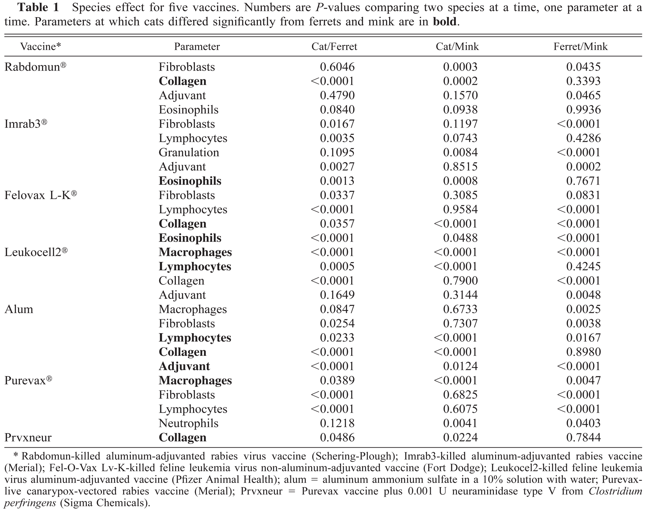
Rabdomun-killed aluminum-adjuvanted rabies virus vaccine (Schering-Plough); Imrab3-killed aluminum-adjuvanted rabies vaccine (Merial); Fel-O-Vax Lv-K-killed feline leukemia virus non-aluminum-adjuvanted vaccine (Fort Dodge); Leukocel2-killed feline leukemia virus aluminum-adjuvanted vaccine (Pfizer Animal Health); alum = aluminum ammonium sulfate in a 10% solution with water; Purevaxlive canarypox-vectored rabies vaccine (Merial); Prvxneur = Purevax vaccine plus 0.001 U neuraminidase type V from Clostridium perfringens (Sigma Chemicals).
Table 2 shows the responses to three killed aluminum-adjuvanted vaccines, one species at a time and one parameter at a time to reveal any vaccine effects. Fibroblast production in cats differed significantly in response to the three killed aluminum-adjuvanted vaccines (P = 0.0010). This finding contrasts with responses in the ferret (P = 0.4156) and mink (P = 0.8499). Cats also differed in collagen production (P = 0.0037), but ferrets (P = 0.0724) and mink (P = 0.2090) did not. Macrophage production in cats differed markedly in response to the three vaccines (P = 0.0002), whereas in ferrets and mink the response was not different (P = 0.1248 and P = 0.8419, respectively). The production of granulation tissue in cats differed (P = 0.044) across the three vaccines from that in ferrets (P = 0.079) but not as much as in mink (P = 0.0001). These results suggest that cats differed in the local tissue response to the three killed aluminum-adjuvanted vaccines when compared with ferrets and mink.
Vaccine effect (P-values) in cats, ferrets, and mink for three killed aluminum-adjuvanted vaccines.∗ Bold parameters exhibit a significant vaccine effect in cats but not in mink or ferrets.
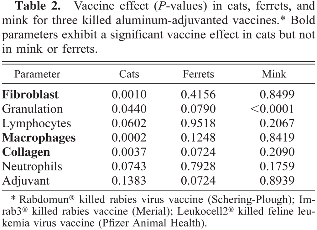
Rabdomun® killed rabies virus vaccine (Schering-Plough); Imrab3 ® killed rabies vaccine (Merial); Leukocell2® killed feline leukemia virus vaccine (Pfizer Animal Health).
Table 3 shows responses to the two FeLV vaccines one species at a time. In cats, the two vaccines produced a significantly different response in five of the measured parameters. Table 4 shows responses to the three rabies vaccines one species at a time. Again, cats demonstrated a vaccine effect in five parameters.
Vaccine effect. Significant differences between two feline leukemia vaccines∗ in cats, ferrets, and mink for given parameters. Numbers are P-values.
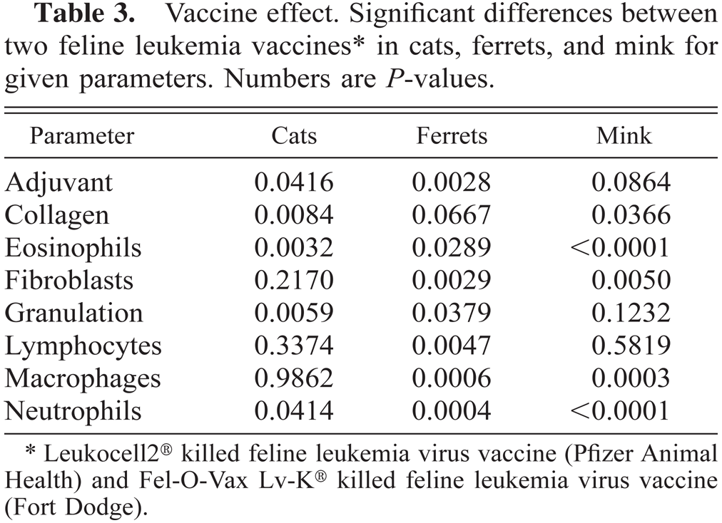
Leukocell2® killed feline leukemia virus vaccine (Pfizer Animal Health) and Fel-O-Vax Lv-K® killed feline leukemia virus vaccine (Fort Dodge).
Vaccine effect (P-values) showing differences among three rabies vaccines∗ in cats, ferrets, and mink.
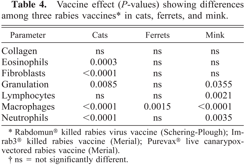
Rabdomun® killed rabies virus vaccine (Schering-Plough); Imrab3® killed rabies vaccine (Merial); Purevax® live canarypoxvectored rabies vaccine (Merial).
ns = not significantly different.
Discussion
In an effort to understand the pathogenesis of feline VAS, many researchers have studied the VAS directly.1,2,4,6,7,11–13,15,16,20,23–25 Early vaccine lesions were examined cytologically and by palpation.34,40 Using histopathology, Hendrick and colleagues examined 10 rabies vaccine-site reactions in cats and dogs.22 The present study is the first to compare the progression of vaccine-associated lesions in cats, mink, and ferrets over a 2–3-week period. We also compared the histologic vaccine-site reactions to five commercial vaccines in cats.
Studies have shown that rabies vaccines more consistently produce granulomatous inflammation at vaccine sites, although leukemia virus vaccines are incriminated more often in the pathogenesis of VAS.5,18,20,29,30,35 The local tissue responses to two killed aluminum-adjuvanted rabies vaccines and one aluminum-adjuvanted leukemia virus vaccine were compared using a SAS ANOVA program. Cats differed from mink and ferrets by responding to each vaccine in a slightly different manner, in contrast to mink and ferrets. These vaccines have the same primary adjuvant component; therefore, the antigen and/or additional components (e.g., preservatives, tissue culture material) may play a role in determining the local response.
In a previous report,34 an aluminum-adjuvanted leukemia virus vaccine produced measurable reactions in five of six cats, whereas a non-aluminum-adjuvanted leukemia vaccine produced a measurable lesion in only one of 12 cats. In the present study at 24 hours postinoculation, histologic reaction was seen in four of four cats vaccinated with aluminum-adjuvanted Leukocell2® and in four of four cats vaccinated with non-aluminum-adjuvanted Fel-O-Vax®. Over the 3-week period, however, Fel-O-Vax® sites had more neutrophils on day 7 (P = 0.0414), more collagen on day 14 (P = 0.0084), and more granulation tissue on day 14 (P = 0.0059) than did Leukocell2® sites (Figs. 2, 3; Table 3). Histologically, therefore, the non-aluminum-adjuvanted FeLV vaccine produced a more robust reaction than did the aluminum-adjuvanted vaccine. Non-aluminum-based adjuvants have been linked to VAS and may be equally provocative of a local reaction.4 It is not currently known how the magnitude of the immediate postvaccination response correlates with oncogenesis in susceptible animals. The increased local tissue reaction could indicate impending malignant transformation secondary to excessive elaboration of cytokines and growth factors. Alternatively, the more robust response could signify rapid clearance of degradable vaccine components and resolution of the vaccine-induced local irritation.
Schultze and colleagues reported that the “postvaccinal reactions were most evident in cats receiving the rabies vaccine.”40 These reactions consisted of palpable lesions and increased numbers of lymphocytes and macrophages in fine-needle aspirates. In the present study, the killed aluminum-adjuvanted rabies vaccines elicited fewer macrophages than did the killed aluminum-adjuvanted leukemia vaccine and fewer neutrophils in the first 3 days in cats (P = 0.0002). By day 14, however, the two adjuvanted rabies vaccines had elicited an equally strong macrophage response in cats, which then surpassed that seen at the FeLV vaccine sites by day 21. Although canarypox-vectored rabies vaccines produced fewer macrophages than did the other rabies vaccines on day 14, there were large numbers of lymphocytes. By day 21, however, the killed adjuvanted rabies vaccine sites surpassed the non-adjuvanted rabies vaccine sites in cellularity. If a persistent presence of lymphocytes and macrophages is required in the pathogenesis of VAS, the canarypox-vectored vaccine would be less likely to play a role in VAS oncogenesis.
Reports of VAS occasionally describe an eosinophilic component.22 In this histologic study, eosinophils were seen in low numbers or not at all at 24 hours postinoculation. Eosinophils were seen at only one saline vaccine site on day 1 and not at any other time. Rabdomun®, Imrab3®, and Fel-O-Vax® sites developed moderate to high eosinophil numbers in one or two cats. Because alum did not elicit an eosinophil response to the same degree that Fel-O-Vax®, Rabdomun®, Imrab3® did, aluminum does not seem to be the stimulus for eosinophil chemotaxis. Because two different antigens elicited an eosinophil response on day 1, the primary antigen also may not be the stimulus. One hypothesis is that certain animals respond to vaccines with an influx of eosinophils. At each time point, some animals had eosinophils to more than one kind of vaccine. Therefore, individual animal variation may account for the eosinophilic response in some cats. Those vaccines that more often elicited eosinophils were the killed aluminum-adjuvanted rabies and the non-aluminum adjuvanted leukemia vaccines. Individual animal variation plus a vaccine that predisposes to eosinophil production could conceivably play a role in oncogenesis.
Schultze et al. found that at day 7 postvaccination, cytologic examination of fine-needle aspirates (FNA) at sites of rabies, FeLV, and feline rhinotracheitis, calicivirus, and panleukopenia virus (FRCPV) vaccination revealed no differences in the relative numbers of lymphocytes, neutrophils, and macrophages. However, all aspirates were more cellular than saline controls.40
Histologic examination of the day 7 sites in the present study revealed low numbers of fibroblasts with a slight increase in collagen at the three rabies and two leukemia vaccine sites. Neutrophils (in low numbers) were observed only at the two killed conventional rabies vaccine sites at day 7. Macrophages and lymphocytes were more abundant in the tissue samples of this study than in the FNA samples of the Schultze et al. study.
Fourteen days after vaccination, Schultze and colleagues found that relative numbers of aspirated eosinophils and neutrophils did not differ at sites of Imrab3®, Fel-O-Vax Lv-K®, and a combined viral rhinotracheitis-calicivirus, panleukopenia vaccination. Lymphocytes aspirated from the Fel-O-Vax® and Imrab3® vaccination sites, however, were more numerous than those from the combination vaccine site. The relative number of macrophages was also greater in the aspirates from the rabies vaccine sites.40
In our study, Rabdomun®, Imrab®, and Leukocell2® elicited a comparable number of macrophages in cats. The feline lymphocyte response to Imrab3® and Rabdomun® exceeded that to Leukocell2® (Table 2; Figs. 3–5). Only two of five Leukocell2® sites in our study had lymphocytes, with a moderate number in one and a large number in the other. Fewer lymphocytes were seen cytologically.40 Comparable to the Schultze et al. study, the number of eosinophils observed at aluminum adjuvanted rabies vaccine sites exceeded that at aluminum adjuvanted leukemia vaccine sites.
In this study, adjuvant decreased over time at sites of the aluminum-adjuvanted leukemia vaccine but varied at sites of the other aluminum-adjuvanted vaccines. Because each cat received the same amount of vaccine, the inconsistent presence of adjuvant at sampling sites suggests that the center of the lesion was occasionally missed when the tissue was excised. It may also reflect variable biologic degradation of the vaccine components. Aluminum is considered inert, but other adjuvant components, such as oxygen, potassium, or sulfur, may be metabolized or removed, which could reduce the amount of adjuvant remaining visible over time. The nonaluminum adjuvant of Fel-O-Vax® was believed to be seen within macrophages on days 14 and 21.
In the Schultze et al. study, by 21 days postvaccination aspirates from the killed aluminum adjuvanted rabies vaccine sites contained “significantly greater relative numbers of eosinophils, lymphocytes, neutrophils, and fibroblasts than did those from other injection sites.”40 The cellularity of the FeLV vaccine sites had decreased to that found at the saline control site. Macrophage numbers aspirated from the combination vaccine site and the Fel-O-Vax® and Imrab3® sites did not differ in that previous study.40 Rabies vaccination consistently produced palpable lesions (generally believed to be granulomatous)14,22 in the Schultze et al. study of nine cats.40
In our histologic study, of the four aluminum-adjuvanted inoculations on day 21, cats had rising numbers of macrophages and lymphocytes in response to the two rabies vaccines and alum but not in response to the leukemia vaccine. In the Shultze et al. aspirates, there were few macrophages by day 21 at the rabies and leukemia sites; the number of lymphocytes continued to climb at the rabies site but fell at the leukemia vaccine site.40
In the present study, cats differed from mink and ferrets, two other small routinely vaccinated carnivores, in their response to commercial vaccines. Cats produced more fibroblasts, collagen, and lymphocytes in response to one aluminum-adjuvanted rabies vaccine (Rabdomun®) but fewer fibroblasts than mink in response to Imrab3®. Because these vaccines both include aluminum hydroxide in the adjuvant, the differential response may be related to some other ingredient of the vaccine. Additionally, cats responded with more neutrophils on day 1 and more lymphocytes on day 14 than did mink or ferrets when vaccinated with a canarypox-vectored rabies vaccine. The differential response of cats to rabies vaccines suggests a sensitivity to vaccine components that is unequaled in mink or ferrets.
In this study, the three species produced similar numbers of macrophages, fibroblasts, and lymphocytes in response to the two leukemia virus vaccines. Cats appeared to produce fewer binucleate cells by day 14 than did the other two species. Fel-O-Vax® elicited more granulation tissue than did Leukocell2® (P = 0.0059) in all three species, which was unexpected given the fact that Fel-O-Vax® is adjuvanted without aluminum.
Cytologic examination of rabies vaccine sites revealed “few activated macrophages and a blue, amorphous material in the cytoplasm … throughout the study.”40 Histologically, adjuvanted rabies vaccines initially elicited few macrophages on day 1. On day 3, Rabdomun® and Imrab3® vaccine sites had low but notable numbers of macrophages. By day 14, moderate to large numbers of macrophages were evident in all three species. Tissue macrophages may not exfoliate well from vaccine sites and thus would be less likely to be collected in the aspirated samples.
It has been reported that rabies virus vaccines elicit a stronger lymphocyte response than do FeLV vaccine or the combination FRCPV vaccine.40 In our study, by day 7, lymphocytes were prominent at most adjuvanted rabies vaccine sites. By day 14, Rabdomun® and Imrab3® vaccine sites had a moderate number of lymphocytes, similar to the Purevax® vaccine sites in cats (none in ferrets or in the three mink without the lymph node-like structures), in contrast to low numbers at FeLV vaccine sites. It has been suggested that the phenotype of lymphocyte (T helper 1 versus T helper 2) may play a role in the predisposition of some cats to tumor formation.19 Because the rise in lymphocytes in cats roughly paralleled the rise in macrophages, the lymphocytes may have been stimulated by adjuvant-activated macrophages. The rabies antigen also may play a role in activating lymphocytes; more lymphocytes are generated in response to the rabies vaccines than in response to leukemia vaccines. However, this finding is not consistent with the suggestion that there is a lower likelihood of carcinogenesis in response to rabies vaccines than in response to leukemia vaccines.4,17
Early lesions in our study showed granulomatous inflammation, with varying degrees of lymphocytic infiltrates. Similarly, the mature VAS examined often consisted of “a dense fibrous capsule and an inflammatory infiltrate of lymphocytes and macrophages.”25,46 The authors of one study stated they have seen histologic lesions that appear to be in transition from the chronic granulomatous inflammatory lesion to neoplasia.25 None of the lesions observed in the present study were neoplastic, but several exhibited signs of chronic active inflammation. This study focused on vaccine site lesions of 2–3 weeks duration to compare initial inflammatory and wound repair response to various vaccines in three species. To better examine the progression of chronic inflammation and its role in oncogenesis, a longer study would be more useful.
The only capsules seen in this study were those surrounding the organized lymphoid follicles in two mink.
Cats respond differently to certain commercial vaccines than do species not known to produce VAS, suggesting that the feline genome plays a role in the pathogenesis of VAS. One way in which cats may respond differently is in the type of inflammatory response. The human tissue reaction to foreign substances has been described as allergic or nonallergic.32 Lever and Schaumberg-Lever described a nonallergic foreign body reaction (when a substance is deposited in the dermis or subcutaneous tissues) as typically showing macrophages and many giant cells around the foreign material, with lymphocytes and plasma cells but few or no epithelioid cells.32
Allergic foreign body granulomas have many epithelioid cells with or without giant cells or caseous necrosis. Slight or no phagocytosis is evident.32 The three species examined here that received vaccines containing an aluminum-based adjuvant had, by day 14, large numbers of epithelioid macrophages and others with copious amounts of phagocytosed amorphous material believed to be adjuvant. Rare giant cells were evident in several sections but were not a consistent feature. Aluminum apparently elicits a mixed response in these small carnivores, with characteristics of both an allergic and nonallergic foreign body reaction. The local reaction to aluminum-adjuvanted vaccines observed in these three species resembles Lever and Schaumberg-Lever's description of an aluminum granuloma: “lymphoid follicles, macrophages with ample cytoplasm, a few giant cells … and large areas of eosinophilic necrosis.”32 Eosinophilic necrosis was not a consistent finding in this study; it was observed in only a few cases. In an earlier study, eosinophilic necrosis was consistently seen in feline lesions examined 2 weeks to 2 months after vaccination with rabies vaccine.22 A period of time greater than 21 days may be necessary for eosinophilic necrosis to develop.
Lymphoid germinal centers with a fibrous tissue capsule and vasculature were observed near the canarypox rabies with neuraminidase and the non-aluminum-adjuvanted leukemia virus vaccine sites in one mink and near the canarypox rabies with neuraminidase vaccine site in another. The structures were identified as reactive deep dermal lymph nodes (H. Steinberg, personal communication). Few authors have described lymph nodes in mink,27,28 and none have mentioned lymph nodes in the subcutaneous tissue along the dorsum.
When cats are compared with mink and ferrets, subtle differences are measurable in how they respond locally to commercial vaccine products. Histologic differences may reflect quantitative and qualitative differences in elaboration of cytokines and growth factors. Subtle differences from other species may relate to the cat's susceptibility to oncogenesis. Additional studies are in progress to test this hypothesis.
Footnotes
Acknowledgements
We thank Fei Zou and Yunfei Chen for valuable assistance in statistical program writing.