
Abstract
In each of seven ferrets (Mustela putorius furo) with leiomyosarcoma, a single dermal mass was identified and biopsied. Each mass consisted of a well-demarcated but nonencapsulated proliferation of large spindle- to strap-shaped cells arranged in interwoven bundles. The cells resembled the smooth muscle cells of the adjacent arrector pili muscles, but with marked nuclear pleomorphism. Immunohistochemical staining for smooth muscle actin, desmin, and vimentin was positive and staining for myoglobin and cytokeratin was negative. Follow-up on three of the ferrets indicates that the prognosis is good following complete surgical excision.
Keywords
There has been a dramatic increase in the number of documented neoplasms found in the domestic ferret (Mustela putorius furo) because of their increased use in laboratory medicine and their growing popularity as household pets. 6 The most commonly affected sites are the endocrine, hemolymphatic, and integumentary systems. 1,3,7,8 In a study of 57 cutaneous neoplasms in ferrets, basal cell tumors and mastocytomas were the most common cutaneous neoplasms, with no mention of leiomyomas or leiomyosarcomas. 8 In the present report, we describe seven leiomyosarcomas arising in the arrector pili muscle, with emphasis on the histopathologic features and the immunohistochemical techniques used to identify them.
Seven biopsies from ferrets with piloleiomyosarcoma were submitted during an 8-year period (1993–2000) to the surgical pathology service at the University of Pennsylvania School of Veterinary Medicine. These tumors were uncommon, representing only 2.4% (7/288) of all ferret submissions during that time. Specimens were fixed in 10% neutral buffered formalin, processed routinely for paraffin embedment, and stained with hematoxylin and eosin (HE).
Additional paraffin-embedded sections were stained immunohistochemically for smooth muscle actin (monoclonal anti-human, Dako Corp.), vimentin (V9 monoclonal anti-porcine, Dako), desmin (monoclonal anti-human, BioGenex), myoglobin (polyclonal rabbit anti-human, Dako), and cytokeratin (AE1/AE3 monoclonal anti-human, Boehringer Mannheim) using an Autostainer Immunostaining System (Dako). The slides to be stained with antibodies to cytokeratin were pretreated with Proteinase K for 5 minutes. The primary antibodies were applied for 30 minutes at the following concentrations: actin 1:25, vimentin 1:30, desmin 1:40, myoglobin 1:2,400, and cytokeratin 1:500. A two-step non-avidin-biotin detection method (ENVISION+, Dako) was used for the vimentin and cytokeratin antibodies. A labeled streptavidin–peroxidase detection method (LSAB2, Dako) was used for the actin, desmin, and myoglobin antibodies. Diaminobenzidine was used as the chromogen, and sections were counterstained with hematoxylin.
Six of the seven ferrets were male, and four of the six males were castrated. The ferrets were 2–6 years of age (○ = 3 years, median = 2 years) at the time of presentation. The masses had been noted 2 weeks to 4 years prior to surgical excision. All of the masses arose on the head or trunk; none were on the limbs. Most of the specimens were from the dorsum of the body, where the normal arrector pili muscles are largest. 9 Tumors were 0.5–2.0 cm in diameter. The surface of the larger masses was ulcerated. None had gross lymph node involvement or evidence of metastasis.
The histopathologic appearance of the seven leiomyosarcomas was very similar and consisted of well-demarcated but nonencapsulated oval to irregular dermal masses (Fig. 1). The masses were always immediately adjacent to a hair follicle and often were continuous with the arrector pili muscle. They were composed of spindle- to strap-shaped cells arranged in interwoven bundles that closely resembled the smooth muscle cells of the adjacent arrector pili muscles. The cells had abundant fibrillar to vacuolated eosinophilic cytoplasm with indistinct cell borders. The nuclei were pleomorphic, with coarse chromatin and large basophilic nucleoli. Mitotic figures were uncommon (1 or 2 per 10 high-power 40× fields), but occasional abnormal mitoses were present (Fig. 2). Four of the tumors had multifocal lymphocyte foci infiltrating the periphery; these tumors were also larger and more irregular in shape.
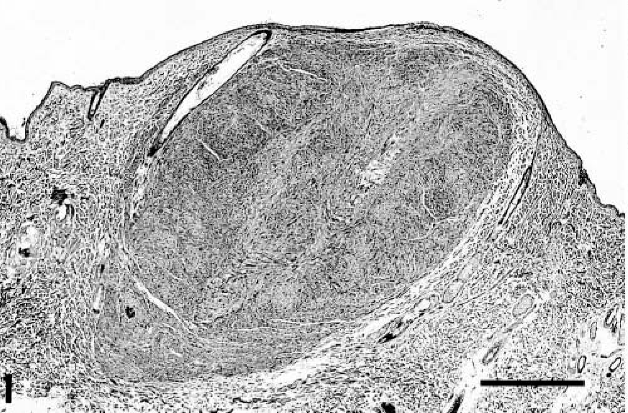
Piloleiomyosarcoma; ferret. The well-demarcated neoplasm is immediately adjacent to a hair follicle and continuous with the arrector pili muscle. HE. Bar = 700 µm.
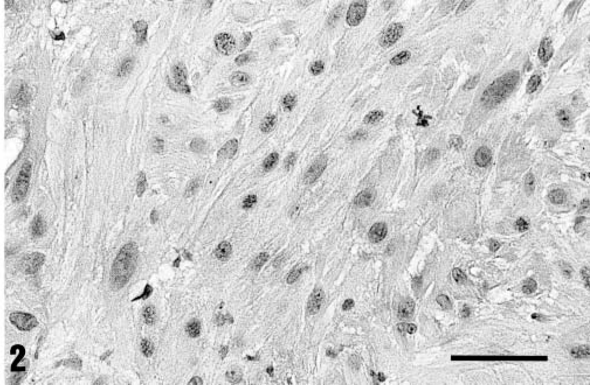
Piloleiomyosarcoma; ferret. The cells have abundant fibrillar to vacuolated cytoplasm, pleomorphic nuclei with coarse chromatin, and occasional abnormal mitotic figures. HE. Bar = 35 µm.
All tumors and the adjacent arrector pili muscles reacted positively with antibodies to vimentin, actin, and desmin and negatively to myoglobin and cytokeratin. This immunohistochemical staining pattern of neoplastic cells is consistent with a tumor of smooth muscle origin. The immunohistochemistry results together with the nuclear pleomorphism and association with the arrector pili muscle led to the conclusion that these were malignant smooth muscle tumors arising from arrector pili muscles, i.e., piloleiomyosarcomas.
Follow-up information was available for three ferrets. One ferret survived 2 years after complete surgical excision without any evidence of recurrence or metastasis but then died of unrelated causes. Another ferret is alive and well 18 months after excision. The third ferret required two surgeries to achieve complete surgical excision, but the ferret is alive and well 5 months after the second surgery. Despite the nuclear pleomorphism found in these tumors, follow-up on these three ferrets indicates that the prognosis is good following complete surgical excision.
In humans, superficial leiomyosarcoma can occur in either the dermis or subcutis. The dermal type originates from arrector pili muscle (piloleiomyosarcoma), and the subcutaneous type originates from vascular smooth muscle (angioleiomyosarcoma). 5 The tumors of arrector pili origin have a 30% incidence of local recurrence, and the superficial angioleiomyosarcomas have a 54% incidence of local recurrence. In addition, metastatic disease has not been reported with piloleiomyosarcoma, whereas the angioleiomyosarcomas have a 39% incidence of metastasis. 10 In human superficial leiomyosarcomas, the degree of nuclear pleomorphism is not correlated with malignant behavior.
A subcutaneous leiomyosarcoma has been reported in a ferret. 2 The histopathology and immunohistochemical findings were similar to those described here. However, follow-up information regarding local recurrence and metastasis was not provided. Leiomyomas of the arrector pili muscle have also been reported in the dog and cat. 4 They are cured by local excision.
Piloleiomyosarcomas are rare cutaneous neoplasms in ferrets. They have a unique histologic appearance and a characteristic immunohistochemical staining pattern. Despite the marked nuclear pleomorphism, the prognosis is presumably good following complete surgical excision.