
Abstract
An outbreak of Fusobacterium necrophorum-induced septicemia occurred in a group of 40 captive wild-caught pronghorns (Antilocapra americana). Primary pododermatitis or necrotic stomatitis progressed to produce fatal septicemia with metastatic lesions in the forestomachs, lung, liver, and cecum in 38 of the animals. Two remaining animals were euthanatized because of chronic pododermatitis. Housing the animals in a pasture previously used by bovids and heavy rains with persistence of ground water pools in the pasture were contributing factors in the pathogenesis of this outbreak.
Fusobacterium necrophorum is associated with a variety of diseases in domestic animals. 1,6,10 The organism usually does not cross intact epidermal surfaces, and the pathogenesis of fusobacterial infections usually involves an injury that breaches an intact epidermal layer. 1,6 Fusobacterial foot abscesses in sheep and pododermatitis in cattle are associated with a wet environment, minor traumatic events, and synergistic infection with other bacteria that enable F. necrophorum to establish an infection. 6,8 Trauma is considered important in the pathogenesis of necrotic stomatitis in calves, laryngitis caused by friction of vocal folds in cattle, and rhinal osteomyelitis and cellulitis (bullnose) caused by cutting of canine teeth in pigs. 1,6 Papillomavirus and parapoxvirus infections also have been associated with secondary necrobacillosis in lambs and calves, and F. necrophorum may participate in infections caused by enteric pathogens such as Bachyspira hyodysenteriae. F. necrophorum is normally present in the rumen, and rumen acidosis with mucosal damage may allow these organisms to invade, become bacteremic, and cause hepatic necrobacillosis. F. necrophorum infection usually does not cause septicemias except in young animals.
The necrobacillary epizootic discussed in this report involved a group of sexually mature pronghorns (Antilocapra americana) captured in Wyoming and translocated immediately to Texas in November 1999. The pronghorns were herded by helicopter into net-sided corrals, physically restrained, and hand loaded onto trailers. Upon arrival in Texas, the animals were administered an oral brucellosis vaccine as they were removed from the trailers and released into a secluded, screened pasture. They were fed a pelleted deer ration, and both alfalfa and coastal bermuda hay were provided free choice. The pronghorns were monitored visually twice daily but were not handled. Initial capture-related mortality was high; however, these losses ended by the end of December. The health status of the remaining 40 pronghorns appeared good, and further deaths were minimal. Forty-three days after the animals arrived, there was a heavy rain, and 7 days after these rains, two animals died and were necropsied. These two were the first of an outbreak of necrobacillosis that caused the death or destruction of the remainder of the herd.
Tissues collected at necropsies were fixed in buffered formalin. For light microscopic examination, tissue in paraffin blocks were sectioned at 5 µm and stained with Gill's No. 3 hematoxylin and eosin (HE), Brown/Hopps tissue Gram's stain, and the silver fungal stain Grocott's ammoniacal silver. Tissues routinely examined included brain, liver, heart, skeletal muscle, tongue, kidney, rumen, reticulum, omasum, abomasum, small intestine, cecum, colon, adrenal gland, spleen, superficial cervical and prefemoral lymph nodes, affected feet, and lung. Periodically during the outbreak, lesions collected from animals presented with little postmortem autolysis were cultured aerobically for bacterial pathogens on blood agar, Maconkey agar, and tryptose broth and anaerobically on blood agar and brain-heart infusion broth (Difco, Detroit, MI) with oxyrase (Oxyrase, Inc., Mansfield, OH). Isolates were identified to genus or to genus and species using the Vitek Auto Microbic System (BioMerieux Vitek, Hazelwood, MO).
Lesions in the first two affected animals were similar. A severe, draining pododermatitis was present in one foot in each animal (Fig. 1). Necrotic tissue and inflammation surrounded the foot subcutaneously and extended along tendon sheaths proximally almost to the level of the fetlock. Viable tissue was sharply demarcated from the necrotic areas. Arthritis occurred in the P2–3 joint, with purulent material exuding from the swollen interdigital area, especially at the heel. Multifocal dry white lesions with dark red borders were observed in the rumen, reticulum, omasum, and, in one animal, liver, indicating systemic spread of the infection (Figs. 2, 3). Based on the characteristic macroscopic and histologic lesions, fusobacterial pododermatitis with systemic fusobacteriosis, “necrobacillosis,” was diagnosed. F. necrophorum was isolated from representative lesions of the feet, forestomachs, and liver. Subsequently, animals were presented on an almost daily basis, with similar lesions. Occasionally, additional lesions of septicemic fusobacteriosis were noted in the lung and tongue (Fig. 4).
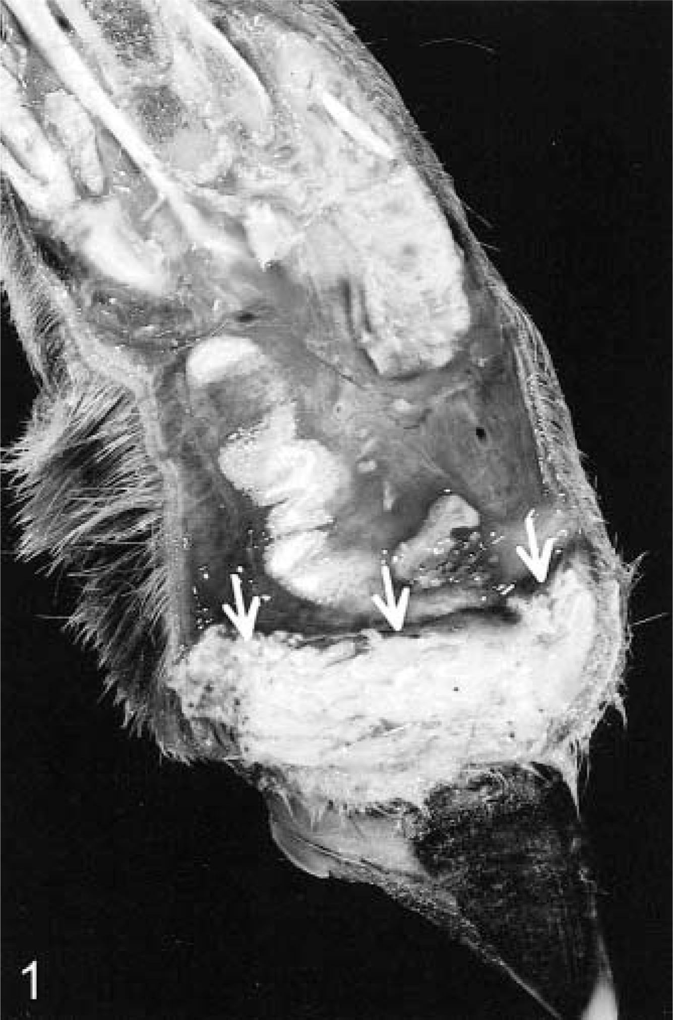
Foot; pronghorn. Sagittally sectioned between digits. Pododermatitis was due to Fusobacterium necrophorum infection. Note light gray area of necrotic tissue with a sharp demarcation (arrows) from moist, inflamed viable tissue.
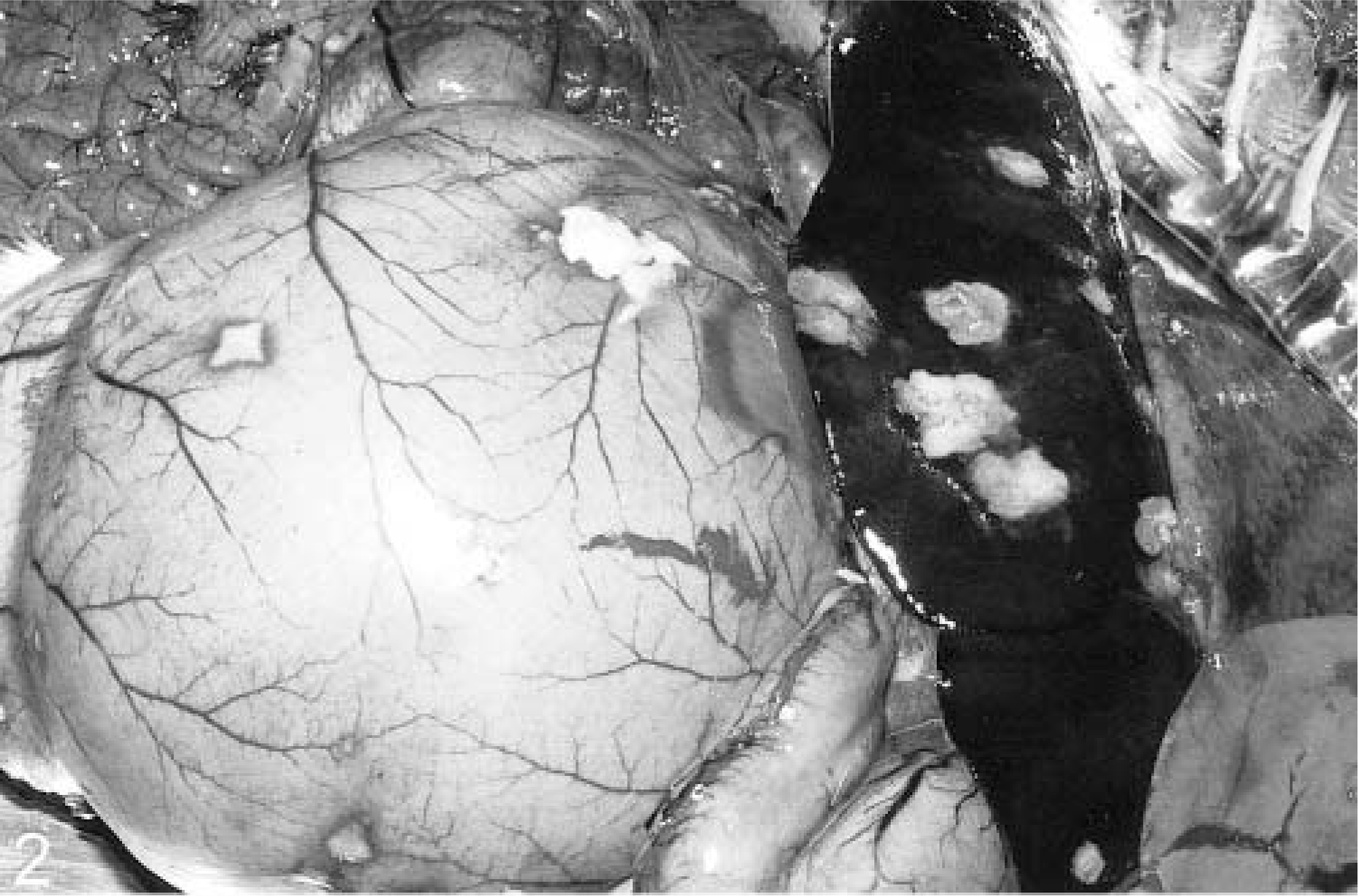
Abdomen; pronghorn. Necropsy revealed multiple foci of necrosis in the rumenal wall and liver due to F. necrophorum septicemia.
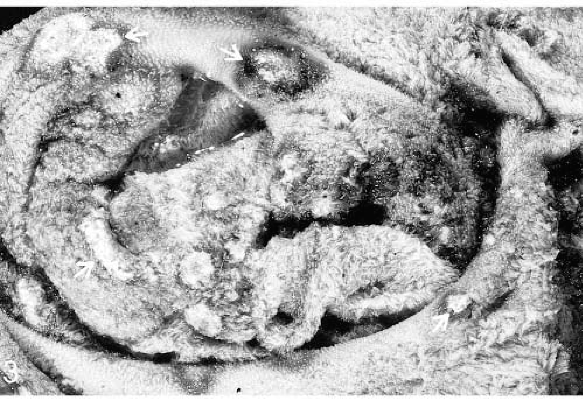
Rumenal mucosa; pronghorn. F. necrophorum septicemia resulted in multiple foci of mucosal necrosis (arrows) covered by fibrin.
Tongue; pronghorn. Focal necrosis due to F. necrophorum septicemia is seen as a discrete area (arrows) of pallor with a dark red edge. The mucosa appears intact.
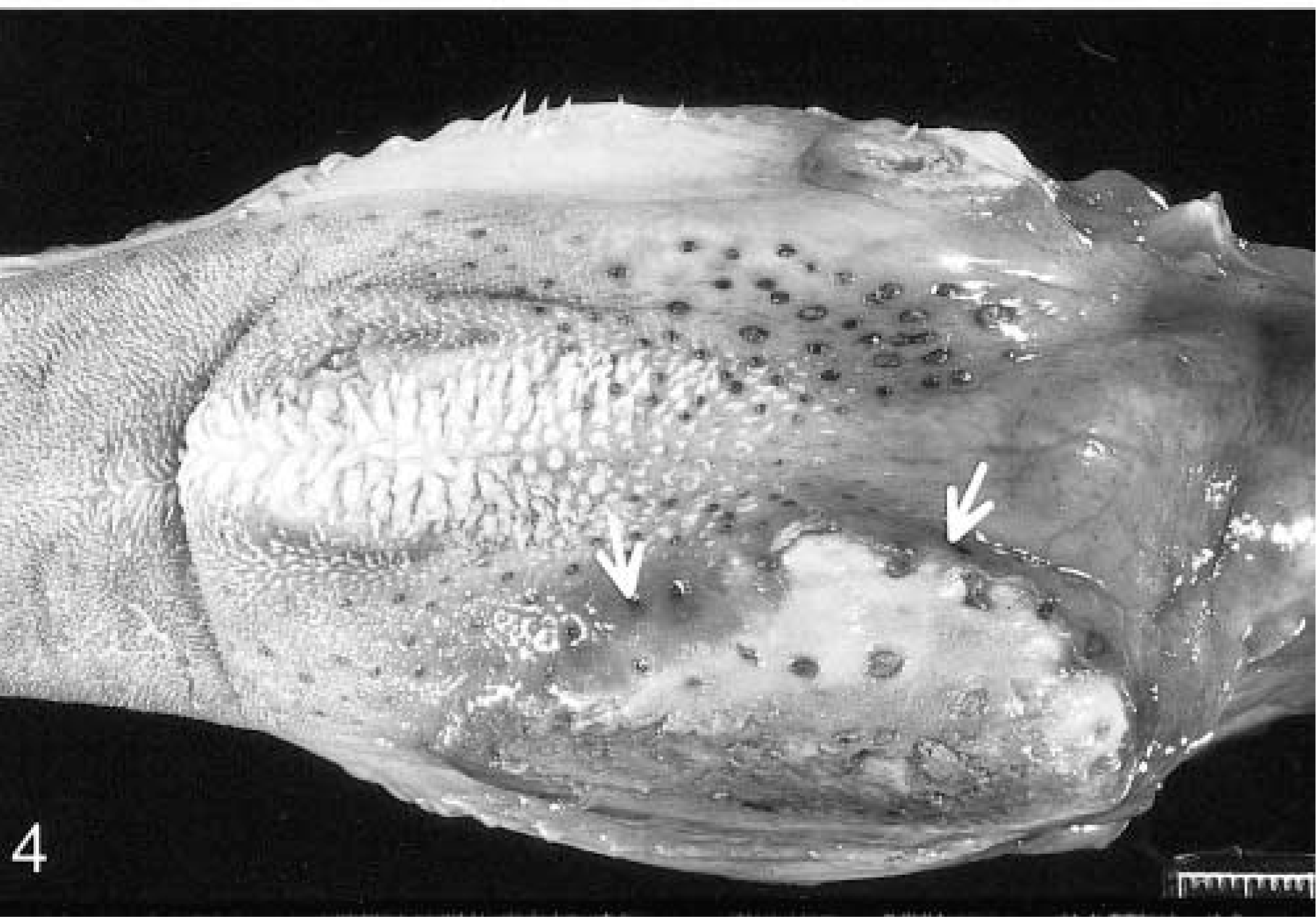
By 18 days after the first deaths, 15 pronghorns had died of typical necrobacillosis. On day 18, an animal was presented without pododermatitis but with lesions typical of fusobacterial necrotic stomatitis in the buccal mucosa at the commissures of the mouth (Fig. 5). In addition, lesions of necrobacillosis were noted in the forestomachs. From day 18 forward, animals were presented with lesions of systemic necrobacillosis with primary fusobacterial lesions in either the oral cavity or a limb. Some animals had systemic fusobacterial lesions and lesions that could be considered primary in both the oral cavity and a limb. Overall, 15 right front feet, seven left front feet, three left rear feet, and one right rear foot were affected. Only two animals had two affected feet. In only two animals was a hoof sloughed from one toe. Occasionally, oral lesions extended to affect large areas of the palate, gums, and cheeks, frequently bilaterally. The animals died quickly after visible evidence of swollen feet or clinical depression. The spectrum of lesions was similar in most animals. In five animals, the forestomach lesions were transmural and induced a fibrinous peritonitis, and in four animals the fusobacterial pneumonia extended to cause a pleuritis. In two animals, fusobacterial lesions were also in the cecum. By 60 days after the first deaths, only two pronghorns were alive. Both of these animals had pododermatitis, and were euthanatized. At necropsy, these animals exhibited only severe pododermatitis.
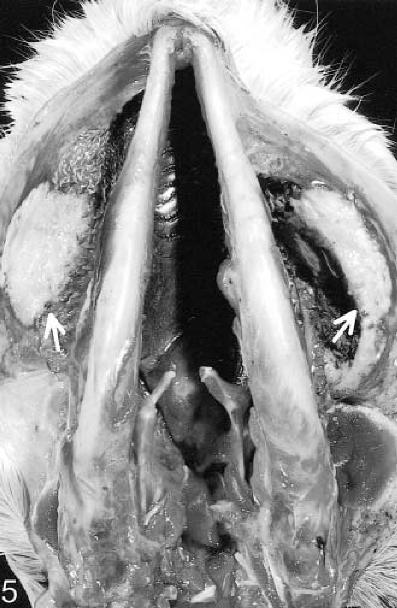
Skull and oral cavity; pronghorn. Ventral view, buccal mucosa reflected. Bilateral foci of F. necrophorum-induced stomatitis and necrosis (arrows) are in the cheeks at the commissures.
Histologically, the lesions presumed to have been the point for entry of the organism into the animal were similar whether they were in the mouth or limb. These lesions consisted of discrete areas of coagulative necrosis surrounded by a narrow border of primarily necrotic neutrophils with some macrophages and a few fibroblasts and neocapillaries. In HE-stained sections, the necrotic tissue adjacent to the viable border formed a basophilic band of numerous filamentous, approximately 80–100-µm bacteria morphologically consistent in appearance with F. necrophorum. These bacteria were gram negative and were easily visible when stained with Grocott's ammoniacal silver (Fig. 6). In focal metastatic lesions in forestomachs, liver, lung, and cecum, the discrete, coagulative necrosis was bordered by little inflammatory cell infiltrate. In these lesions, numerous filamentous bacteria also formed a band at the border with viable tissue. When the lesion extended through the pleura or serosa into the thorax or abdomen, large amounts of fibrin with necrotic neutrophils coated the surfaces of organs. No fungal organisms were noted, and mixed bacterial populations were seen on the rumenal mucosa over the necrotic lesions. In the lungs of animals with severe oral lesions, plant material and numerous bacterial colonies were in airways, suggesting terminal aspiration bronchopneumonia.
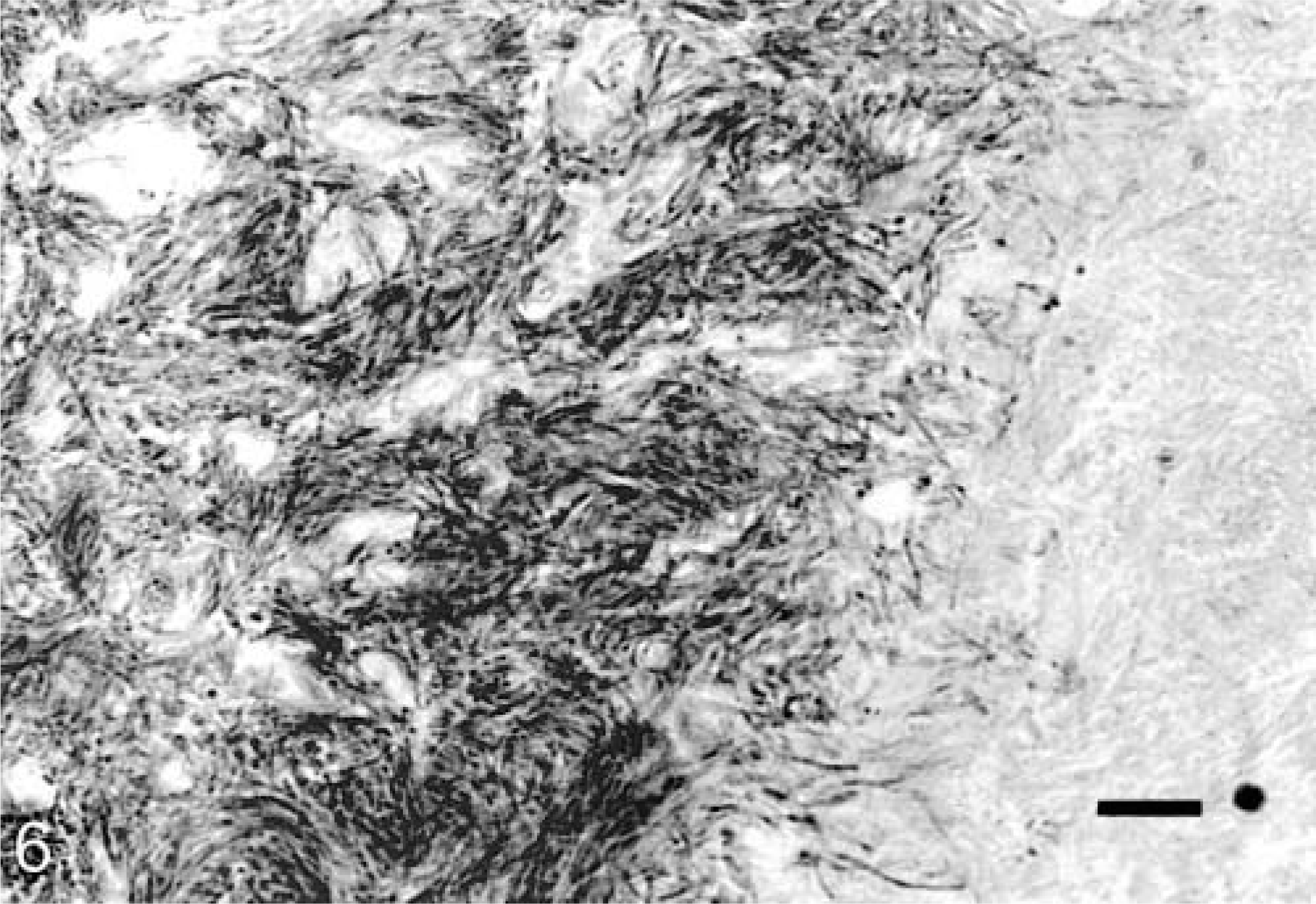
Buccal mucosa; pronghorn. Numerous filamentous organisms with morphology consistent with that of F. necrophorum proliferate near the edge of the necrotic oral lesion (necrobacillosis). Grocott's ammoniacal silver. Bar = 80 µm.
At 4 weeks into the outbreak, the pasture had not dried because of drainage from an adjacent pasture. There were no hoofprints around the waterers. The pronghorns apparently preferred to stand in muddy low spots and drink from pools on the ground or from water caught within hoofprints. The pasture remained wet for about 40 days after the rains.
A variety of organisms, including many species of coliforms, Arcanobacterium pyogenes, Bacillus sp., and Corynebacterium sp. were isolated from aerobic cultures of limb lesions. F. necrophorum sometimes in combination with another Fusobacterium, Streptococcus sp., or Clostridium sp., were isolated from most anaerobic cultures of foot lesions. In those lesions from which F. necrophorum was not cultured, many organisms with the morphology of F. necrophorum usually were observed in histologic sections of the lesions. Likewise, F. necrophorum was isolated from anaerobic cultures of lesions of parenchymal organs and the oral cavity, whereas a variety of coliforms were isolated in aerobic cultures of these lesions.
The severe mortality of captive wild ruminants with necrotizing oral lesions was clinically similar to that in a recently reported outbreak of adenoviral disease in mule deer in California. 13 The adenoviral disease was characterized by extensive areas of oral necrosis subsequent to infarction due to virus-induced vasculitis. Although there was necrosis and thrombosis of vessels subjacent to the necrotic foci in the pronghorns, no adenoviral inclusions were observed in tissues, and it was assumed that the vessel lesions were the result of the proximity of vessels to the extensive necrosis and the bacterial toxins.
Necrobacillosis has been reported in several wildlife species, including pronghorns, where infection with F. necrophorum induces both lethal stomatitis and pododermatitis. These reports represent both sporadic cases and large outbreaks in free-ranging animals. Outbreaks of lethal fusobacterial infections in pronghorns, elk (Cervus canadensis), mule deer (Odocoileus hemionus), and white-tailed deer (Odocoileus virginianus) have occurred in instances when these wild ruminants have been crowded. 2,7,10,12 Three of 17 pronghorns found dead in Canada were necropsied, and two (a yearling and a 2-year-old) had stomatitis or pododermatitis with histologic features of necrobacillosis, including organisms seen in Gram-stained tissue sections. In elk that congregated on the winter feeding grounds of the National Elk Refuge, Jackson, Wyoming, necrobacillosis was identified as the principal cause of 409 deaths. The syndrome in elk primarily involved necrotic stomatitis, and calves were affected in two-thirds of these cases. Oral lesions involved the tongue, cheek, and gingiva and were thought to originate from the ulcerated, periodontal gingiva. Lesions of the forestomachs, liver, spleen, and lung were observed. Epizootics of necrobacillosis in deer have been reported in California and Canada. 10 In California, hundreds of mule deer with lesions of necrobacillosis were found dead near mud holes, which served as their only water source. In Canada, groups of white-tailed and mule deer with lesions of necrobacillosis were found in areas associated with feeding cattle. These deer had necrotic stomatitis. The disease in wildlife is probably more prevalent than is reflected in published reports. The effects of autolysis and predation on carcasses often preclude accurate documentation in free-roaming ruminants.
Epizootics of necrobacillosis have been reported in wild ruminants maintained in refuges or research facilities. Necrotizing balanoposthitis has been associated with infection by F. necrophorum and A. pyogenes in European bison (Bison bonasus bonasus) in Poland. 4 The lesions were similar to those reported in feedlot steers in the USA, 5 and the pathogenesis was presumed to involve minor trauma of the preputial mucocutaneous border and exposure of the mucosa to the contaminated soil when the bison lay down in feeding areas. Similarly, 12 blue duikers (Cephalophus monticola fusicolor) in a research facility developed mandibular osteomyelitis and facial abscesses from which F. necrophorum and A. pyogenes were cultured. 9 Eight of the duikers were <2 years old. Although there were no identifiable lesions in their oral mucosa, it was presumed that a subclinical oral necrobacillosis, perhaps associated with tooth eruption in young animals, initiated the infections. An outbreak virtually identical to that of the present report occurred in an experimental herd of 25 captive wild-caught Columbian black-tailed deer (Odocoileus hemionus columbianus). 3 Most of the animals affected were fawns and yearlings, and the majority were affected with oral lesions (necrotic foci of the cheeks, palate, and tonsil), whereas only five had lesions of interdigital cellulitis. F. necrophorum and A. pyogenes were the most frequent isolates from lesions. In this outbreak, the heavy rains, mild climatic conditions, and persistence of muddy conditions in the enclosure were considered key factors contributing to the epizootic.
Necrobacillosis of wild ruminants appears to be a disease seen primarily when animals are brought together in areas where the soil may be contaminated by the feces of domestic ruminants, and it is further predisposed by wet conditions. 3,6,10 Pronghorns are native to arid grasslands and can survive on the water provided in their native forage. The grouping of these pronghorns in a pen that previously housed cattle was important in the pathogenesis of the outbreak. The pen was contaminated with F. necrophorum, and the wet conditions predisposed the animals to infection by softening the hooves. The initial infections resulted in draining foot lesions, which amplified the pathogen population. The habit of drinking from and standing in the ground pools modified the infection such that primary lesions began to appear commonly in the oral cavity. The cause of the initial oral lesions was not identified, but the lesions may have been induced by the change in diet (coarse stems of alfalfa hay). In addition, the captive pronghorns often were noted to grind their teeth, and that habit may have induced minor buccal damage. Other underlying factors that would promote entry of F. necrophorum were not identified. The facility is in an endemic bluetongue disease area; however, lesions were not typical of bluetongue disease, and the outbreak occurred at a time of year when bluetongue virus vectors were not active. The possibility of a toxin in the feed was considered, but the same feed was used with other animals on the farm without incident, and histologic lesions suggesting toxins were not observed. The water that ran onto the pasture came from adjacent pastures that housed other animals without disease problems; thus, the possibility of toxic runoff was discounted.
In domestic ruminants, necrobacillosis does not commonly extend to a septicemia from the oral and pedal lesions. 1,6 In this outbreak, the disease resembled Lemierre's syndrome in human beings. 11 Lemierre's syndrome is a septic, systemic F. necrophorum infection that extends from a primary oropharyngitis. In the period before antibiotics were available, Lemierre's syndrome was seen primarily in young adults or adolescents and resulted in jugular thrombophlebitis and metastatic infections of the lung, joints, muscles, liver, and brain. Necrobacillosis is a major health consideration for herds of captive wild-caught ruminants. Wet pastures, temperate ambient temperatures, and containment of these animals in pastures contaminated by cattle feces greatly increases the probability of inducing fatal infections that gain entry through oral or pedal epithelial defects.