
Abstract
The mes rat is from an inbred mutant colony of rats with spontaneous eosinophilia. In order to investigate the pathogenesis of the mes rat, the histopathology and hematology for 76 mes rats were examined at several weeks of age. Tissue eosinophilia developed at 8 weeks of age when the blood eosinophil was 500 cells per microliter or more. Subsequently, eosinophilia progressed with age, and splenic eosinophilopoiesis and erythropoiesis appeared simultaneously. Many inflammatory lesions were induced after 10 weeks of age when the blood eosinophils became 1,000 cells per microliter or more. Gastroenteritis and mesenteric lymphadenitis were seen in 44 of 47 (94%) and 31 of 47 (66%) rats, respectively, after 10 weeks of age. Aortitis that deteriorated with age was found in 19 of 39 (49%) rats after 12 weeks of age. Hepatic fibrosis was found in four rats that exhibited severe eosinophilia and anemia. These results are comparable to the features of a hypereosinophilic syndrome in humans and other animals.
Eosinophilia develops in reactive diseases such as allergic and parasitic diseases, some malignant diseases, and in idiopathic hypereosinophilic syndrome (HES). 1,11,14 The eosinophilia is accompanied by a wide variety of inflammatory lesions because toxic inflammatory mediators are released from eosinophils. 1,11,14 In particular, systemic and chronic hypereosinophilia, as in HES, is known to result in severe organ damage. 1,14 HES is a systemic disorder defined as chronic hypereosinophilia accompanied by significant organ system dysfunction without apparent etiology. It has been reported in humans, ferrets, cats, horses, and dogs. 1,3–6,9,10,14 However, details of the pathology and pathogenesis of HES are unclear.
In 1999, Matsumoto et al. found mutant rats with spontaneous eosinophilia that were derived from pregnant specific pathogen free Slc:Sprague-Dawley (SD) rats. This colony was maintained by brother-sister matings and was named the Matsumoto Eosinophilia Shinshu (mes) rat. 8 This inbred colony has been maintained for 10 generations. The mes rats have hematologic features resembling HES, and eosinophil infiltration in various organs has been previously reported. 7,8 Therefore, it is probable that the mes rat could be a useful animal model for eosinophilia, HES, or both. In this paper, we present the histopathologic and hematologic changes in mes rats with age and compare them with HES in humans and other animals.
In this study, 38 male and 38 female mes rats of the F2 to F4 generations were used. The rats were housed in a controlled environment barrier-system room, fed a commercial diet, and given water ad libitum. Four to five rats of each sex were examined at 4, 6, 8, 10, 12, 16, 20 and 25 weeks old, respectively. For clinical pathology examinations, heparinized blood was collected from the jugular vein. Routine hematologic examinations were preformed, and blood immunoglobulin E (IgE) was measured by using an IgE ELISA kit (Morinaga & Co., Ltd., Kanagawa, Japan). For macroscopic and histopathologic examinations, all rats were anesthetized with ether and euthanized by cervical dislocation. Necropsies were performed and the tissues were collected, fixed with 10% neutral buffered formalin, embedded in paraffin, sectioned at 4 µm, stained with HE and examined routinely. Selected tissues were stained with Gram stain, Giemsa stain, and periodic acid–Schiff (PAS) stain. This study was carried out in accordance with the “Guidelines for Animal Care and Experimentation” of the Shinshu University School of Medicine.
In the clinical pathology examinations, apparent increases in blood eosinophils were found at 8 weeks of age (Table 1). Subsequently, numbers of blood eosinophils increased with age, and the degree of increase in females was greater than that in males. The nuclei of eosinophils were generally coiled to form a ringlike or figure-eight pattern, and the granules were normal and acidophilic. Blasts and abnormal eosinophils were not detected on blood films. There were no differences in blood IgE levels between the mes and normal SD rats. Macroscopically, enlargement of the mesenteric lymph nodes and spleen were found in most rats after 4 and 10 weeks of age, respectively. Moreover, enlargement of the liver, with elevated, indurated, and hemorrhagic lesions in the stomach and small intestine, were found in many rats after 10 weeks of age.
Blood eosinophils and eosinophilic lesions in mes rats.
Mean values.
Percent eosinophils.
Not found.
No. rats with lesions.
Histopathologically, eosinophilic proliferation and infiltration in the bone marrow, gastrointestinal tract, mesenteric lymph node, and spleen was found in some rats initially at 8 weeks of age. After 10 weeks of age, eosinophilic proliferation in the bone marrow was found in all rats. The bone marrow was hypercellular primarily because of eosinophilic proliferation. All maturation stages were present, but mature eosinophils predominated. Infiltration of mature eosinophils was found severely in the red pulp of the spleen; the lamina propria, submucosa, and muscularis of the gastrointestinal tract; and the medulla of the mesenteric lymph nodes in most rats. It was also found in the lung, thymus, liver, and uterus of some rats. After 12 weeks of age, proliferations of erythroblasts and immature eosinophils in the red pulp of the spleen were found in many rats, and the incidence and severity of these findings increased age dependently. HE staining revealed normal eosinophils were typical morphology in each stage.
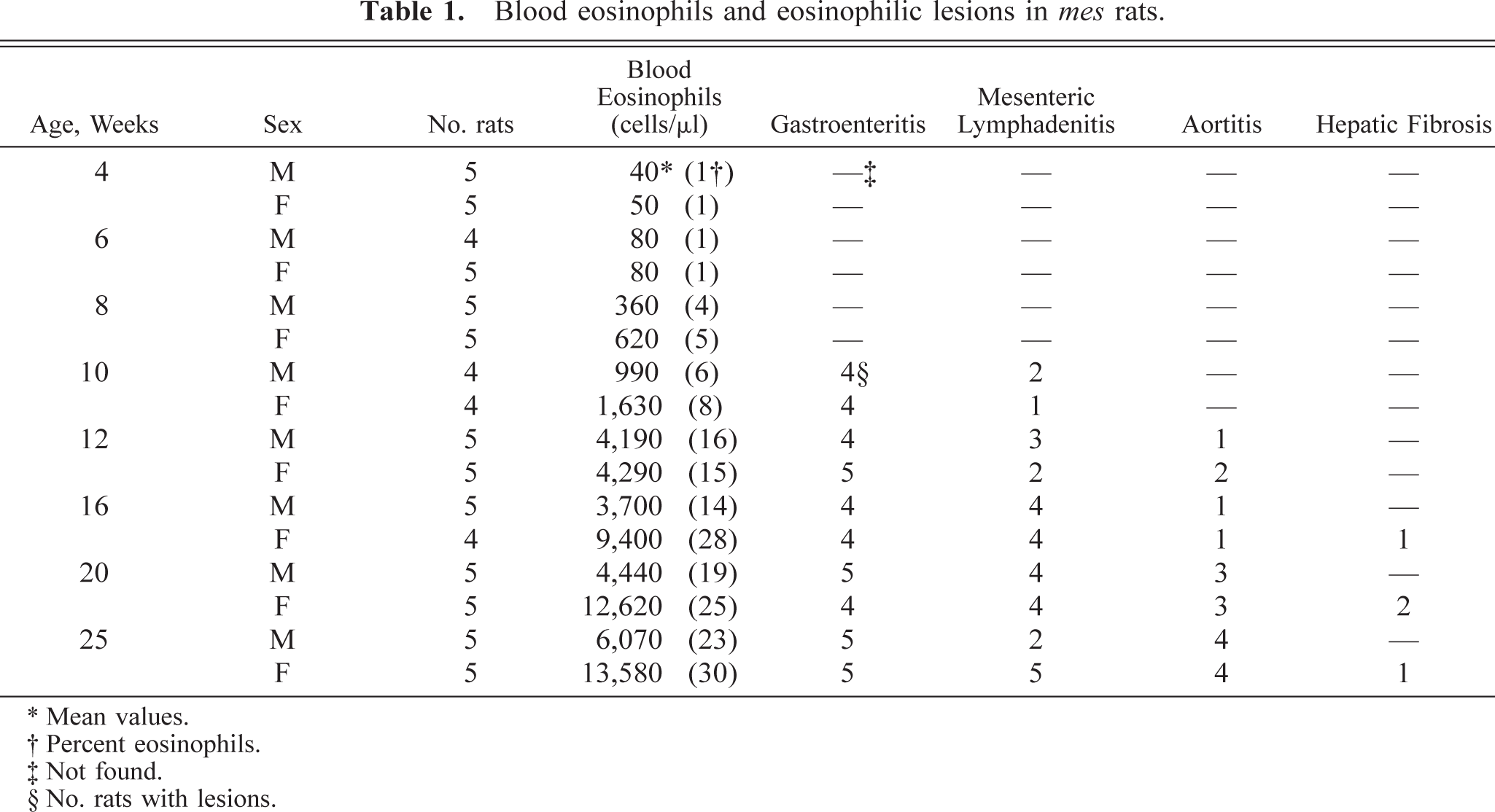
In the gastrointestinal tract, diffuse fibrosis with inflammatory response was found in the lamina propria, submucosa, and muscularis in 44 of 47 rats after 10 weeks of age (Table 1, Fig. 1); it was prominent in the lamina propria of the stomach and the muscularis in the jejunum and ileum. Fibrosis was accompanied by infiltration of numerous eosinophils and, occasionally, deposition of granular eosinophilic material associated with macrophages. Additionally, focal granulomas in the lamina propria and submucosa of the stomach were found in 8 of 47 rats (Fig. 1). The granulomas were composed of degenerated eosinophils and homogeneous or granular eosinophilic material surrounded by macrophages. Eosinophilic infiltration often disrupted the normal architecture, atrophy, and compensatory metaplasia of the gastric glands, and ulceration was confirmed in the more severe cases (Fig. 1).
Glandular stomach; 20-week-old male rat. Fibrosis and granuloma consisting of eosinophils and eosinophilic materials in the lamina propria and submucosa; ulceration and eosinophil infiltration in the lamina propria and submucosa. HE stain. Bar = 60 µm.
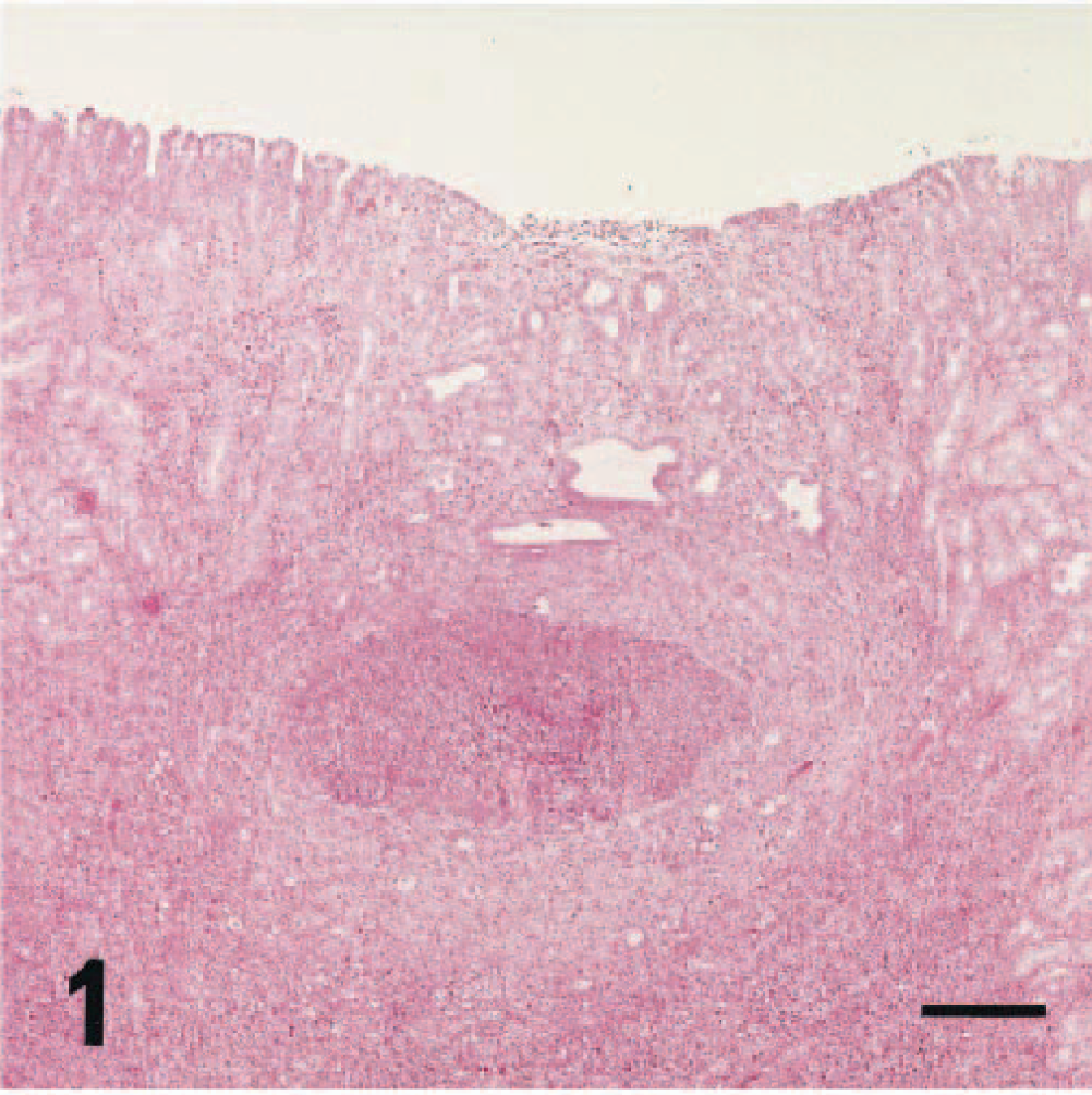
In the mesenteric lymph nodes, multifocal granulomas in the medulla were found in 31 of 47 rats after 10 weeks of age (Table 1, Fig. 2). The granulomas were composed of degenerated eosinophils and homogeneous or granular eosinophilic materials surrounded by macrophages and multinucleated giant cells. Additionally, infiltrations of numerous plasmacytes and macrophages in the paracortex were found in most rats after 8 weeks of age, and the severity correlated with the degree of macroscopic enlargement.
Mesenteric lymph node; 20-week-old male rat. Granuloma consisting of eosinophils and multinucleated giant cells in the medulla. HE stain. Bar = 10 µm.
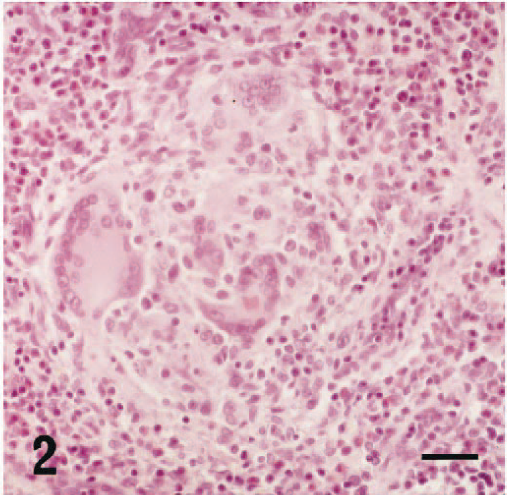
In the thoracic aorta and pulmonary artery, inflammation characterized by thickening of the intima was found in 19 of 39 rats after 12 weeks of age (Table 1, Fig. 3), and the incidence and severity of these findings increased age dependently. Eosinophilic infiltration was limited to both the intima and adventitia. Furthermore, in the thoracic aorta, deposition of bandlike eosinophilic material associated with macrophages and multinucleated giant cells accumulated between the intima and media (Fig. 3). In the liver, centrilobular fibrosis and periportal eosinophil infiltration were found sporadically in four rats that exhibited severe eosinophilia and anemia (Table 1, Fig. 4). Centrilobular eosinophil infiltration was not present. In the lung and thymus, granulomas similar to those described in the mesenteric lymph nodes were found sporadically in some rats.
Thoracic aorta; 25-week-old male rat. Thickening of the intima, eosinophil infiltration at both the intima and adventitia, and deposition of eosinophilic material between the intima and media. HE stain. Bar = 25 µm.
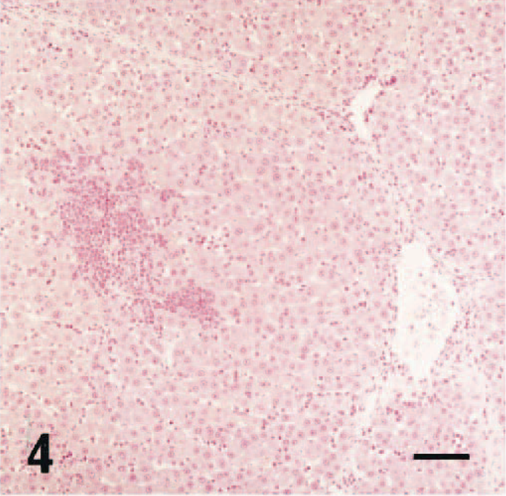
Liver; 20-week-old female rat. Centrilobular fibrosis and periportal eosinophil infiltration. HE stain. Bar = 25 µm.
When sections of the gastrointestinal tract, mesenteric lymph nodes, and thoracic aorta were stained with Giemsa stain and PAS stain, the eosinophilic material was pink with Giemsa stain and reacted positively with PAS stain. Organisms were not detected in Gram, Giemsa, and PAS staining.
The onset of systemic eosinophilia in the mes rats started by 8 weeks of age, and the eosinophilia progressed thereafter. In particular, blood eosinophil counts increased dramatically and exceeded the level for diagnosis of human HES (>1,500 eosinophils per microliter) by 12 weeks of age, 1,14 achieving the level of “severe” on the diagnostic scale for human eosinophilia (>5,000 eosinophils per microliter) by 25 weeks of age. 1,11 The reasons for the sex difference of the eosinophilia are still unclear because it has not been reported in other species. It may be induced by sex hormones such as estrogen, which regulates uterine eosinophilia. 13 Splenomegaly, with the extramedullary eosinophilopoiesis and erythropoiesis, is thought to be a result of hypersplenism induced by saturation of bone marrow eosinophilopoiesis and correlated with the anemia in the mes rats. Hypersplenism is a feature of human hypereosinophilia, including HES. 1,14 Many inflammatory lesions were induced in various organs after 10 weeks of age when the blood eosinophils showed 1,000 cells per microliter or more and depending on the severity of the eosinophilia.
Gastroenteritis, mesenteric lymphadenitis, and aortitis are thought to be eosinophil-derived inflammatory lesions because deposition of eosinophilic material was found in the lesions. This eosinophilic material is thought to be Splendore-Hoeppli material because the characteristics are similar to those described in ferrets. 3 It is suggested that inflammation is caused by the cytotoxicity of mediators such as the eosinophilic granule proteins that are released from the eosinophils. 11,14 In particular, the diffuse forms of gastroenteritis and mesenteric lymphadenitis are similar to the lesions described in ferrets, cats, horses, and dogs. 3,4,9,10
Many eosinophilic inflammatory lesions were found in the mes rats, yet cardiac lesions could not be found. Cardiac lesions are the most common feature of the chronic hypereosinophilia, including HES seen in humans. 1,14 Cardiac damage may be induced by the progression of thrombosis induced by the eosinophilic granule proteins. 1,14 However, there were no changes that could have been associated with progressive thrombosis in the mes rats. Furthermore, there was no elevation of blood IgE levels that is seen in many patients with HES. 14
The relationship between the hepatic fibrosis and the eosinophilia is unclear because the fibrous lesion was not correlated with eosinophil infiltration. Hepatic fibrosis may be induced by anemia because it was only found in rats showing severe anemia. These specific increases of blood eosinophils and mesenteric lymphocytosis and plasmacytosis suggest the participation of interleukin-5 (IL-5) in the pathogenesis of the eosinophilia in mes rats. IL-5 is the cytokine stimulating the growth and differentiation of eosinophils in humans and mice and B lymphocytes in mice. It plays a major pathogenic role in HES and various eosinophilic conditions in humans. 1,11,12,14 Transgenic mice excessively expressing IL-5 develop systemic and chronic hypereosinophilia similar to that in the mes rats. 2,6 In support of this theory, a decrease in blood eosinophils followed the administration of antimurine IL-5 antibody in a recent study in the mes rats (data are not shown).
From these results, it is concluded that mes rats could be a useful animal model for research on HES because the hypereosinophilia, with many associated eosinophilic lesions seen in the mes rats, is comparable to the features of HES in humans and other animals.
Footnotes
Acknowledgements
We thank M. Uehara and K. Matsumoto for their technical assistance.