
Abstract
Angiokeratoma is described by various authors as a rare variant of the hemangioma in the dog, characterized by a vascular component, like all types of vascular neoplasms, but also by an epithelial component. A case of angiokeratoma is described in a male 8-year-old dog. The tumor was located in heavily pigmented skin on the anterior surface of the front limb and not in the more usual previously described locations, eyelid and conjunctiva. Microscopic examination revealed a well-circumscribed mass with irregular hyperplasia of the epidermis and dilated vascular spaces filled with blood in the superficial dermis.
Vascular tumors of the skin are classified as hemangiomas and hemangiosarcomas. Hemangiomas (also named angiomas) are benign neoplasms arising from the endothelial cells of blood vessels and are uncommon in dogs and rare in cats. 2,4
In dogs, hemangiomas are more common than hemangiosarcomas and occur at an average age of 10 years, with no sex predilection and with an incidence that ranges from 2 to 3% of skin and subcutaneous tumors. 2,4,6 Breeds at risk include Boxer, Golden Retriever, German Shepherd, English Springer Spaniel, Dalmatian, English Pointer, and others. 4
Hemangiomas are usually well circumscribed, firm to fluctuant, rounded, bluish to reddish-black, range from 0.5 to 4 cm in diameter, and have a dermal to subcutaneous location. 2,4,5
Dermal and subcutaneous hemangiomas can occur at any anatomic site, but the dermal vascular tumors have a predilection for the ventral abdominal and inguinal skin in dogs with nonpigmented skin and short-hair coats. 2
Histologically, hemangiomas are characterized by the proliferation of blood-filled vascular spaces lined by single layers of well-differentiated endothelial cells. They can be subclassified into cavernous or capillary, depending on the size of the vascular spaces and the amount of intervening fibrous tissue. 4,5
Angiokeratoma is a rare variant of hemangioma in dogs that contains both vascular and epithelial components. It is typically a small, well-circumscribed mass that is elevated above the skin surface and occurs most often on the eyelid and conjunctiva but can also arise in the superficial dermis at any site. There is no age or breed predisposition for this tumor type. 2,6
In humans, the angiokeratoma is better studied and is also characterized as a rare, wart-like vascular lesion of the skin that occurs at multiple sites, is more common in males than in females, and is rarely seen in individuals with pigmented skin. 1,3 However, the human angiokeratoma is classified as either an acquired type (Mibelli, Fordyce, solitary, and multiple papular) or a congenital type (angiokeratoma circumscriptum and angiokeratoma corporis diffusum), which have different anatomical, age, and sex predilection. 1
A biopsy sample of skin from a male 8-year-old Boxer was submitted to the pathology service of the Universidade de Trás-os-Montes e Alto Douro. The sample included a 1-cm diameter, ovoid, well-circumscribed, bluish mass that was elevated above the skin surface. This mass was removed from the anterior surface of the left front limb of the dog. The skin at this location was heavily pigmented. No previous masses had been reported in this dog.
Fragments of the mass were fixed in neutral buffered formalin and embedded in paraffin, sectioned at 3 µm, and stained with hematoxylin and eosin (HE).
Microscopic examination revealed irregular hyperplasia of the epidermis (Fig. 1). The superficial dermis was thickened by dilated, blood-filled vascular spaces (Fig. 2). Each vascular space was lined by a single layer of cells that resembled normal endothelial cells, aligned on thin collagenous septa. Epithelial trabeculae extended downward to separate and partially surround some of the vascular structures (Fig. 3). No mitotic figures were identified and the borders of the neoplasia were totally contained within the limits of the tissue section observed (Fig. 1).
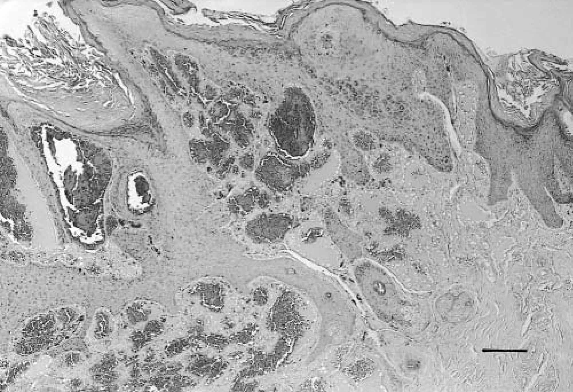
Skin. Irregular hyperplasia of the epidermis. Neoplastic border. HE. Bar = 20 µm.
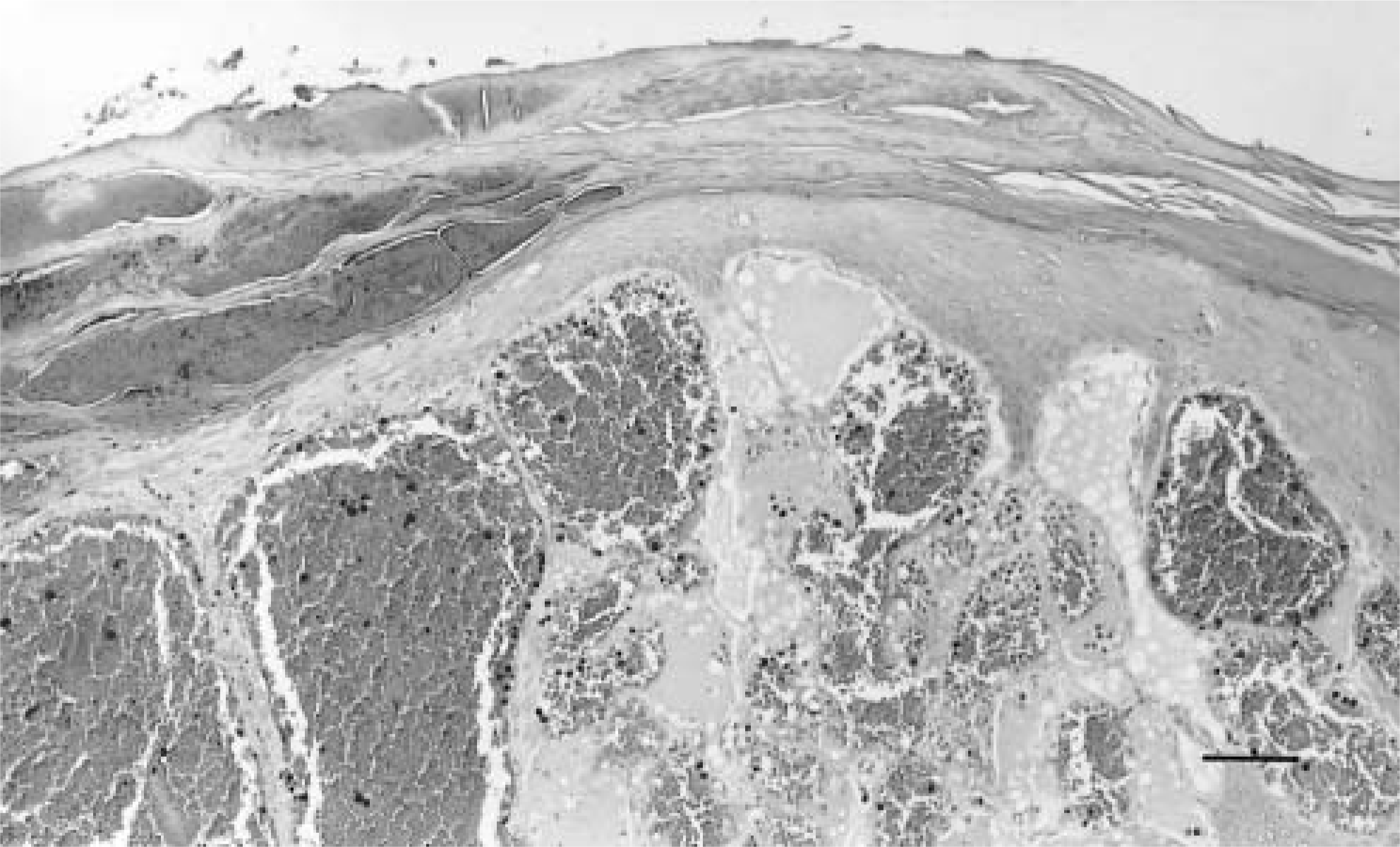
Skin. Dilated vascular spaces in the superficial dermis. HE. Bar = 50 µm.
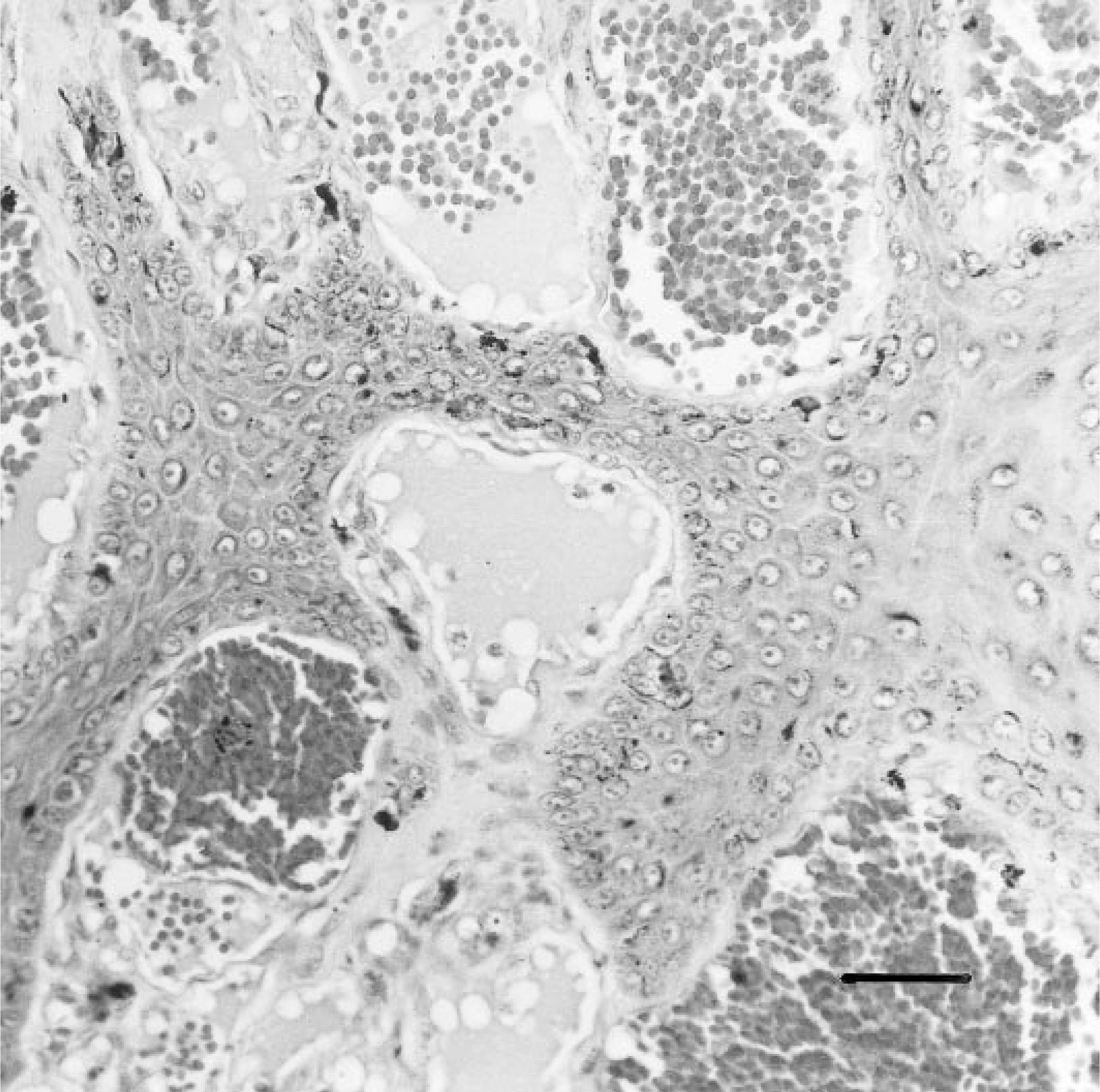
Skin. Note the epithelial trabeculae extending downward to separate and partially surround some of the vascular structures. HE. Bar = 100 µm.
The histologic characteristics were similar to those of hemangiomas except for the presence of this unusual marked hyperplasia of the overlying epidermis, which appeared to invade between the vascular spaces of the superficial dermis. This is similar to descriptions of angiokeratomas made by other authors. 2,6
The mass observed in this dog had the vascular and epithelial components that suggested the lesion was a rare subtype of hemangioma, the angiokeratoma.
This case is unusual in that the mass involved heavily pigmented skin of the anterior surface of the front limb; most reported cases of angiokeratoma have occurred on the conjunctiva and eyelids, while hemangioma occurs most often on the abdominal and inguinal skin in dogs. 2,6
Footnotes
Acknowledgements
Thanks are due to Dr. Anabela Alves, Dr. Dolors Fondevila, and Prof. Conceição Peleteiro for their contribution to this manuscript.