
Abstract
Kaposiform hemangioendothelioma is a rare vascular tumor that almost exclusively occurs in children. It is a low-grade malignant neoplasm that is locally aggressive but without proven metastatic potential. Herein, we describe a case of Kaposiform hemangioendothelioma in a 10-year-old male dog. To the best of our knowledge, this is the first report of this vascular tumor occurring in a nonhuman animal species. The tumor was located on the ventromedial surface of the posterior limb. Microscopic examination of the lesion revealed a multinodular tumor with sheets of spindled endothelial cells forming vascular slits similar to Kaposi's sarcoma and peripheral tumor lobules resembling capillary hemangioma.
Skin vascular tumors are common in dogs. 4,8 Occasionally they are seen in cats, but are rare in other domestic or wild animals. 4,8 The most frequent vascular tumor is the hemangioma. 5 This benign tumor as well as it malignant counterpart, the hemangiosarcoma (known in human pathology as angiosarcoma), usually occur in older dogs. 5 There is evidence that these tumors can be caused by increased exposure to solar radiation because tumors occur more frequently in fair skinned, short-haired breeds. 5,9
Hemangiomas usually present as solitary well-circumscribed, slow-growing nodules and are cured by complete excision. 8,11 The skin is one of the most common sites of occurrence for this lesion. Histologically, the vascular proliferation has a lobular configuration. 10 It consists of capillary or cavernous vascular channels lined with normal endothelial cells. Usually capillary hemangiomas appear more superficially in the dermis, whereas cavernous hemangiomas are located in the deeper dermis or subcutis.
Hemangiosarcomas commonly present as multicentric disease. 8,11 They are most commonly found in spleen and heart followed by the skin. 1,3 The skin can be both the primary site of the tumor or the site of metastasis from the spleen or heart. These tumors are aggressive, with a high rate of recurrence and metastasis. Histologically, lesions are poorly circumscribed, with the tumor infiltrating into the subcutis. They are made of a meshwork of anastomosing, dilated vessels extending between preexisting collagen bundles. The vessels are irregular and are lined by endothelial cells that range from virtually normal-looking to plump, atypical protuberant cells with enlarged hyperchromatic nuclei. Kaposi's sarcoma, considered a variant of hemangiosarcoma, does not normally occur in animals. Kaposi-like sarcoma was only described in mice injected with 1,2-dimethylhydrazine di-hydrochloride. 7 The tumor was shown to develop after chemical induction only in the liver and not in the skin. 7
One case of epitheliod hemangioendothelioma, low-grade malignant vascular neoplasm, was also reported in the lung of a dog. 6 That lesion was characterized by tumor cells with abundant eosinophilic cytoplasm and the presence of intra-cytoplasmic vacuoles.
Kaposiform hemangioendothelioma is a rare, low-grade malignant vascular neoplasm. 12 It was mostly reported as a soft-tissue mass or skin tumor in children younger than 10 years. 2 Some lesions were reported in conjunction with lymphagiomatosis or were associated with the Kasabach-Merritt syndrome. 2 These tumors are usually locally aggressive, but distant metastases have not been reported.
This case represents the first report of this entity occurring in any domestic or wild animal. An otherwise healthy 10-year-old male Dalmatian dog was noted to develop a cluster of 1- to 3-mm red, well-circumscribed papules on his left ventromedial posterior limb (Fig. 1). The lesions were noted to be slowly enlarging over the months. The dog presented for examination after an episode of bleeding secondary to excoriation of one of the papules. No lymphadenopathy or hepatosplenomegaly was appreciated during the examination. The clinical impression on presentation was hemangioma versus hemangiosarcoma.
Skin. Clustered red papules on posterior limb. Clinical photograph. Bar = 1 cm.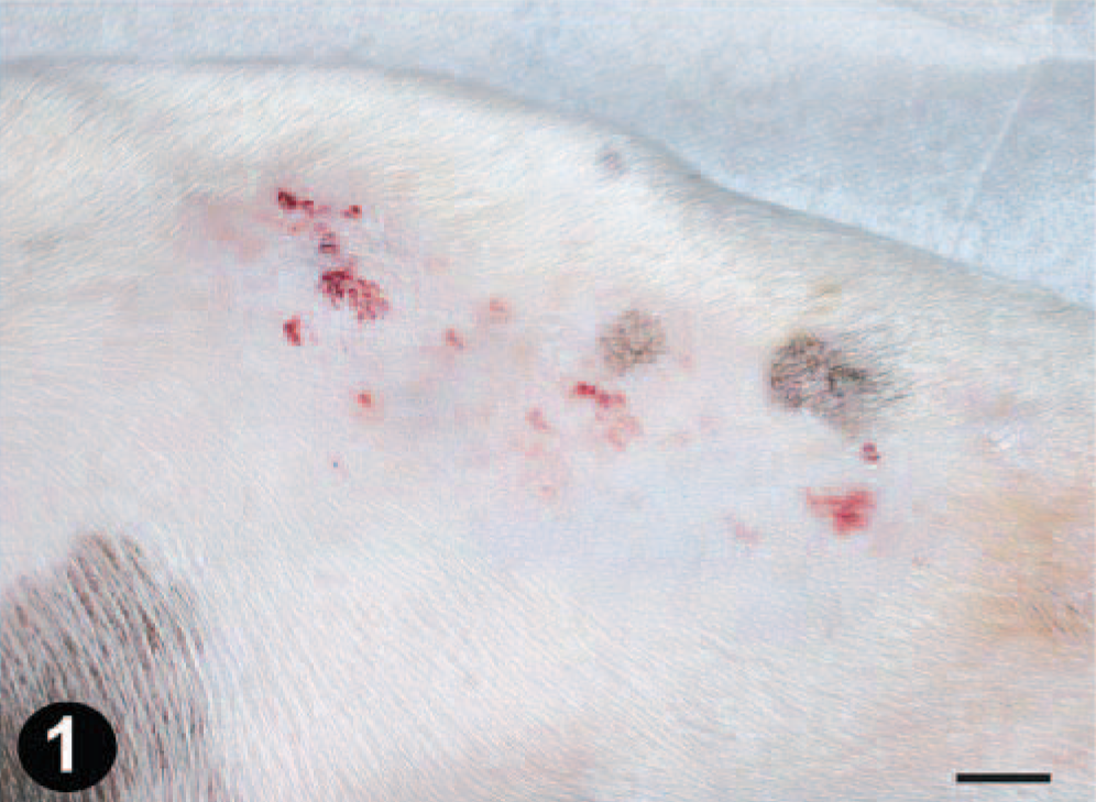
The biopsy of the lesion revealed multinodular (Fig. 2), focally infiltrating tumor. Deeper dermal nodules were made of spindle cells forming vascular slits reminiscent of the histology seen in Kaposi's sarcoma (Fig. 3A)). More superficially located dermal nodules consisted of small blood vessels, with histology resembling capillary hemangioma (Fig. 3B)). The spindle cells and capillaries were strongly positive for endothelial cell marker CD31 (Fig. 4). Intravascular platelet trapping and periodic acid-Schiff (PAS)–positive hyaline globules were not seen.
Skin. Multinodular dermal vascular proliferation. HE. Bar = 100 µm.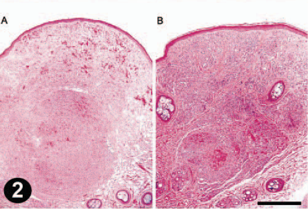
Skin dermis. Kaposiform vascular proliferation. HE. Bar = 250 µm. 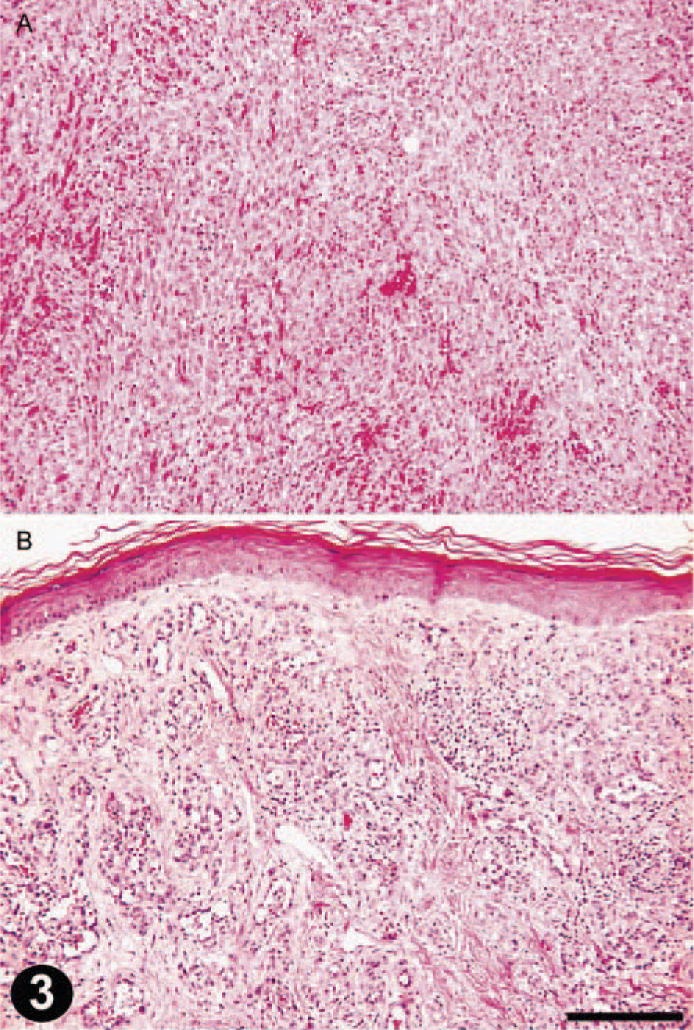
Skin dermis. Immunoperoxidase staining with CD31 endothelial marker. Bar = 100 µm.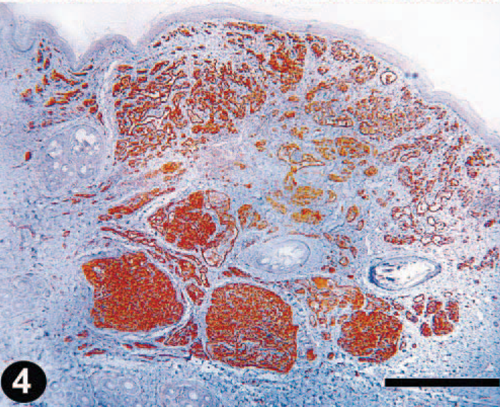
Differential diagnosis included Kaposi's sarcoma, capillary hemangioma, spindle cell hemangioendothelioma, and epithelioid hemangioendothelioma. Kaposi's sarcoma has a characteristic lymphoplasmacytic infiltrate and a more diffusely infiltrative pattern not seen in this case. It also contains PAS-positive hyaline globules not seen in our case.
Capillary hemangioma was excluded because of the presence of spindle cells with slitlike channels. The spindle cell hemangioendotheliomas are characterized by the cavernous vascular spaces present usually at the periphery of the lesion, but these spaces were not present in our case. The epithelioid hemangioendothelioma was excluded because of absence of intracytoplasmic vacuoles containing red blood cells.
The diagnosis of Kaposiform hemangioendothelioma was rendered. Because this tumor in humans was reported to be associated with the Kasabach-Merritt syndrome, coagulopathy associated with thrombocytopenia, platelet count was determined but was found to be within the normal range. The lesion was completely reexcised, and the dog remained healthy 6 months later without evidence of recurrence or metastasis.
To the best of our knowledge, this unusual case is the first report of Kaposiform hemangioendothelioma in any non-human animal species. In contrast to human cases that were reported mostly in children, this case was found in an older animal.