
Abstract
A nodule was identified within the right mammary gland of a 16-year-old male squirrel monkey (Saimiri sciureus). The mass was excised and diagnosed as a mammary adenocarcinoma. The monkey developed congestive heart failure 1.5 years later and was euthanatized. At necropsy, a subcutaneous mass was found in the right axillary region. Histologically, the mass was identified as a lymph node whose architecture was effaced by neoplastic cells resembling those of the mammary tumor. Metastasis to internal organs was not observed. This is the first reported case of a mammary tumor in a New World primate and the only known case of mammary cancer in a male nonhuman primate.
Keywords
Breast carcinoma is the most common and second most lethal form of cancer in women in the United States. 15 In the domestic animals, mammary tumors are common only in the dog and cat. In laboratory species, these tumors are most commonly observed in mice and rats. 16 The reported cases of mammary adenocarcinoma in nonhuman primates have occurred in female Old World primates such as macaques. 1 2 Here, we describe for the first time a case of mammary adenocarcinoma in a New World primate, a squirrel monkey (Saimiri sciureus); it is also the first reported case in a male nonhuman primate.
An elevated, approximately 1.0-cm-diameter subcutaneous nodule was noted in the right mammary gland of an otherwise normal, approximately 16-year-old adult male squirrel monkey. The mass partially incorporated the right nipple, and the overlying skin was focally ulcerated. The mass was not attached to the underlying tissues. The right axillary lymph node was enlarged to about 8 mm in diameter. No other clinical abnormalities were noted, and thoracic radiographs were within normal limits. The mammary mass was removed by wide excisional biopsy and placed in 10% neutral buffered formalin for processing for histologic examination. It was 10 × 8 × 11 mm and was bordered by a thin connective-tissue capsule. The mass was firm and its cut surface was varying shades of red, yellow, and white.
Unexpected complications during surgery prevented removal of the draining right axillary lymph node. A needle aspirate of the lymph node was performed prior to anesthetic recovery. Results of the aspirate were inconclusive. No further treatment was elected, and the monkey was returned to the colony.
Physical examinations over the next 15 months were within normal limits. No evidence of pulmonary metastasis was noted on thoracic radiographs taken 15 months after the mass was excised. Approximately 18 months after the removal of the mass, the animal developed congestive heart failure and was euthanatized. At necropsy, a 3-cm ovoid subcutaneous mass was noted in the right axillary region. The mass was encapsulated, partially reddened, and multifocally adhered via thick fibrous strands to the surrounding connective tissue.
The axillary region mass and sections from major organs were fixed in 10% neutral buffered formalin. Fixed tissues were trimmed, processed, embedded in paraffin, sectioned at 5 μm, and stained with hematoxylin and eosin (HE) for light microscopy.
Histologically, an expansile, infiltrative, roughly ovoid mass that compressed the surrounding connective tissues was present in the dermis and subcutis of the mammary region. The mass was composed of a mixture of sheets of cells, tubules, and acini supported by a fine fibrovascular stroma. The tubules were lined by single to multiple layers of pleomorphic epithelial cells surrounding a central lumen. The lumina contained variable amounts of amorphous eosinophilic material (Fig. 1). The epithelial cells within the sheets and tubules had indistinct borders and contained scant to moderate amounts of amphophilic cytoplasm. The oval nuclei contained finely stippled to coarsely clumped, evenly distributed chromatin and generally one eccentric, eosinophilic nucleolus. Mitotic figures averaged <1/400× field. Scattered areas of necrosis and hemorrhage were present within the parenchyma of the mass. The mass was bordered by multifocal aggregates of lymphocytes and plasma cells. The overlying adnexa were atrophic, sweat gland lumina were dilated, and the epidermis was focally ulcerated. No tumor cells were observed in adjacent blood and lymphatic vessels.
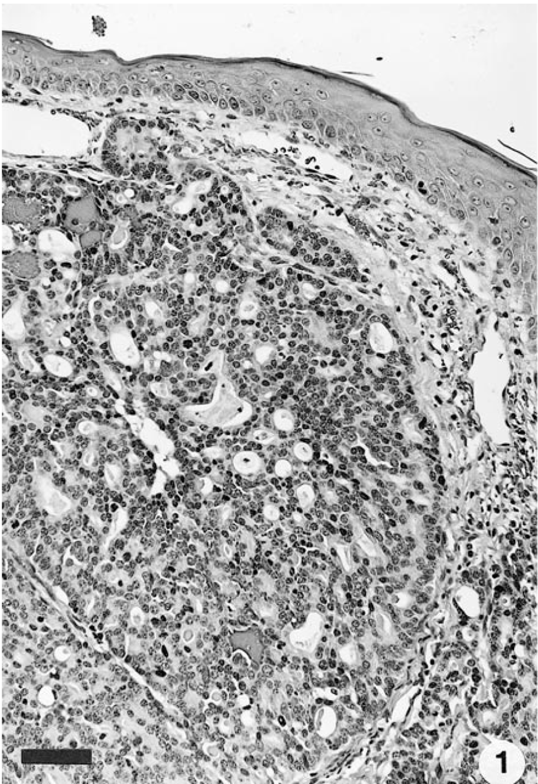
Mammary gland; squirrel monkey. Mammary adenocarcinoma composed of tubules and acini, infiltrating into adjacent dermis. HE. Bar = 80 μm.
The axillary mass was composed of epithelial cells in solid and tubular array supported by a variably thick fibrovascular stroma. It was similar in appearance to the previously excised mammary tumor and had effaced the architecture of the underlying lymph node (Fig. 2). No evidence of further metastasis was observed.
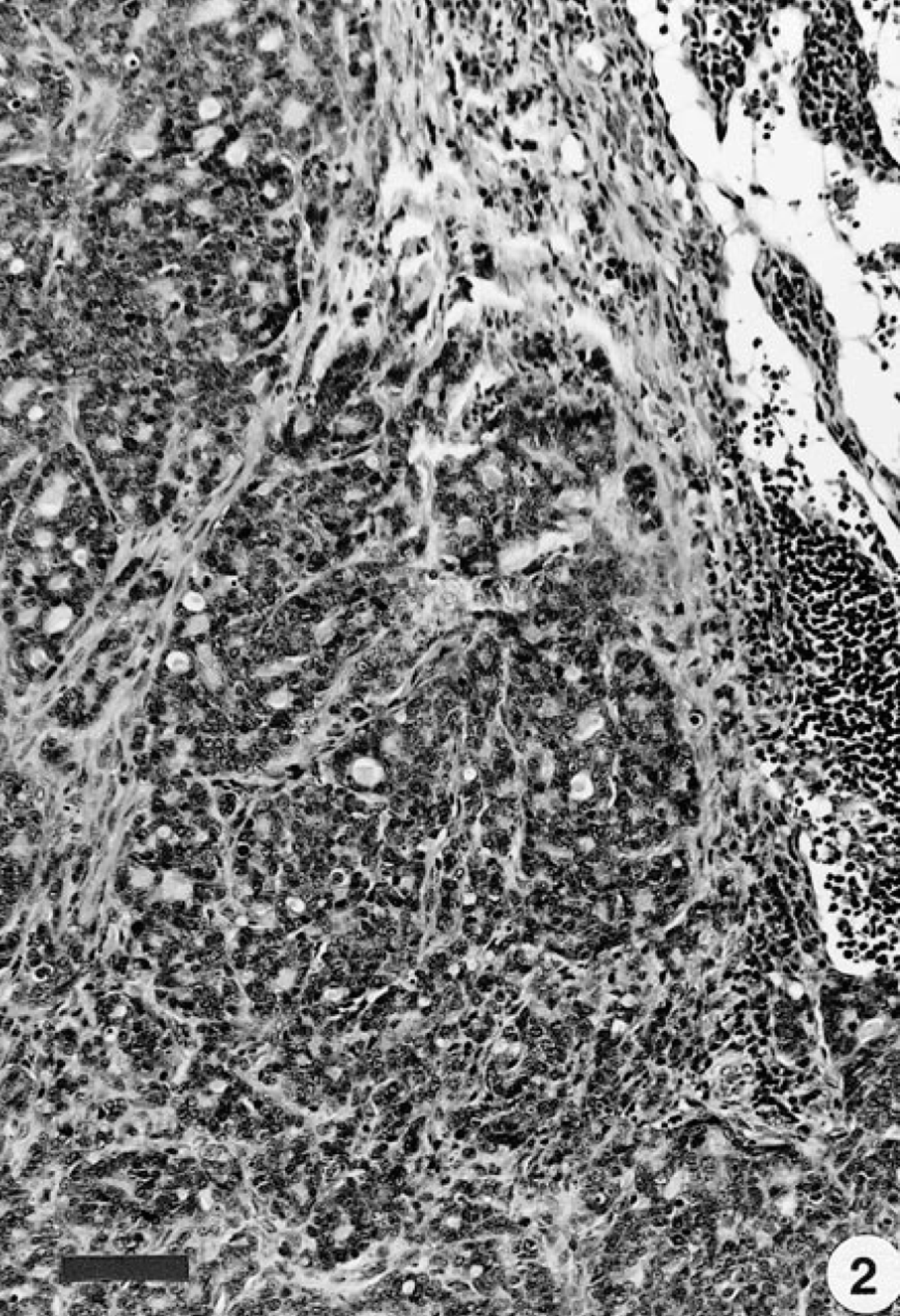
Lymph node; squirrel monkey. Normal architecture has been effaced by mass histologically similar to that found in mammary gland. HE. Bar = 110 μm.
The microscopic appearance of the neoplasm in this squirrel monkey is compatible with a low-grade infiltrating ductal carcinoma of humans. The tumor in humans has no consistent microscopic features. It may be composed of tubules, acini, or solid sheets of cells and may contain varying amounts of desmoplastic stroma. 5 Combinations of these patterns are most commonly observed. Infiltrating ductal carcinoma accounts for approximately 60% of all breast carcinomas and 70% of the invasive breast carcinomas in women. 15 Breast cancers in humans occur at a frequency of 1 male:100 female tumors. Affected men generally are of middle to advanced age. 21 The tumors in both men and women spread via the lymphatic and blood vascular systems. At the time of diagnosis, ductal carcinomas have spread to draining lymph nodes in approximately half of all patients. Hematogenous spread to nearly every organ in the body can occur. 5 The tumor of the monkey of this report is similar to human breast carcinomas in that it occurred in an aged male and had probably spread to the axillary lymph nodes at the time of diagnosis.
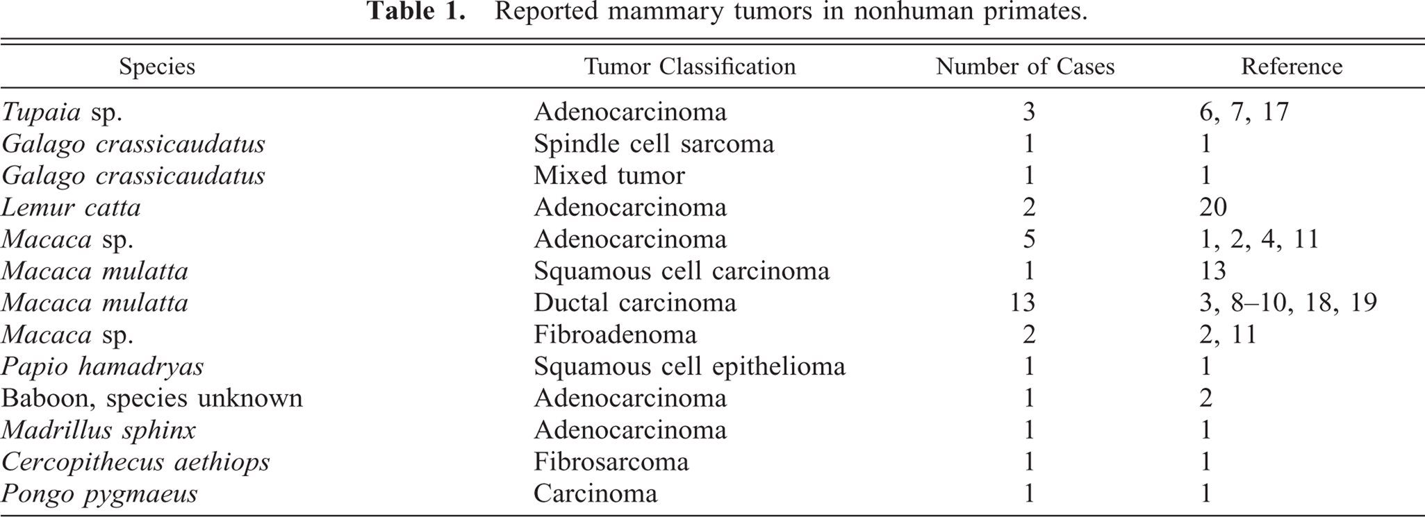
In contrast to the disease in humans, mammary adenocarcinomas in nonhuman primates are extremely rare. A literature search revealed only 33 cases, all of which occurred in female Old World primates (Table 1). The majority of the tumors arose spontaneously. Mammary tumors occurred in four macaques that were either treated with hormones or exposed to radiation. 3 4 8 10 However, a direct cause-and-effect relationship was not definitively demonstrated in any of these cases.
Reported mammary tumors in nonhuman primates.
The paucity of reports of mammary adenocarcinomas in nonhuman primates may be because the majority of the animals used in research are not kept for their entire life span. Of the previously reported cases in which ages were known, the majority of mammary tumors occurred in animals that were of middle age or older. The squirrel monkey of this report was an aged individual; the reported life span for this species is 20 years. 14 A low incidence of mammary tumors has been noted in long-term retrospective studies of necropsy records from zoological parks and primate centers, facilities that tend to house species for longer terms. 6 11 12 Thus, nonhuman primates may be genetically less susceptible than humans to development of mammary tumors.
Footnotes
Acknowledgements
We thank Dr. Gerald Berry (Department of Pathology, Stanford University School of Medicine) for histologic review of the glass slides. Financial support was provided in part by the National Institute of Mental Health (MH47573).