
Abstract
Mucopolysaccharidosis VII was diagnosed in a domestic shorthair cat from California. The cat was small and had multiple abnormalities, including a small body disproportionate to the size of the skull, angular deformities of the ribs, abnormally short forelimbs, luxating patellas, generalized epiphyseal dysplasia involving the vertebrae and long bones, cuboidal vertebrae, pectus excavatum, subluxation of both hips, osteosclerosis of the tentorium cerebelli and left petrous temporal bone, tracheal hypoplasia, and corneal clouding. b-Glucuronidase activity was markedly decreased in peripheral blood leukocytes. The cat died at 21 months of age, and a complete necropsy was performed. Tissues were examined by light and transmission electron microscopy. Large clear, round vacuoles representing distended lysosomes were present in many epithelial and connective tissue cells, including fibrocytes, chondrocytes, smooth muscle cells, hepatocytes, astrocytes, and macrophages.
The mucopolysaccharidoses (MPS) are a group of lysosomal disorders, each characterized by deficiency of one specific or multiple lysosomal enzymes that are necessary for degradation of glycosaminoglycans (GAGs). Enzyme deficiency leads to accumulation of GAG products in lysosomes of multiple organs. MPS VII (Sly syndrome) is characterized by a deficiency of β-glucuronidase activity, with accumulation of dermatan sulfate, heparan sulfate, and chondroitin sulfate. This disease has been described in humans, dogs, mice, and a single cat from Switzerland. 2–8, 12 Canine and murine animal models have been used to investigate enzyme replacement, bone marrow transplantation, and gene therapy as possible treatments of the disease. 1 9–11 Here, we describe the clinical and postmortem findings of MPS VII in a cat.
A male domestic shorthair cat from a feral litter in California was presented for evaluation at approximately 9 weeks of age. Multiple skeletal abnormalities were observed on physical examination. The kitten had a small body in proportion to the size of the skull, angular deformities of the ribs, abnormally short forelimbs, and luxating patellas. The kitten had mucopurulent rhinitis and conjunctivitis and was treated with antibiotics. When examined again at 16 weeks of age, the kitten had hindlimb weakness and difficulty walking. Whole body radiographs revealed generalized epiphyseal dysplasia involving the vertebrae and long bones, cuboidal vertebrae, pectus excavatum, subluxation of both hips, osteosclerosis of the tentorium cerebelli and left petrous temporal bone, and tracheal hypoplasia (Fig. 1). Corneal clouding developed in the left eye at 4 months of age and in the right eye by 9 months of age. The kitten was paraparetic at 6 months of age.
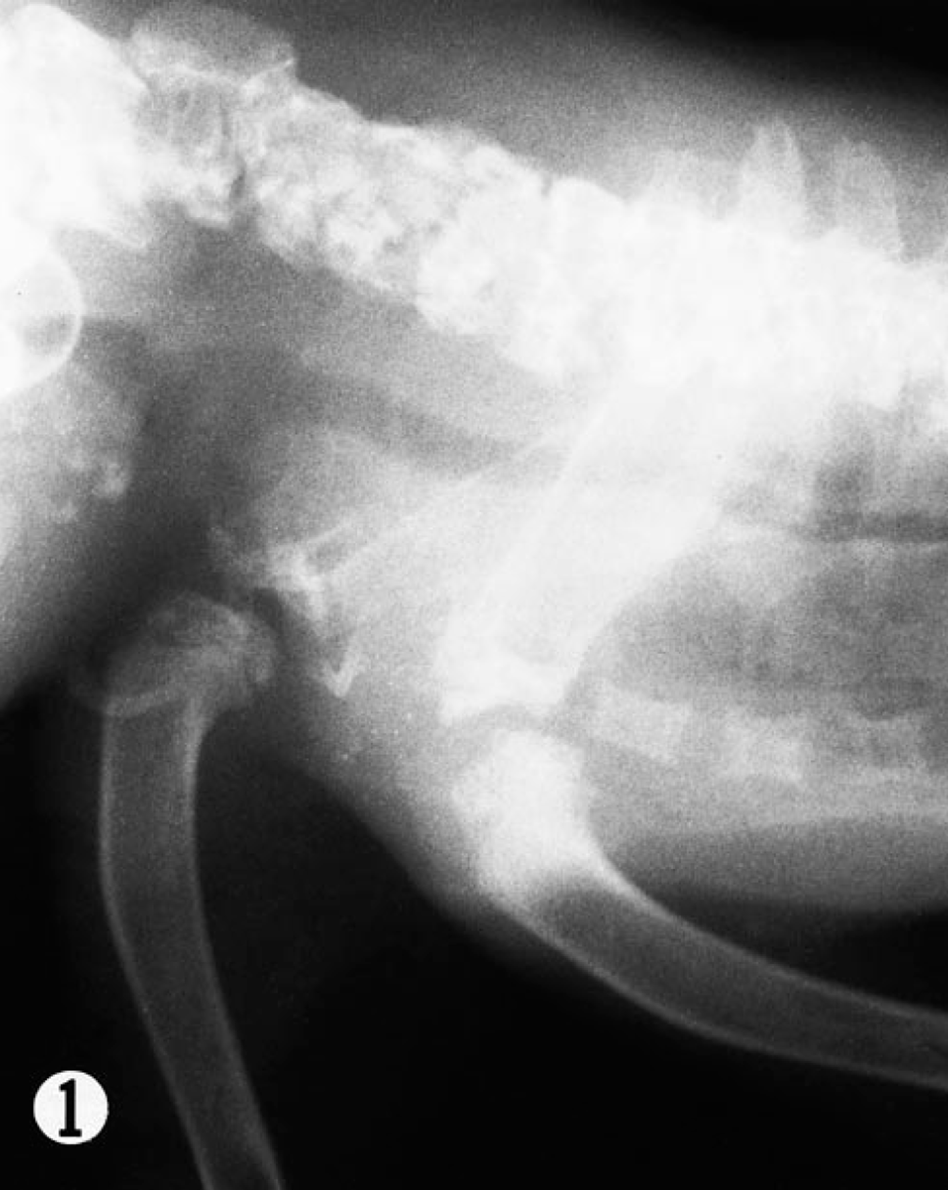
Cervical spine and shoulders; cat. Radiograph of 9-month-old cat with MPS VII. The cervical and thoracic vertebrae are cuboidal in shape and have irregular fragmented endplates. The proximal humeral epiphyses are hypoplastic, and both the humeral heads and their respective glenoid cavities are secondarily malformed. The humeral physes and sternebrae are also irregular. The trachea is hypoplastic and irregular in contour.
At 9 months of age, the cat weighed only 1 kg and had complete ankylosis of both tarsi and stifles, ventroflexion of the neck due to ankylosis of the cervical spine, corneal clouding in the right eye, and complete corneal opacification of the left cornea. At 12 months of age, the cat weighed 1.2 kg and corneal opacification had partially resolved. At 21 months of age, the cat was presented for decreased appetite and lethargy. The corneal clouding had nearly resolved but body weight was still only 0.9 kg. Two weeks later the cat died.
Examination of blood smears obtained at 8 months of age revealed granules in neutrophils and monocytes consistent with MPS VI, MPS VII, or GM2 gangliosidosis (Fig. 2). A complete blood count and biochemistry analysis performed at 1 year of age showed mild mature neutrophilia and lymphopenia (stress leukogram) and slightly increased blood urea nitrogen and serum cholesterol concentrations.
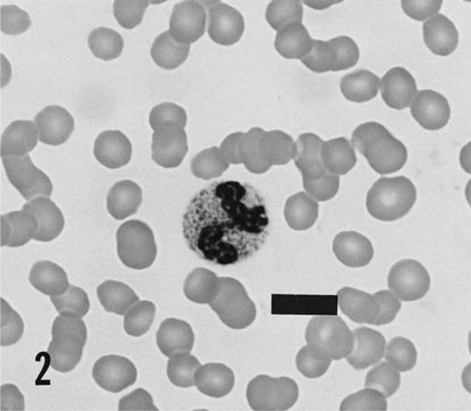
Blood smear; cat. Granules in the neutrophil are typical of mucopolysaccharidosis. Wright's stain. Bar = 7 μm.
Because the clinical features suggested a storage disease, enzyme activity was analyzed to make a definitive diagnosis. β-Glucuronidase activity was markedly decreased to 2.5 nmol/hour/mg protein in leukocytes (about 1% of normal), which identified the storage disease specifically as MPS VII. Activities of β-galactosidase, β-mannosidase, α-l-fucosidase, α-mannosidase, β-hexosaminidase, arylsulfatase A, α-l-iduronidase, α-glucosaminidase, and arylsulfatase B and sialic acid content of leukocytes were within normal limits.
Postmortem evaluation included a gross necropsy plus light and electron microscopic examinations. Tissues for routine light microscopic examination were fixed in 10% neutral buffered formalin, processed routinely, and stained with hematoxylin and eosin. Additional samples of liver, bone marrow, rib, lung, trachea, lymph node, heart valve, brain, ear, skin, cornea, and conjunctiva were fixed in 5% buffered glutaraldehyde, postfixed in 2% osmium tetroxide, and embedded in Spurr's plastic (Ted Pella, Redding, CA). For light microscopic examination of the plastic-embedded tissues, sections were cut at 1 μm, dried on a glass slide on a hot plate, and stained on 2 hot plate for 1 minute with 0.8% toluidine blue–0.2% pyronin Y dye in 0.8% sodium tetraborate. For electron microscopy, plastic-embedded brain, cornea, liver, lung, and trachea samples were sectioned to 900 Å on 200-mesh copper grids, stained with 1% uranyl acetate for 10 minutes and Reynold's lead citrate for 10 minutes, and examined using a Zeiss EM 109 transmission electron microscope.
Gross lesions at necropsy were consistent with the clinical and radiographic findings. Short forelegs, deformed ribs, lax coxofemoral joints, and stiff stifle, tarsal, and elbow joints were identified. The trachea was variably narrowed, the liver was enlarged, and the atrioventricular heart valves were thickened. Serous atrophy was present in the epicardium, mesentery, and other fatty connective tissues.
Light microscopic examination revealed swollen epithelial and connective tissue cells with cytoplasm that was pale and foamy or contained discrete clear, round vacuoles. The stroma of atrioventricular valves had a myxomatous appearance. Fibrocytes in the valves were swollen and had small clear, round cytoplasmic vacuoles. Fibrocytes in the dermis, corneal stroma, and bone marrow stroma had similar vacuoles. Smooth muscle cells in the aorta, coronary arteries, renal arcuate arteries, and other arteries had extensive vacuolization. Scattered smooth muscle cells in the muscularis layer of the jejunum contained vacuoles.
Chondrocytes in trachea, bronchi, costochondral junction, and ear cartilage were large round or oval cells with closely packed and distinct clear, round vacuoles filling the cytoplasm (Fig. 3). Perichondral fibroblasts also were vacuolated. The ground substance of the cartilage had a normal, deeply basophilic appearance.
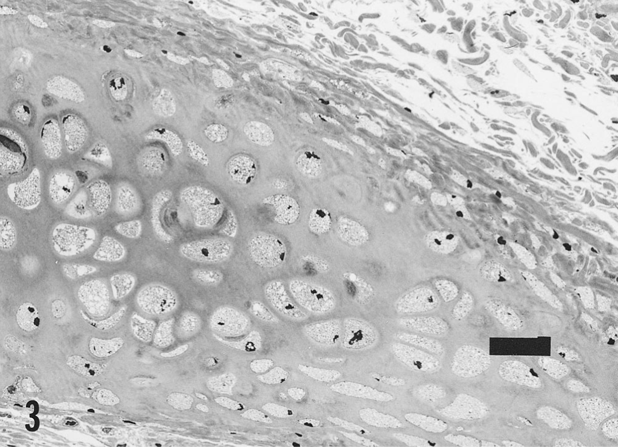
Tracheal cartilage; cat. Chondrocytes are swollen and the cytoplasm has many clear, round vacuoles, which represent enlarged lysosomes. The change is seen in the round central chondrocytes and the spindle-shaped perichondral chondrocytes. Toluidine blue. Bar = 40 μm.
All hepatocytes and Kupffer cells contained many distinct clear, round vacuoles (Fig. 4). Bile ducts had a normal histologic appearance. In the lungs, vacuoles were present in alveolar lining cells and alveolar macrophages. Vacuoles also were present in macrophages in bone marrow, lymph nodes, and subepithelial locations.
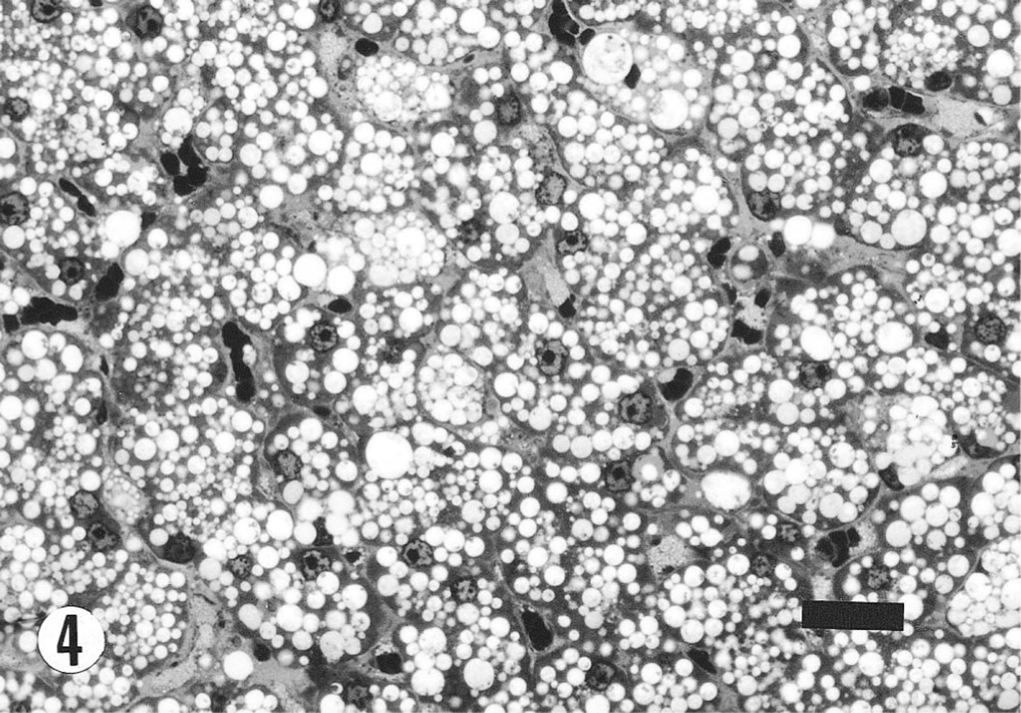
Liver; cat. Hepatocytes and Kuppfer cells contain many clear membrane-bound vacuoles. Toluidine blue. Bar = 40 μm.
In the cervical spinal cord, astrocytes were enlarged and had large clear vacuoles filling the cytoplasm. Similar vacuoles were present around small blood vessels in gray and white matter and between fibers of the nerve roots. The sciatic and other peripheral nerves had many large vacuoles in Schwann cells. In the brain stem, vacuoles were found only in fibrocytes around capillaries.
Seminiferous tubules of testes were uniform in diameter. Spermatogonia and spermatocytes were present, but no spermatids were observed. Tubules of the epididymis were lined by cuboidal epithelial cells, and the lumina contained no spermatozoa. No abnormalities were detected in pituitary gland, thyroid and parathyroid glands, or pancreas.
Electron microscopic examination of vacuolated cells revealed round membrane-bound structures scattered throughout the cytoplasm of affected cells (Fig. 5). These spaces were mostly electron lucent, with a slight amount of electron-dense fibrillar material scattered through them.
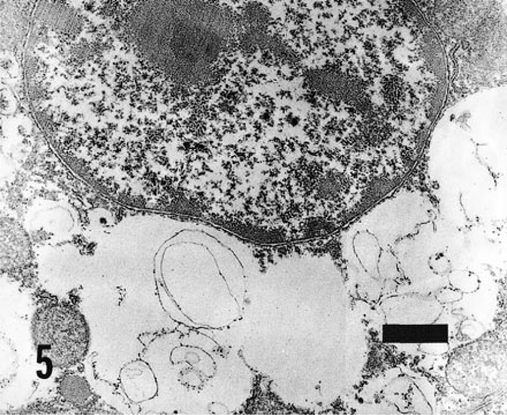
Electron micrograph. Hepatocyte; cat. Membrane-bound vacuoles in the cytoplasm, adjacent to the nucleus, are lysosomes distended with abnormal glycosaminoglycans. Uranyl acetate and lead citrate. Bar = 0.8 μm.
The ancestry of this feral kitten is unknown, but a similar feral kitten was identified earlier in the same geographic location. The other kitten was a female that was stunted, became nonambulatory at 5 months of age, and had corneal clouding and recurrent upper respiratory infections. That kitten died at 7 months of age, weighing approximately 1 kg. After MPS VII was diagnosed in the second kitten, it was suspected that the first kitten had the same disease, but this suspicion could not be confirmed. Because MPS VII is an autosomal recessive disease in other species, this may also be the case in cats. Inbreeding of a cat population that carried the genetic defect may have produced these two kittens.
If additional cats with MPS VII could be identified, they would be valuable models of an important human disease. The similarity of clinical signs, pathologic features, and enzyme activity level to MPS VII in humans would allow development of a feline model useful for studies of possible therapies, including gene therapy.
The definitive diagnosis in this cat was based on the decreased activity of β-glucuronidase. The combination of radiographic, clinical, and hematologic findings led to a presumptive diagnosis of MPS, but none of the findings in these three categories are specific enough to lead to a final diagnosis. Differential diagnoses of the radiographic findings include MPS, epiphyseal dysplasia, hypervitaminosis A, and congenital hypothyroidism. Corneal clouding may be present in various lysosomal storage diseases, and corneal edema or corneal dystrophy appear similar but are not caused by accumulation of storage products. The presence of metachromatic granules within neutrophils and lymphocytes is suggestive of MPS or GM2-gangliosidosis. This case is consistent with MPS VII in other species 6 and with the previous report of MPS VII in a single cat in Switzerland. 2 Although MPS I and MPS VI have been identified in cats in the United States, 5 7 this is the first report of MPS VII.