
Abstract
Primary cardiac neoplasms are rare in humans and animals. In humans, the most common primary cardiac tumor is the myxoma, which is frequently found in the left atrium. Cardiac myxoma has been reported in the dog but not in the cat. We describe the gross, immunohistochemical, and light microscopic examination of a myxoma in the right atrium of a 6-year-old domestic shorthair cat. Histologically, the tumor consisted predominantly of mesenchymal cells with several foci of bone and cartilage present. The tumor was encapsulated and benign.
Primary cardiac neoplasms are rare in humans and animals. 3 5 In cats, primary tumors of the heart are extremely rare. 6 Myxomas are the most common primary cardiac tumor in humans and are predominantly found in the atria, particularly the left atrium. 4 5 Cardiac myxoma has been reported in the dog 2 but not in the cat.
A 6-year-old male domestic shorthair cat was presented for anorexia and lethargy of 2 days duration. He died suddenly at the onset of physical examination. At necropsy, a 6 × 11 ×18-mm firm multilobulated mass was found originating from the endocardial free wall of the right atrium and almost completely obliterating the right atrial chamber (Fig. 1). The mass had a smooth, glossy, pale gray-white surface, and on cut section it was white and gritty. The remainder of the heart and all other organs were grossly normal.
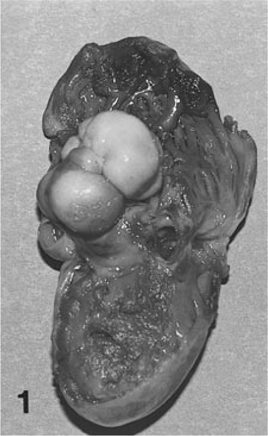
Right atrium; cat. Multilobulated mass attached to the right atrial endocardium.
Tissue samples from all major organs were fixed in 10% neutral buffered formalin, embedded in paraffin, sectioned at 5 μm, and stained with hematoxylin and eosin (HE). Sections of the mass from the right atrium were also stained with alcian blue. Immunohistochemical procedures for the identification of actin, desmin, cyokeratin, vimentin, and α−1 antichymotrypsin were also performed.
Histologically, large areas of the mass were composed of mesenchymal cells, many stellate and others spindloid, in a matrix of loosely arranged collagen fibers and mucin (Fig. 2). Several foci of osteoid, cartilage, and bone were present. There was partial mineralization of cartilage. The bone was both intramembranous and endochondral (Fig. 3). The areas of cartilage formation were surrounded by loose areolar tissue in which the stellate cells morphologically resembled primitive mesenchymal cells. In some areas, there were dense bands of collagen, which divided foci of loose areolar tissue. The mass was surrounded by an incomplete fibrous capsule, which varied in thickness and from which smaller bundles of fibrous tissue extended into the mass (Fig. 3). The mass did not invade the adjacent myocardium. No mitotic figures, cellular or nuclear pleomorphism, or necrosis were present.
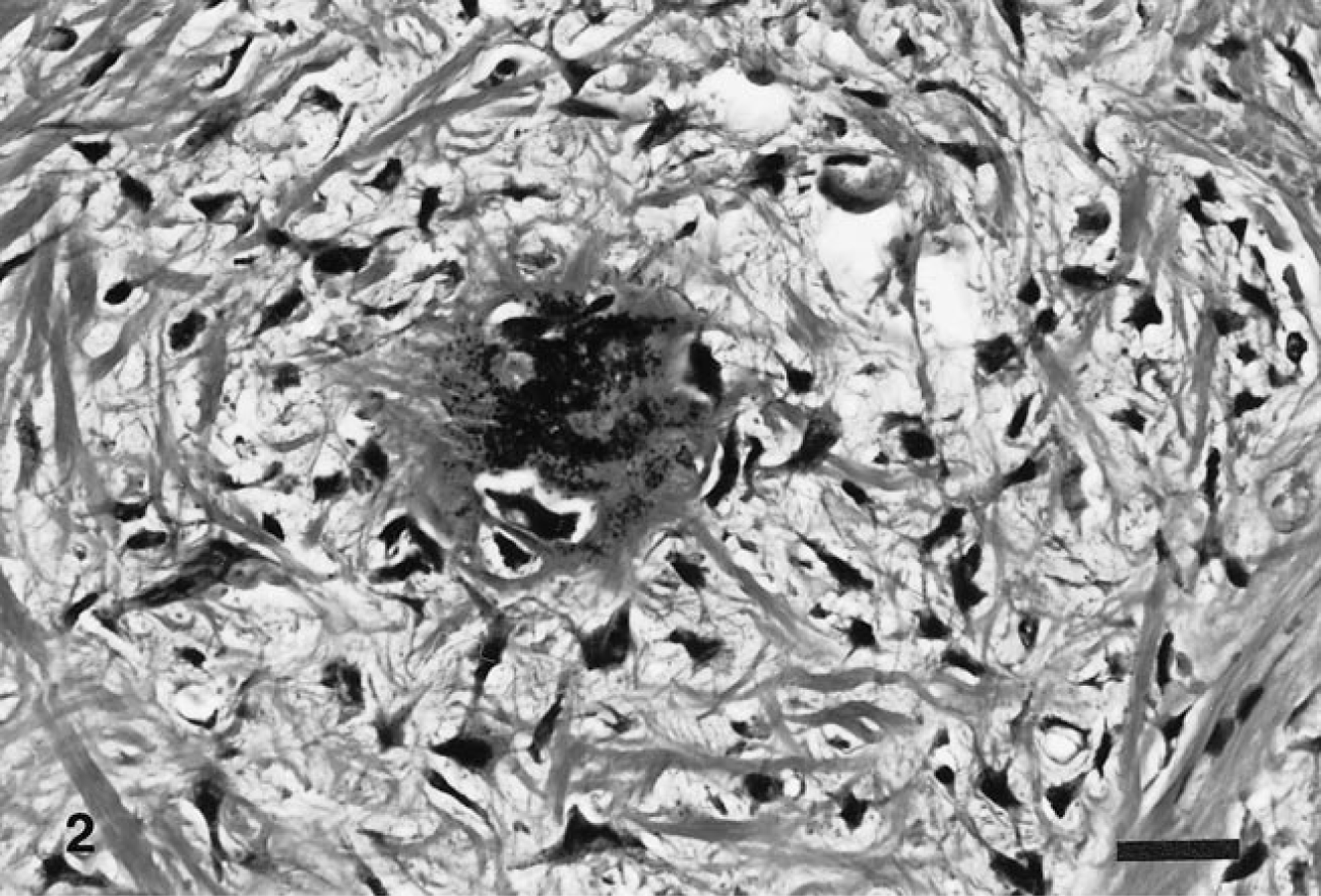
Right atrial mass; cat. Mesenchymal cells in a matrix of collagen fibers and mucin with a focus of mineralization. HE. Bar = 25μm.
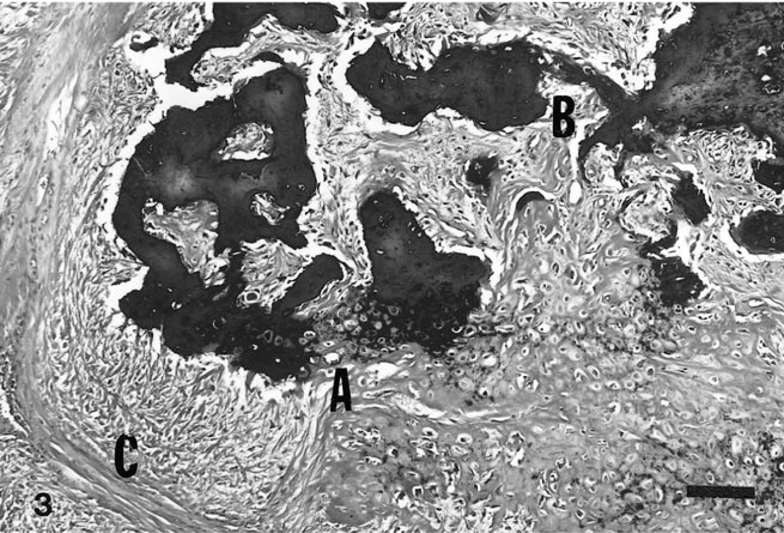
Right atrial mass; cat. Note cartilage with endochondral ossification (A) and intramembranous ossification (B) and subdivision of the tumor by collagen (C). HE. Bar = 100 μm.
The tumor stained intensely with alcian blue (pH 2.5); staining intensity was markedly diminished with hyaluronidase. Cells within the tumor were positive for actin, vimentin, and α-1 chymotrypsin and negative for cytokeratin and desmin, consistent with mesenchymal cells. Based on the histologic features, staining properties, and the presence of cartilage and bone, a diagnosis of ossifying myxoma was made.
Differential diagnoses included chondroma, osteochondroma, and chondromyxoid fibrosarcoma. All of these tumors, however, arise from bone. Extraskeletal chondromas and myxoid chondrosarcomas in humans arise in the soft tissues of the appendicular skeleton and thus were eliminated from consideration in this cat. Likewise, chordomas arise from notochord remnants along the spine or from the intervertebral discs and are strongly positive for cytokeratin.
Ultrastructural and histochemical studies have led to the suggestion that myxomas arise from a primitive mesenchymal cell with the potential for multidirectional differentiation. 4 Although these tumors are generally benign and are successfully removed in humans, they will occasionally metastasize.
Myxomas in humans are among the most common primary tumors of the heart, comprising 50% of primary cardiac neoplasms. In addition to stellate cells in an acid mucopolysaccharide–rich ground substance, they may contain cartilage, bone, foci of mineralization, Gamna-Gandy bodies, hematopoietic tissue, thymic and foregut remnants, and occasionally mucous glands. 4 They may be sporadic or familial. 1 The sporadic form occurs principally in middle-age women as a single tumor. The familial form occurs in younger persons with a nearly equal sex distribution. One third of familial tumors are multicentric. Unlike the sporadic form, familial cardiac myxomas are associated with a variety of extracardiac intercurrent conditions, including other tumors and Cushing's disease termed, Carney's syndrome.
Footnotes
Acknowledgements
The technical support provided by Mr. Richard Spence and Ms. Jane Chladny is appreciated.