
Abstract
Histopathologic and immunohistochemical examinations were performed on localized amyloidosis associated with mammary tumors in two dogs. These tumors were identified as adenoma and adenocarcinoma. An acellular, amorphous pale eosinophilic material (amyloid) was observed in the lumina of acini lined by neoplastic cells and in the stroma of the tumors. Concentrically laminated pale eosinophilic bodies (corpora amylacea) were also found in the lumina of the acini. Amyloid and corpora amylacea stained positively with Congo red with and without 5% potassium permanganate pretreatment and revealed a green birefringence under polarized light. Corpora amylacea showed an occasional Maltese-cross pattern. Immunohistochemically, amyloid and corpora amylacea usually stained positively with anti-bovine α-casein antibody but negatively with anti-human amyloid AA, anti-bovine κ-light and λ-light chains, anti-human lactoferrin, anti-human transferrin, anti-human secretory component, and anti-human polyglucosan antibodies. These findings suggested that the amyloid deposition in these canine mammary tumors was related to lactating casein.
Amyloid is a pathologic proteinaceous substance that produces a collection of diverse clinical syndromes characterized by the extracellular deposition of amorphous, congophilic protein within tissues. Amyloid proteins are distinguished by their twisted β-pleated sheet-fibrillar ultrastructure and are made up of nonbranching fibrils of indefinite length and a width of approximately 7.5–10 nm.3 Amyloidosis is divided into systemic and localized forms based on the localization of the amyloid.3 The systemic form is comprised of three types: AL amyloid associated with primary amyloidosis or immunoglobulin dyscrasias, AA amyloid associated with chronic inflammatory processes, and AF amyloid associated with heredofamilial amyloidosis. The two most common types of localized amyloidosis are AE amyloid in the endocrine organs either associated or not associated with neoplastic conditions and AS amyloid associated with senile cardiomyopathy and cerebral plaques (paired helical filaments; β-protein).3 Furthermore, localized nodular deposits of amyloid have been described in such sites as the human mammary gland,8 gastrointestinal tract,15 genitourinary tract,4 and skin.17 In animals, localized amyloidosis associated with a neoplastic condition is uncommon but has been described in pancreatic endocrine tumor,10 medullary thyroid carcinoma,6 calcifying epithelial odontogenic tumor,11 ameloblastoma,5 and plasmacytoma.14
A description of localized amyloidosis associated with canine mammary tumors was published in 198516 and included the histochemical and ultrastructural characteristics of amyloid deposited in the stroma of tumors and the corpora amylacea containing amyloid. Here, we describe the histochemical and immunohistochemical nature of amyloid associated with mammary tumors in two dogs.
An 11-year-old mixed-breed female dog (No. 1) had a neoplastic mass (2 × 3 × 2 cm) in the left mammary gland. The mass was surgically excised for routine histopathologic examination. A 12-year-old mixed-breed female dog (No. 2) had two neoplastic masses (3 × 4 × 2 cm, 1 × 2 × 2 cm) in the left mammary gland. These tumors were surgically excised for histopathologic examination. Six months later, the tumors reccurred in the left mammary gland, and metastasis in the lungs was observed by radiographic analysis. This dog was necropsied immediately after natural death.
Macroscopically, the neoplastic mass in dog No. 1 was grayish white, somewhat firm, and well circumscribed. In dog No. 2, two primary masses and a recurrent neoplastic mass were somewhat firm and poorly circumscribed. On cut surface, the masses were well lobulated and grayish white and contained multiple small cysts. Metastatic neoplasms in the lungs were unencapsulated, hard, and homogeneously white. No significant changes were observed in the other main visceral organs.
Tissue samples were collected from the mammary neoplasm of dog No. 1 and from the primary and recurrent mammary neoplasms and metastatic neoplasms in the lungs of dog No. 2. Tissues were fixed in 10% neutral buffered formalin and embedded in paraffin.
Histopathologically, the neoplasm of dog No. 1 was diagnosed as adenoma of the mammary gland. In dog No. 2, two primary neoplasms were diagnosed as a papillary adenocarcinoma and a benign mixed tumor. A recurrent neoplasm identified as a papillary adenocarcinoma resulted in metastasis to the lungs.
The adenoma of dog No. 1 consisted of a well-demarcated proliferation of well-differentiated cuboidal or columnar epithelium in an acinar or lobular pattern. In some areas, an acellular amorphous pale eosinophilic material (amyloid) was seen in the lumina of the acini lined by neoplastic cells and in the stroma of the tumors (Fig. 1). Concentrically laminated pale eosinophilic bodies (corpora amylacea) were found in the lumina of the acini.
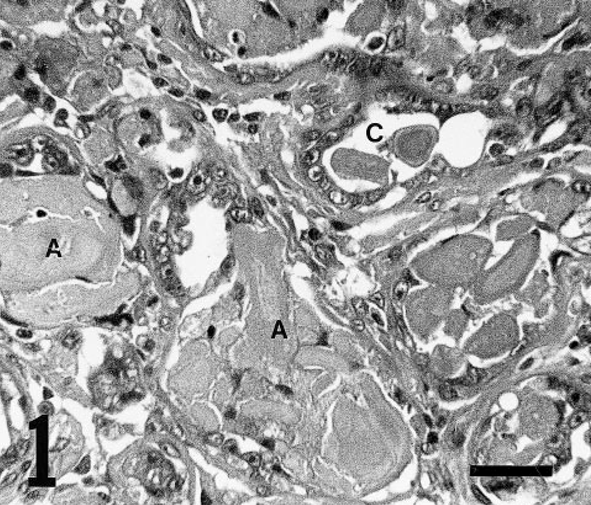
Mammary adenoma; dog No. 1. Adenoma consists of a well-demarcated proliferation of well-differentiated cuboidal or columnar epithelium in an acinar pattern. An acellular amorphous pale eosinophilic material (A) is seen in the lumina of the neoplastic acini and stroma of the tumors. Corpora amylacea (C) are found in lumina of the acini. HE. Bar = 30 μm.
In dog No. 2, a primary and recurrent adenocarcinoma was characterized by papillary, tubular, partly solid proliferations of atypical cuboidal to columnar epithelium. Amyloid was seen in the lumina of the acini and in the stroma of the tumors (Fig. 2). Corpora amylacea were also found in the acini of the tumors. Amyloid, however, was not observed in a metastatic neoplasm in the lungs. In the mixed tumor of dog No. 2, neither amyloid nor corpora amylacea was observed.
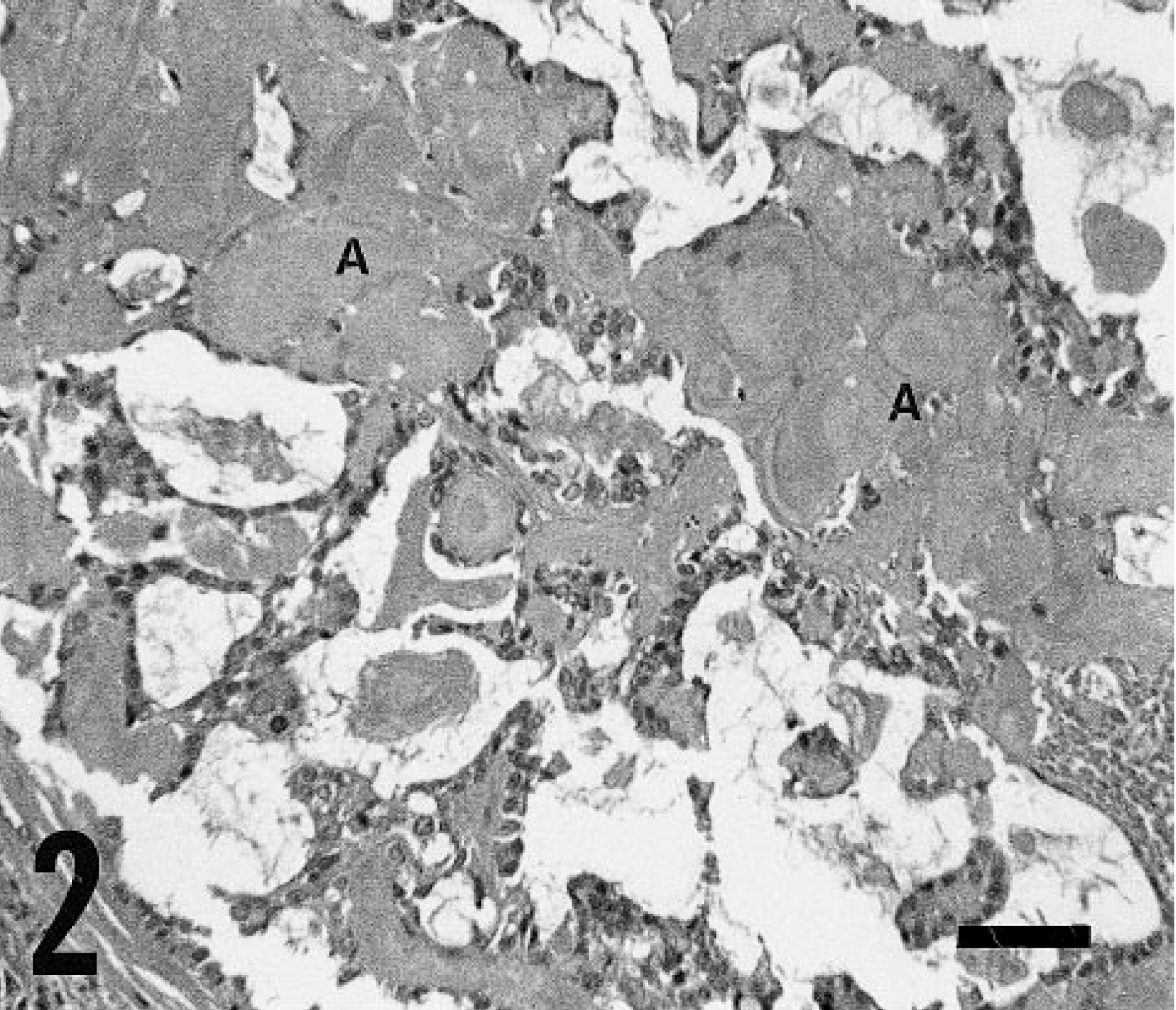
Mammary carcinoma; dog No. 2. Adenocarcinoma is characterized by a papillary proliferation of atypical cuboidal to columnar epithelium. An acellular amorphous pale eosinophilic material (A) is seen in the stroma of tumor. HE. Bar = 50 μm.
Amyloid in the lumina and stroma of both the adenoma and adenocarcinoma stained positively with periodic acid–Schiff (PAS) with and without diastase digestion. Amyloid also stained positively with Congo red with and without 5% potassium permanganate pretreatment and had a green birefringence under polarized light. The corpora amylacea were also positive with the PAS reaction with and without diastase digestion and also stained positively with Congo red, revealing a green birefringence in an occasional Maltese cross pattern. Amyloid was not detected in the liver, kidneys, spleen, heart, adrenal gland, or small and large intestines of dog No. 2.
Histologic sections were used for immunohistochemical analysis with the avidin–biotin–peroxidase complex (ABC) procedure (Vectastain Elite ABCkit, Vector Laboratories, Burlingame, CA) and counterstained with hematoxylin. Control incubations using excess antigen against immune or nonimmune sera as the first antibody and omitting the first antibody resulted in the absence of specific staining.
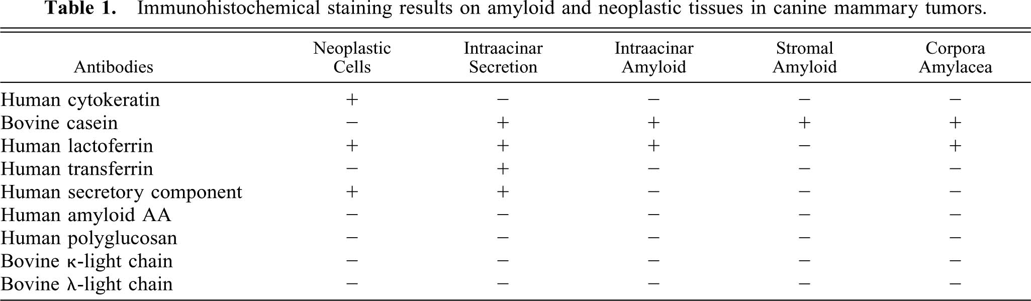
The immunohistochemical properties of the neoplastic cells, the amyloid, and the corpora amylacea are prevented in Table 1. Neoplastic epithelial cells of adenoma and adenocarcinoma stained positively with anti-human lactoferrin (hLF; Advanced Immuno Chemical, CA, USA), anti-human cytokeratin (DAKO, Glostrup, Denmark), and anti-human secretory component (hSC; DAKO) antibodies. Secretions in the lumina of normal mammary glands reacted to anti-hLF, anti-human transferrin (hTF; Bethyl Lab., USA), anti-hSC, and anti-bovine α-casein (bα-CA; Cosmo Bio, Tokyo, Japan) antibodies. Amyloid in the lumina of neoplastic acini in both adenoma and adenocarcinoma stained positively with anti-hLF and bα-CA antibodies. Amyloid in the stoma, however, reacted to anti-bα-CA antibody (Fig. 3). The corpora amylacea were stained with anti-hLF and bα-CA (Fig. 4) antibodies. Amyloid and corpora amylacea did not react to anti-amyloid AA (Kyowa Medix, Tokyo, Japan), anti-bovine κ-light and λ-light chains (bκ-LC, bλ-LC; VMRD Inc., USA), or anti-human polyglucosan (hPG; donated by Dr. T. Yokota, Yamaguchi, Japan) antibodies.
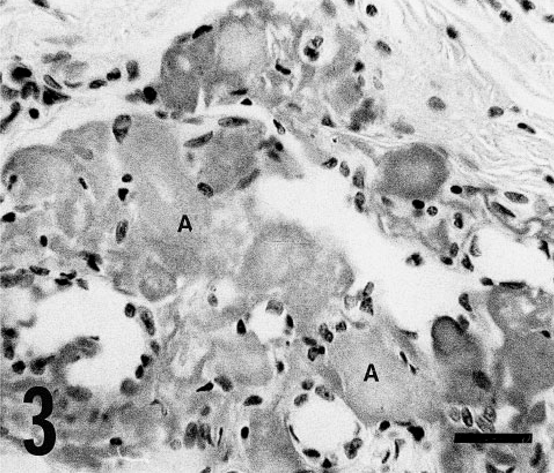
Mammary carcinoma; dog No. 2. An acellular amorphous pale eosinophilic material (A) in the stroma of the tumor is positive against anti-bovine α-casein antibody. Avidin–biotin–peroxidase complex (ABC) method, Mayer's hematoxylin counterstain. Bar = 30 μm.

Mammary carcinoma; dog No. 2. Corpora amylacea (C) in the lumina of the acini are stained positive against anti-bovine α-casein antibody. Avidin–biotin–peroxidase complex (ABC) method, Mayer's hematoxylin counterstain. Bar = 30 μm.
Immunohistochemical staining results on amyloid and neoplastic tissues in canine mammary tumors.
The histopathologic and histochemical findings of four canine mammary neoplasms that contained amyloid, either as amorphous deposits between neoplastic cells or in the tumor stroma or as amyloid-containing corpora amylacea, have been previously described.16 These neoplasms were two solid and two tubular carcinomas. Acellular amorphous pale eosinophilic material was seen both between neoplastic cells and in the surrounding connective tissue in two tubular carcinomas. The material also stained positively with Congo red and showed a green birefringence, consistent with the properties of amyloid proteins. Amyloid-containing corpora amylacea in an occasional Maltese-cross pattern were found in both tubular neoplasms. However, amyloid was not detected in the solid carcinomas. Amyloid was not of the AA type because of its resistance to pretreatment with potassium permanganate. Histochemically, positive staining with Adam's stain suggested that tryptophan may be contained in the amyloid.
In the present study, a mammary adenoma (dog No. 1) and primary and recurrent papillary adenocarcinomas (dog No. 2) contained an acellular amorphous material showing positive staining with Congo red and a green birefringence under polarized light. This material was resistant to pretreatment with potassium permanganate and was localized in the lumina of the neoplastic acini and in the stroma of the tumors. Thus, localized amyloid deposits in the present mammary tumors were presumably not AA-type amyloid proteins. Immunohistochemically, amyloid material in the lumina of neoplastic acini and in the stroma reacted positively to anti-bα-CA antibody. Immunohistochemical analysis has revealed that transferrin and lactoferrin are present in the epithelial cells and milk of human mammary glands.9 However, stromal amyloid in these dogs was not stained with anti-hTF and anti-hLF antibodies.
The amyloid in the previously described mammary tumors contained tryptophan.16 Tryptophan is a very well known aromatic amino acid contained in casein, which is a major component of lactating proteins. Casein consists of fractions of α-, β-, γ, and κ-casein. α-Casein is easy to precipitate in the presence of calcium ion.1,2 Therefore, the histochemical data16 may indirectly indicate that the amyloid proteins in those canine mammary tumors were related to the lactating casein, as was the case in the present study.
The concentrically laminated pale eosinophilic bodies in the tumors from dog Nos. 1 and 2 had the characteristic features of amyloid-containing corpora amylacea, as revealed by histologic and immunohistochemical examinations. Corpora amylacea of bovine mammary glands are formed with an aggregation of casein micelles and are positively stained with Congo red.2 Therefore, amyloid-containing corpora amylacea and stromal amyloid may also be derived from the lactating casein. Bovine and rat mammary glands have frequently shown corpora amylacea composed of amyloid protein.1,2,13 However, neuronal corpora amylacea are common in human beings and dogs and are composed of polyglucosan.7 Using anti-hPG and anti-bα-CA antibodies in the present study, immunohistochemical staining revealed that the corpora amylacea did not contain polyglucosan but rather contained casein of an amyloid protein.
In human beings, primary localized amyloid has been reported within a breast tumor.12 Amyloid tumor (amyloidoma) in the breast was reported in a few patients.8 However, these amyloidomas were thought to be unrelated to neoplastic conditions because such “tumors” consisted of AL amyloid. The pathogenesis underlying solitary amyloid tumors is still unknown. Chronic inflammation with infiltration of lymphocytes, plasma cells, and multinucleated giant cells are postulated to be symptomatic of amyloid tumors.8 In the mammary tumors in dog Nos. 1 and 2, amyloid deposition occurred with no relationship to mild infiltrates of lymphocytes, which were found in some areas of the tumors. Furthermore, immunohistochemical examination for AL amyloid did not demonstrate immunoreactivity to anti-bκ-LC and anti-bλ-LC antibodies. In a localized form of amyloidosis associated with endocrine tumors, amyloid usually consists of part of a hormone or prohormone product.3,10 Abnormalities in hormone synthesis, processing, secretion, or degradation by neoplastic cells may play a role in the pathogenesis of amyloidosis.10 In the present canine mammary tumors, amyloid was localized in the neoplastic tissues but not in normal mammary glands. Therefore, we presume that the development of amyloid protein was induced by abnormalities in synthesis, processing, secretion, or degradation of lactating casein in neoplastic epithelial cells.