
Abstract
Pheochromocytomas are uncommon neoplasms of the adrenal medulla that are most frequently reported in rats and select mouse strains. In many cases, especially those in man, pheochromocytoma is associated with familial tumor syndromes, because of inherited mutations in a variety of proto-oncogenes and tumor suppressor genes. Nonhuman primates are valuable animal models for a variety of human diseases because of their similar anatomy and physiology; however, cases of pheochromocytomas have only rarely been reported in nonhuman primates. Herein, we characterize the gross, histologic, and immunohistochemical features of pheochromocytoma in 6 cotton-top tamarins (Saguinus oedipus). Pheochromocytomas represented 6 of 114 of the total causes of death in the studied population (5.3%) and corresponded to 16% of the total number of neoplasms. The average age of affected animals was 17.9 years. On histologic examination, all cases were defined by tight bundles, nests, and cords of neoplastic chromaffin cells. All cases had concurrent myocardial degeneration and fibrosis of varying severity and chronicity. Three of the cases (50%) also had hyalinization and medial thickening of coronary arteries consistent with hypertension. On immunohistochemical examination, 6 of 6 of the cases (100%) stained positively for chromogranin A, synaptophysin, N-CAM (or CD56), and protein gene product 9.5. None of the cases stained for glial fibrillary acidic protein. Pedigree analysis revealed inter-relatedness in 4 of 6 animals, with progenitor animals also affected with pheochromocytomas. The tumors in this population illustrate comparable histologic and immunohistochemical staining patterns with cases in other laboratory animals and humans, and, therefore, may indicate common underlying genetic alterations that precipitate tumor development.
Keywords
The adrenal gland is an anatomically simple organ divided into a cortex and medulla, with the latter populated predominately by specialized chromaffin cells that are derived from the neural crest and a small population of support (sustentacular) cells.10 The chromaffin cells synthesize and secrete various catecholamines, including epinephrine and norepinephrine, which lead to a variety of physiologic effects, including increased cardiac contraction and vasoconstriction.10 The most common pathology associated with the adrenal medulla is the genesis of tumors, including pheochromocytomas and neuroblastomas.5 Pheochromocytomas are variably functional and can secrete epinephrine, leading to a variety of hypertensive and myocardial degenerative effects.16 Microscopic findings typically include an alveolar or trabecular pattern, and, in general, cells resemble normal medullary chromaffin cells. Tumors can contain small numbers of ganglion cells, neuroblasts, and melanin-containing cells; however, these cell types are not predominate in cases of pheochromocytoma.14
Although pheochromocytomas are considered rare in humans (estimated incidence of 1/300,000), considerable interest and research has been focused on developing a suitable animal model to study pheochromocytomas.11, 17 Both knockout mice and rats have been extensively used in mechanistic studies of the genesis of spontaneous and genotoxic pheochromocytoma.17, 18 Nonhuman primates serve as an excellent model for many human diseases because of their similar physiology and pathology; however, the incidence of pheochromocytomas in nonhuman primates is not currently known. A solitary case of an angiomatous pheochromocytoma was reported in a Rhesus macaque (Macaca mulatta).19 Endocrine neoplasia has been reported slightly more frequently in New World primates, with tumors arising within the adrenal gland reported most commonly. These include pheochromocytomas in 3 golden lion tamarins (Leontopithecus rosalia rosalia), 2 mantled howler monkeys (Aloutta villosa), 1 cotton-top tamarin (Saguinus oedipus), and 1 brown spider monkey (Ateles fusciceps).2, 3 Only the brown spider monkey and the cotton-top tamarin were reported to have concurrent myocardial degeneration, presumably from epinephrine excess. Cases of solitary adrenal cortical adenoma have been reported in a single black-tailed marmoset and a single cotton-top tamarin.3, 4
Immunohistochemistry is commonly used to differentiate adrenal medullary tumors from cortical or extra-adrenal tumors.11, 12, 16, 20 Markers commonly used to differentiate pheochromocytomas include chromogranin A (CGA), protein gene product (PGP) 9.5, synaptophysin (SYN), CD56 (also known as neural cell adhesion molecule or N-CAM), and glial fibrillary acidic protein (GFAP). CGA is one of the most specific neuroendocrine immunohistochemical markers currently available, is a part of catecholamine-containing secretory granules, and reliably stains adrenal medullary cells.4, 7, 11, 12, 20 PGP 9.5 forms a portion of the ubiquitin-proteasome system and is highly specific to neurons and cells of the neuroendocrine system.7, 11, 13, 20 SYN is a glycoprotein expressed in the presynaptic vesicles of neurons as well as diffusely in the neuroendocrine system.11, 12, 20 N-CAM is involved in the morphogenesis of neural epithelial cells and is a cohesive cell adhesion molecule found in peripheral and central nervous system cells.8, 11 GFAP is a class III intermediate filament and, although considered relatively specific for astrocytes and neural stem cells, has rarely been used as an immunomarker that stains a subpopulation of sustentacular cells. It is not universally positive in cases of pheochromocytoma.1, 11
Herein, we characterize a cohort of 6 cotton-top tamarins that developed spontaneous pheochromocytomas and that had histologic evidence of myocardial degeneration and hypertension. In addition, we use immunohistochemistry for CGA, SYN, PGP 9.5, N-CAM, and GFAP to compare these tumors with published human cases.
Materials and Methods
A relational database of necropsy records at the New England Primate Research Center (NEPRC) was searched from 2000 to 2008 for adrenal pathology in cotton-top tamarins. From the cases chosen, the case criteria was further refined to the diagnosis of pheochromocytoma, which resulted in 6 recognized cases. There were no tumors other than pheochromocytoma recorded. All animals were necropsied within 2 hours of death, and representative sections of all major organs were collected, fixed in 10% neutral buffered formalin, embedded in paraffin, sectioned at 5 μm, and stained by using HE. Additional sections were prepared for immunohistochemistry. A pedigree analysis to examine the relationship between affected animals was performed by using Pedigraph, a software program developed by the Department of Animal Science, University of Minnesota.
To characterize the neoplasms by using immunohistochemical methods, we used standard immunoperoxidase staining for SYN, CGA, PGP 9.5, NCAM, and GFAP. Formalin-fixed, paraffin-embedded sections were deparaffinized, rehydrated, and subsequently blocked with hydrogen peroxide. Pretreatment for GFAP involved using proteinase K, whereas pretreatment for PGP 9.5, SYN, NCAM, and CGA involved microwaving for 20 minutes in 0.01 M citrate buffer followed by 20 minutes of cooling. All steps were followed by a tris-buffered saline solution wash. Before the application of primary antibodies, all slides were treated with Dako protein block for 10 minutes. Sections were incubated with anti-human CGA (Dako, Carpinteria, CA, USA; polyclonal, 1:1000, 30 minutes at room temperature), anti-mouse SYN (Dako; monoclonal, 1:50, overnight in a refrigerator), anti-cow GFAP (Dako; polyclonal, 1:500, 30 minutes at room temperature), anti-human NCAM (Zymed, San Francisco, CA, USA; monoclonal, 1:500, overnight in a refrigerator), and anti-human PGP 9.5 (Biomeda, Foster City, CA, USA; monoclonal, 1:400, overnight in a refrigerator). Slides were then incubated with secondary antibody biotinylated goat anti-rabbit (Vector Laboratories, Burlingame, CA, USA; 1:200, 30 minutes at room temperature) for CGA and GFAP and biotinylated horse anti-mouse (Vector Laboratories, 1:200, 30 minutes at room temperature) for SYN, NCAM, and PGP 9.5. This was followed by 30 minutes of incubation at room temperature with Vectastain ABC Elite (Vector Laboratories) (CGA, GFAP, and SYN) or Vectastain ABC standard (Vector Laboratories) (NCAM, PGP 9.5). All the slides were developed with DAB chromagen (Dako) and counterstained with Mayer's hematoxylin. In all cases, step sections were incubated with isotype-specific irrelevant antibodies for negative controls. Positive controls consisted of sections of small intestine (PGP 9.5), brain (GFAP), and adrenal gland (SYN, CGA, NCAM) from age-matched cotton-top tamarins.
Results
Animals
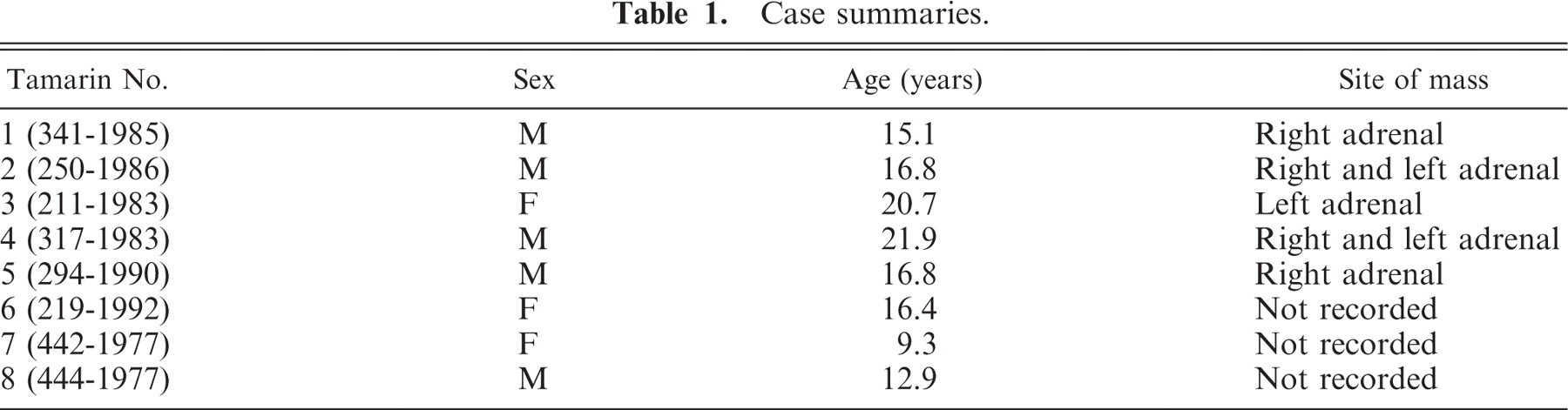
From 2000 to 2008, 114 adult (older than 1 year old) cotton-top tamarins were necropsied at the NEPRC. Of these, 37 had malignant neoplasms, with pheochromocytomas corresponding to 6 of the total number of neoplasms recognized (16%) (Table 1). Pheochromocytomas represented 5.3% of the total number of deaths in this population during the defined time period. There were 4 male and 2 female animals affected. The average age at the time of death was 17.9 years (range, 15.1–21.9 years). After identification of affected animals, a pedigree map was developed to ascertain any possible relationships between the affected animals (Fig. 1). The pedigree analysis revealed that 4 of 6 of the affected animals (67%) were derived from offspring of one mating (442-1977 × 441-1977). Subsequently, all animals in the pedigree were analyzed for adrenal pathology, and 2 additional animals 442-1977 (tamarin No. 7) and animal 444-1977 (tamarin No. 8) were found to have unilateral adrenal masses consistent with pheochromocytomas. The relevant clinical data for these animals are presented at the end of Table 1. These 2 affected progenitor animals were significantly younger compared with the study cohort; however, these 2 animals were not included in the current study group because of the time elapsed since necropsy and the lack of appropriate tissues for immunohistochemistry.
Case summaries.
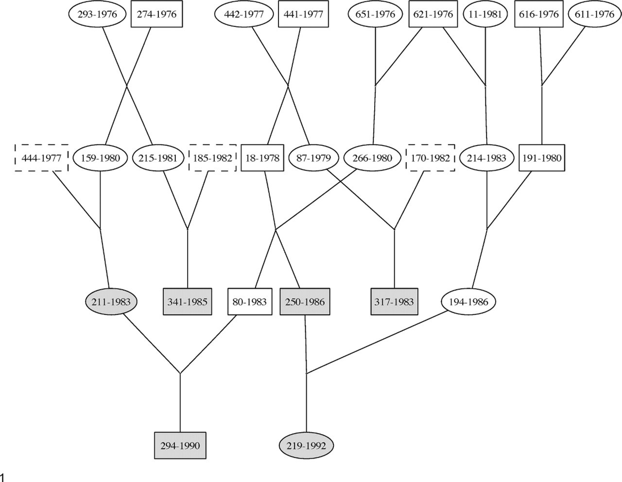
Pedigree for all affected tamarins. The present cohort is highlighted in gray boxes. Animal Nos. 442-1977 and 444-1977 represent earlier progenitor animals that also had histologically confirmed pheochromocytomas.
Clinically, all animals had gradual weight loss, and palpation of an intra-abdominal mass was recorded in 2 animals. One animal had an arrhythmia and systolic ejection murmur, with skipped beats and an abnormal QRS complex on electrocardiogram examination. Complete blood count and serum biochemistry analysis were within normal limits in all animals.
On gross examination, 5 of the 6 animals had variably sized, ovoid masses arising at the cranial aspect of the right kidney (tamarin Nos. 1 and 5), left kidney (tamarin No. 3), or involving both left and right kidneys (tamarin Nos. 2 and 4). Tamarin No. 6 had no noticeable mass detected. There was no vascular invasion noted grossly in any of the animals.
Histopathology
The adrenal neoplasms were expansile and partially to completely replaced the normal cortical architecture (Fig. 2). The neoplastic cells were arranged in tight bundles, cords, and occasional pseudorosettes that were oriented in close approximation to blood-filled capillaries (Fig. 3). Cells were embedded in a small amount of fibrous connective tissue stroma. Generally, the cells had abundant, finely granular, eosinophilic cytoplasm with large, basally situated nuclei, finely stippled-to-marginated chromatin, and a single prominent nucleolus. Cellular and nuclear atypia was mild in all cases, with an average of 1–2 mitotic figures per 10 hpf. Three cases (tamarin Nos. 2, 5, and 6) had a small proportion (<5%) of neoplastic cells with megalokaryocytic nuclei or multinucleate cells. In 3 animals, there were rafts of neoplastic cells located within extracapsular lymphatics (Fig. 4); however, no evidence of distant metastasis was noted in any of the animals. The neoplastic cells were interspersed with hemosiderin laden macrophages in 3 cases (tamarin Nos. 1, 3, and 6), extramedullary hematopoiesis in 4 cases (tamarin Nos. 1, 2, 4, and 6), and in 3 cases intratumoral sinusoids were dilated and partially to completely occluded by fibrin thrombi (tamarin Nos. 1, 2, and 6) (Fig. 5).
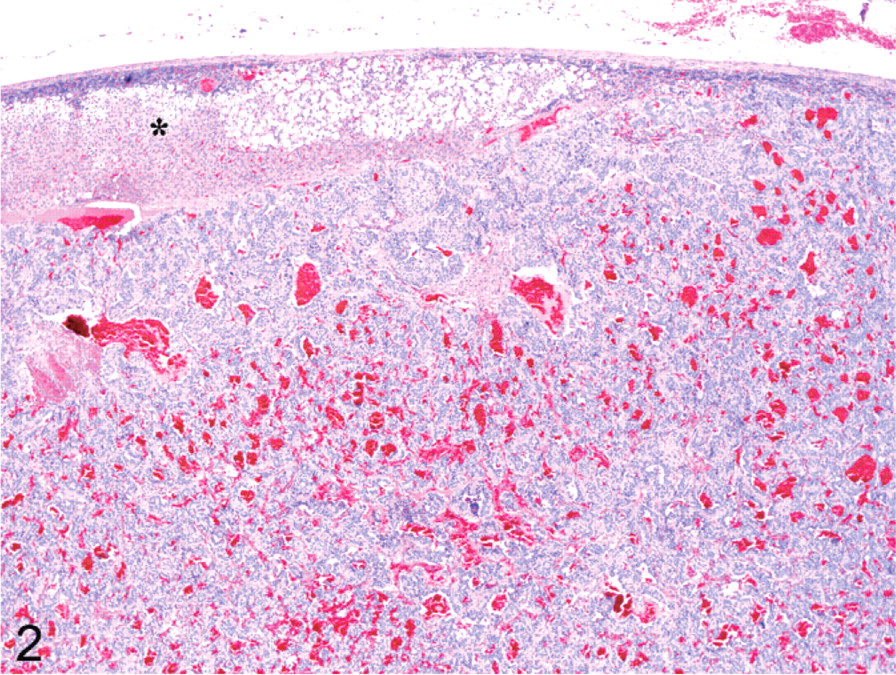
Adrenal gland; cotton-top tamarin No. 2. A large, unencapsulated mass arising from the adrenal medulla compresses the adjacent adrenal cortex (asterisk). HE.
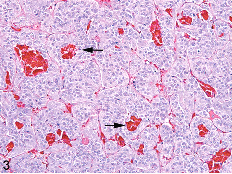
Adrenal gland; cotton-top tamarin No. 6. The neoplastic cells are arranged in tight clusters, nests, and pseudo-rosettes that are oriented around small blood vessels (arrows). HE.
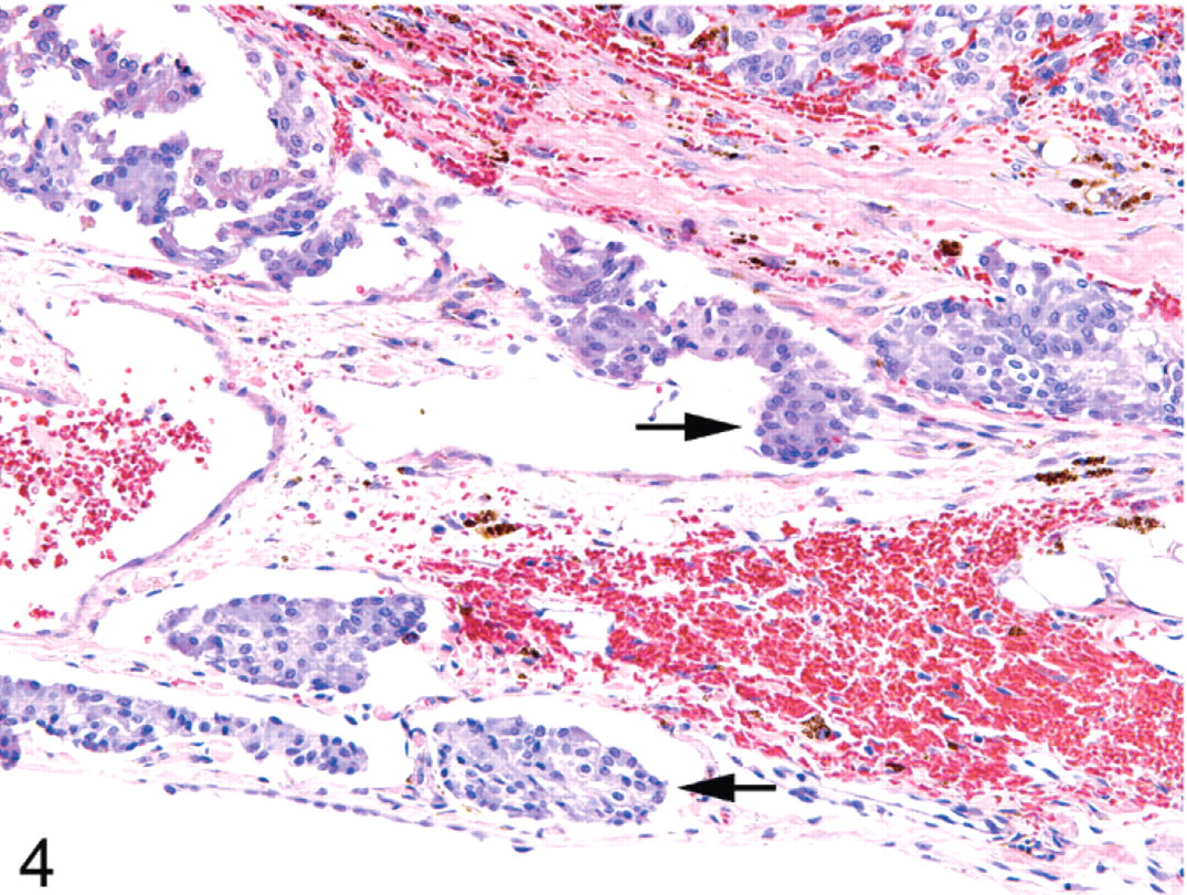
Adrenal gland; cotton-top tamarin No. 2. Rafts of neoplastic cells partially occlude peri-adrenal lymphatics (arrows). HE.
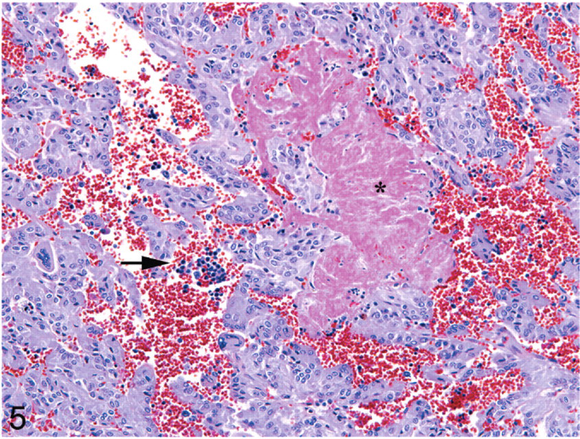
Adrenal gland; cotton-top tamarin No. 1. Within the neoplasm there are several loose aggregates of extramedullary hematopoiesis (arrow) and a single large fibrin thrombus (asterisk) partially fills a small blood vessel. HE.
In the myocardium of all affected animals were multifocal regions of myofiber cytolysis characterized by sarcoplasmic hypereosinophilia, sarcoplasmic vacuolation, loss of cellular detail, and myofiber shrinkage (Fig. 6). All animals also had areas of interstitial fibrosis that entrapped and replaced variable numbers of myofibers; this fibrosis was extensive in two of the affected animals. Two of the animals also had marked interstitial fibrosis in the lung, coupled with increased numbers of hemosiderin laden macrophages (heart-failure cells) (Fig. 7). Three of the cases (tamarin Nos. 1, 2, and 4) had marked hyalinization of the coronary arteries, with thickening of the tunica media and an increased number and size of smooth muscle cells.
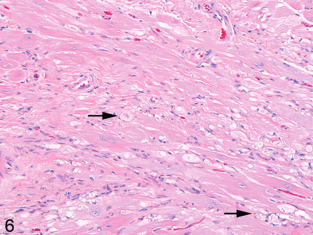
Heart; cotton-top tamarin No. 3. Multifocally, the myocardiocytes are swollen, vacuolated, and degenerate with loss of striations (arrows). HE.
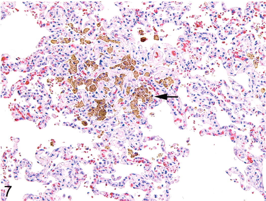
Lung: cotton-top tamarin No. 4. The alveoli contain large numbers of macrophages that are filled with hemosiderin (arrow). HE.
Two of the affected animals (tamarin Nos. 3 and 4) had unilateral thyroid cystadenomas characterized as expansile, encapsulated cystic neoplasms that had projections of neoplastic follicular epithelium forming papillary fronds within the cystic cavities. Additional comorbid histologic findings were chronic lymphoplasmacytic and fibrosing nephritis (6/6), vacuolar hepatopathy (5/6), multifocal hepatic necrosis (2/6), moderate lymphoplasmacytic enterocolitis (1/6), adrenal myelolipoma (1/6), and jejunal lymphangiectasia (1/6).
Immunohistochemistry
Immunohistochemical analysis of the 6 cases of pheochromocytomas was similar for all antibodies studied. SYN immunoreactivity was strong, diffusely intracytoplasmic, and confined solely to the neoplastic chromaffin cells (Fig. 8). Cortical tissue exhibited no immunoreactivity, with scattered capsular nerves strongly positive. CGA exhibited diffuse, granular, cytoplasmic immunoreactivity within the neoplastic chromaffin cells but spared the cortical cells (Fig. 9). PGP 9.5 exhibited diffuse cytoplasmic immunoreactivity in the neoplastic cells as well as staining occasional interstitial cells in the adrenal cortex and the capsular nerves (Fig. 10). N-CAM immunoreactivity was strongly membranous in all of the neoplastic chromaffin cells (Fig. 11). Cells in the zona glomerulosa and the zona fasiculata had scattered, less-intense membrane staining for N-CAM. GFAP was universally negative in all cases examined, staining only occasional solitary interstitial cells in the cortex and capsular nerves (Fig. 12).
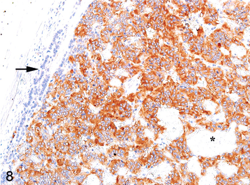
Adrenal gland; cotton-top tamarin No. 2. The neoplastic cells exhibit strong cytoplasmic immunoreactivity for SYN. Intratumoral blood vessels (asterisk) and compressed cortical cells (arrow) have no immunoreactivity. Immunoperoxidase staining, DAB chromagen, Mayer's hematoxylin counterstain.
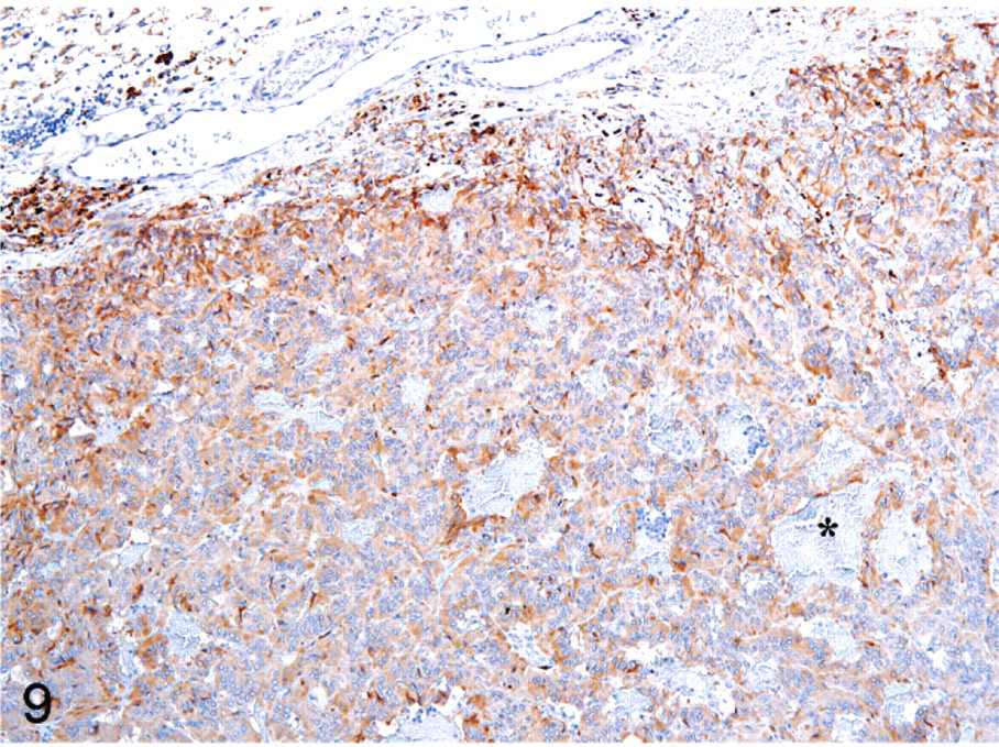
Adrenal gland; cotton-top tamarin No. 6. The neoplastic cells exhibit moderate cytoplasmic immunoreactivity for CGA. Intratumoral blood vessels (asterisk) have no immunoreactivity. Immunoperoxidase staining, DAB chromagen, Mayer's hematoxylin counterstain.
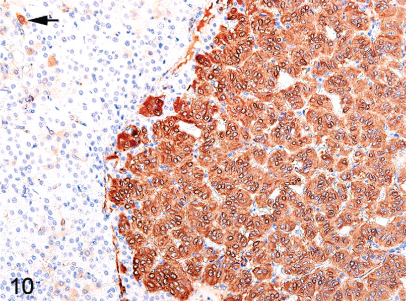
Adrenal gland; cotton-top tamarin No. 5. The neoplastic cells exhibit strong cytoplasmic immunoreactivity for PGP 9.5. Rare interstitial cells in the adjacent cortex (arrow) are also positive. Immunoperoxidase staining, DAB chromagen, Mayer's hematoxylin counterstain.
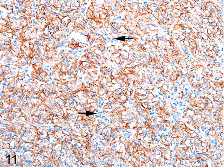
Adrenal gland; cotton-top tamarin No. 5. The neoplastic cells exhibit strong membranous immunoreactivity for N-CAM. All intratumoral blood vessels (arrows) have no immunoreactivity. Immunoperoxidase staining, DAB chromagen, Mayer's hematoxylin counterstain.
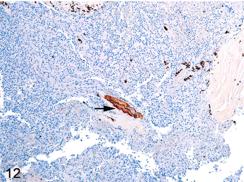
Adrenal gland; cotton-top tamarin No. 3. The neoplastic cells exhibit no immunoreactivity for GFAP. A single nerve (arrow) is strongly positive, and there are scattered hemosiderin laden macrophages (asterisk). Immunoperoxidase staining, DAB chromagen, Mayer's hematoxylin counterstain.
Discussion
Herein, we report the clinical, histologic, and immunohistochemical properties of 6 cases of pheochromocytoma in cotton-top tamarins (Saguinus oedipus). Pheochromocytomas are neuroendocrine tumors derived from the chromaffin cells of the adrenal medulla. Complications typically associated with these tumors include myocardial degeneration that results from excess catecholamine release and systemic hypertension typified by vascular medial hypertrophy. In this study, all animals with pheochromocytomas had concurrent myocardial degeneration and fibrosis. Random myocardial degeneration and fibrosis is not a common age-associated lesion in cotton-top tamarins housed at the NEPRC, and, therefore, an association with increased circulation catecholamines is plausible in this cohort of animals. In addition two of these animals also had alveolar septal fibrosis and hemosiderin laden macrophages within the alveoli, indicative of long-standing cardiac dysfunction. Three animals had marked hyalinization and thickening of the tunica media of intramural coronary arteries, a common finding associated with hypertension. Although other organs (i.e., stomach, eye, and kidney) did not have any histologic evidence of vascular disease, the lesions in the heart are highly suggestive of hypertension, a common adverse effect of pheochromocytomas.10
In humans, pheochromocytomas are considered an uncommon neoplasm, with an average annual incidence of 1 per 300,000 individuals.11 Although rare, considerable interest has been spent attempting to define an appropriate animal model for this tumor.17 Pheochromocytomas are uncommon in all domestic and laboratory animals, except for some strains of rats and various transgenic mouse models.14, 17 In the former, tumors typically arise either de novo in aged animals or are associated with exposure to a variety of genotoxic compounds.17 They are typically observed in males and can be either bilateral or unilateral.17 Similar murine neoplasms were studied in a variety of transgenic models, including mice expressing polyoma viral T-antigens and c-mos, in addition to Nf1 knockout mice.17, 18 Although both mice and rats provide valuable information into the pathogenesis of pheochromocytomas, their different physiologies make comparisons with human cases of pheochromocytoma problematic. Nonhuman primates are physiologically similar to humans and could potentially provide a more concise model to study the disease. Although there are several reports of pheochromocytomas in New World primates, the neoplasms appear to be rare or underreported.2, 3 In the current study, 5.3% of the cotton-top tamarins that died were found to have pheochromocytomas and resultant cardiac pathology sufficient to have led to death. This is a significantly higher percentage than is indicated in the literature for other species and may indicate an increased propensity to develop these tumors in this population of cotton-top tamarins.
In humans, most cases are unilateral and solitary, with fewer than 10% of the tumors considered malignant.11, 15–17 In the present study, two thirds of the cases were unilateral, and a third were bilateral. No prognostic significance exists for unilateral versus bilateral tumors; however, bilateral tumors are more commonly associated with inherited, familial tumor syndromes.16 Many cases of pheochromocytoma in humans are associated with multiple endocrine neoplasia type 2, von Hippel-Lindau (VHL) syndrome, and neurofibromatosis type 1.6, 9 Pheochromocytomas are present in roughly half of the cases of multiple endocrine neoplasia type 2 and result from a mutation in the RET proto-oncogene.6, 15, 16 In fact, bilateral pheochromocytomas are a strong factor in screening for RET mutations in affected human patients.6, 15 In cases of the VHL syndrome, the absence of the VHL protein leads to stabilization of hypoxia inducible factor 1α, thereby potentiating cell growth and angiogenesis.9 In the current study, the increased incidence of pheochromocytomas in this population suggests an effect beyond background tumor development, and genetic studies into the population are being pursued. In addition, the presence of concurrent thyroid cystadenomas in several of these tamarins raises the specter of a multiple endocrine neoplasia-like syndrome, possibly occurring in several of the affected animals. Indeed, the pedigree map illustrates the interrelatedness of the affected animals. Especially intriguing is that one of the original progenitor animals (442-1977) also died from a pheochromocytoma, albeit at a much younger age than the other animals. Although no distinct inheritance pattern can be discerned from the pedigree, the appearance of the tumor occurs in both sexes and in most cases skips a generation.
Immunohistochemistry is considered an invaluable aid in diagnosing tumors of the adrenal medulla. Neuroendocrine markers like CGA, PGP 9.5, N-CAM, and SYN are typically positive in cases of pheochromocytoma, particularly with CGA, which is reported to stain 100% of pheochromocytomas.1, 8, 11, 12, 20 Chromogranins are the major secreted protein of the adrenal medulla and play an important role in binding and aggregating intracellular calcium.4 In addition, there are defined roles for chromogranins in functioning as prohormones, molecular chaperones, and modulators of gene expression.4, 20 PGP 9.5 is a part of the ubiquitin-proteasome system, and, although originally isolated from the brain, expression has now been found in many human tissues, including the diffuse neuroendocrine system, female and male reproductive organs, and hematopoietic cells.7, 11, 13, 20 To our knowledge, this is the first study to use PGP 9.5 in cotton-top tamarin tissue and further illustrates the utility of the marker in diagnosing neuroendocrine tumors. SYN is a membrane glycoprotein found in prejunctional neuroendocrine granules and is used as a specific marker of neuroendocrine tumors.11, 20 N-CAM (also known as CD56) is a common marker also used in diagnostic immunopathology and has specificity to cells of the nervous system, neuroendocrine system, and skeletal muscle.8, 11 In human adrenal glands, N-CAM immunoreactivity has been reported in both cortical and medullary cells, thereby making it a poor marker to differentiate primary tumors arising in each site.8 Immunostaining in the present case series, however, showed much more intense staining in the neoplastic medullary chromaffin cells compared with the cortical cells, suggesting that NCAM is a better marker for chromaffin cells. GFAP is also occasionally used in the diagnosis of pheochromocytomas and is used to stain a subpopulation of the support cells of the adrenal medulla (sustentacular cells).1
In conclusion, pheochromocytomas are uncommon tumors in all domestic and laboratory animals except for rats and select mice strains. Although there are scattered reports of these tumors developing in New World primates, the incidence is not known, although is presumed to be sporadic. The 6 cotton-top tamarins described herein had uni- or bilateral pheochromocytomas. In addition, all animals had acute, multifocal myocardial degeneration and fibrosis consistent with chronic, ongoing myocardial injury, presumably from released epinephrine. On immunohistochemical examination, CGA, SYN, N-CAM, and PGP 9.5 strongly stained the neoplastic cells, whereas GFAP was uniformly negative. Pheochromocytomas in this colony of cotton-top tamarins represented 5.3% of the total causes of death in the studied time frame, and, because of genetic interrelatedness of the affected animals and the identification of affected progenitor animals, genetic studies are ongoing in this population.
Footnotes
Acknowledgements
We thank Kristen Toohey for assistance with photography, Greg Charest for assistance in developing the pedigree map, and Liz Curran for tissue procurement. This research was funded, in part, by National Institutes of Health grants RR00168 and RR07000.