
Abstract
Purpose
We investigate the thenar and plantar sesamoids as markers of skeletal maturity, and grade appearance using two scales, a binary system (absent or present), and an analogue system that relies upon judging regular changes in morphological appearance.
Methods
We studied 94 healthy children (49 female and 45 male patients) between ages three and 18 years who had approximately 700 serially acquired sets of radiographs and physical examinations. The children had at least annual radiographs taken of the left hand and left foot. Velocity of growth was calculated and curves were fit to a cubic spline model to determine age of maximum height velocity, or peak height velocity (PHV). Appearance of the plantar and thenar sesamoids was recorded using a binary system classifying the sesamoids as absent or present and an analogue system classifying the sesamoid as absent, present as a small ossification centre or larger than a small ossification centre.
Results
The plantar sesamoids appear 1.67 years before PHV and reach mature size 1.02 years after PHV. The thenar sesamoids appear 0.32 years before PHV and reach mature size 2.25 years after PHV. The plantar sesamoids are present and thenar sesamoids are absent at a mean 1.5 years prior to PHV. No patients had the thenar sesamoids present while the plantar sesamoids were absent.
Conclusion
As binary markers, when the plantar and thenar sesamoids are considered together it is possible to localize maturity. As analogue markers, they offer more information. The sesamoids also allow clarification of the calcaneal and Sanders stages.
Level of Evidence
Not Applicable.
Introduction
Accurate determination of skeletal maturity is at the heart of paediatric orthopaedic decision-making when predicting the likelihood of scoliosis curve progression or the timing of epiphysiodesis.1–5 The majority of skeletal maturity systems such as the Greulich and Pyle hand atlas, or the Tanner-Whitehouse III hand scores, rely on accurately judging a series of morphological changes of bones to establish skeletal maturity.6,7 The sequence of regular morphological changes of an ossification centre can be considered an analogue radiographic feature. Analogue radiographic features can create potential for interrater disagreement due to varying judgment of the morphological appearance. Depending on the complexity of the skeletal maturity system and the familiarity of the rater, this can result in large interrater errors, as described by Cundy et al 8 for the Greulich and Pyle system.
Certain skeletal maturity systems also feature binary radiographic features; bony features that are either absent or present. 9 Many bones can serve as binary systems of assessment, in that they are absent and later appear, or certain parts of them such as the epiphyses appear. Binary markers are simple, and can sometimes be used by themselves in the absence of other radiographic features. However, most ossification centres appear early during development. The plantar and thenar sesamoids may be more suited for binary assessment method because they develop later, around the time of peak height velocity (PHV). The time period around the adolescent growth spurt is often of most interest to the paediatric orthopaedist, as this is the time period most associated with scoliosis curve progression, slipped capital femoral epiphysis (SCFE) and epiphysiodesis.10,11
Although binary distinctions are simple, they may not be specific enough for exclusive use. They can be difficult to localize temporally since they are either present or absent. When they first appear, it can be difficult to tell how long they have been present unless one has serial radiographs. They oftentimes need to be attached to analogue distinctions to have value. For instance, in the skeletal maturity system classifying ossification of the calcaneal apophysis, the appearance of the plantar sesamoids on an anteroposterior radiograph can help clarify whether a calcaneus is stage 2 or 3. 12 By themselves the plantar sesamoids are not specific enough to delineate a stage 2 calcanei from a stage 3 calcanei, and their presence should be relied upon secondary to the other characteristics of a stage 3 calcaneus. However, for an ambiguous calcaneal apophysis, they can be helpful in deciding the stage of the calcaneus.
The purpose of this study was to examine the information difference conveyed by binary and analogue maturity methods and to describe the appearance of the plantar and thenar sesamoids relative to time from PHV. We also examine the relationship of the plantar sesamoids to the calcaneal apophyseal stages and the thenar sesamoids to Sanders hand scores12, 13.
Materials and methods
We studied 742 serial foot and 694 serial hand radiographs from 94 children (49 female and 45 male patients) aged three to 18 years. These children were part of a prospective, longitudinal assessment of growth conducted at Western Reserve University from 1926 to 1942 under the direction of Dr. T. Wingate Todd of the Brush Foundation.6,14 The children were from the metropolitan area of Cleveland, Ohio and had at least annual radiographs taken of many regions of the body, including the left hand and left foot, along with height, weight and other physiologic data. This compendium was used by Greulich and Pyle to create their bone age atlas of the hand. 6 The families participating in this study were above average in economic and educational status and children were selected based on absence of gross physical or mental defects. 14 In all, 92.2% of the subjects were white and 7.7% of them were black. 14
We identified 94 subjects with consecutive radiographs, at least yearly, from age ten to 15 years with height data recorded at each radiograph. These subjects were the same as those used in describing calcaneal apophyseal ossification, modified Oxford Hip Scores and Sanders hand scores.12,15,16 This age range was chosen because it is the typical age range when children present with adolescent idiopathic scoliosis or for epiphysiodesis for leg length equalization procedures. Velocity of growth was calculated and curves were fit to a cubic spline model to determine age of maximum height velocity or PHV after the approach of Tanner and Davies. 17
The appearance of the plantar and thenar sesamoids was recorded using two grading scales as shown in Figs. 1 and 2. For the binary grading scale, sesamoids were graded based only on their absence or presence. For the analogue grading scale, the sesamoids were classified as either absent, present as a small ossification centre on an anteroposterior radiograph (< 2 mm) or only visible on a lateral radiograph, or larger than a small ossification centre (> 2 mm). The calcaneal scores and Sanders hand scores have been described previously and are shown in Figs. 3 and 4.12,13 Each hand radiograph was graded with a Sanders hand score and each foot radiograph was graded with a calcaneal score.
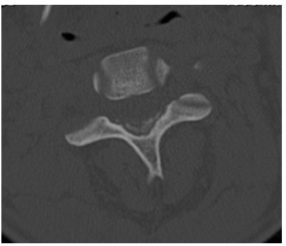
The maturity schemes for judging the plantar sesamoid. Plantar sesamoids are indicated by the red arrows. Grading scheme 1- to 2-point binary system. The plantar sesamoid is either absent (
The maturity schemes for judging the thenar sesamoid. Thenar sesamoids are indicated by the red arrows. Grading scheme 1- to 2-point binary system. The thenar sesamoid is either absent (
Ossification of the calcaneal apophysis. Reprinted with permission from Nicholson et al:
12
(
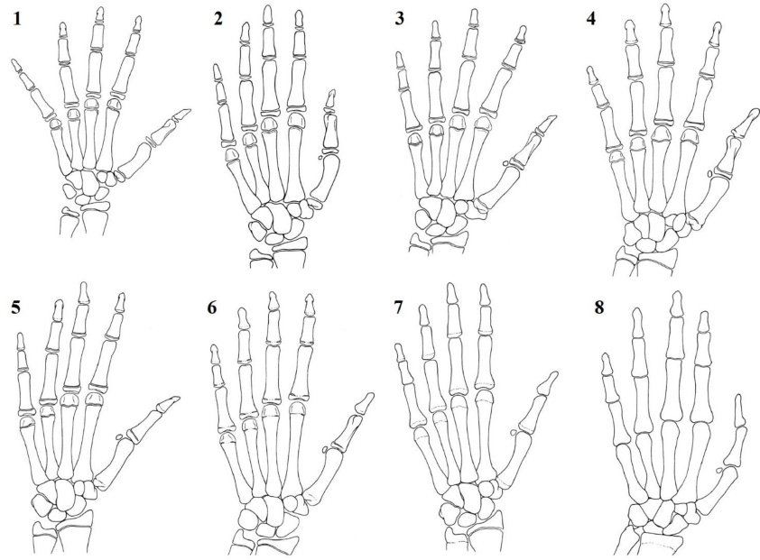
Sanders hand scores. Reprinted with permission from Sanders et al. 13 Stage 1 – all of the binary epiphysis of the hand are not covered; stage 2 – all binary epiphysis fully cover the metaphysis; stage 3 – the preponderance of digits are capped and the second through fifth metacarpal epiphyses are wider than their metaphyses; stage 4 – any of the distal phalangeal physes are closing; stage 5 – all distal phalangeal physis are closed but the other physes remain open; stage 6 – the middle or proximal phalangeal physes are closing; stage 7 – all physes are closed except for the distal radial physis; stage 8 – all physes are closed (ignoring the ulna).
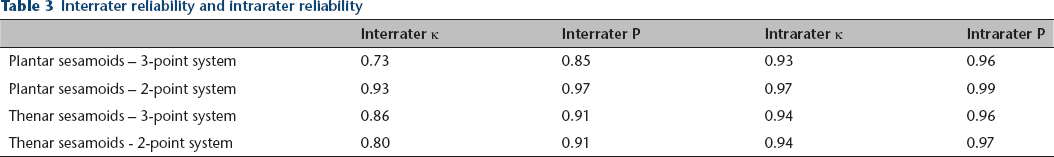
Two observers, a fellowship-trained paediatric orthopaedist (DRC) and an orthopaedic resident (ADN) evaluated 75 randomly selected hand and foot radiographs for evaluation of the plantar and thenar sesamoids. Confidence intervals (CI) and unweighted κ were calculated using Microsoft Excel 2013 (Microsoft, Redmond, Washington).
Results
The plantar sesamoids first appear 1.35 years (95% CI 1.61 to 1.10) before PHV and the thenar sesamoids first appear 0.12 years before (95% CI 0.27 before to 0.04 after) PHV. The plantar sesamoids are visible on a lateral radiograph of the foot before they are visible on an anteroposterior radiograph. The plantar sesamoids reach their mature size at a mean 1.02 years (95% CI 0.80 to 1.24) after PHV. The thenar sesamoids reach their mature size at a mean 2.25 years (95% CI 2.11 to 2.39) after PHV. Both plantar and thenar sesamoids ossify over a similar length of time. However, the plantar sesamoids ossify approximately 1.2 years earlier than the thenar sesamoids. In addition, the plantar sesamoids are often not visible on an anteroposterior radiograph when they first appear.
For the binary grading system, the mean age from PHV for the absence or presence of the sesamoids is shown in Table 1 and Fig. 5a. As shown in Fig. 5, this system provides limited information unless one has serial radiographs showing when the transition from absence of sesamoids to presence of sesamoids occurs. In all, 108 radiographs from 66 patients had the plantar sesamoids present while the thenar sesamoids were absent, as shown in Table 1 and Fig. 5a. No patients had the thenar sesamoids present while the plantar sesamoids were absent.
Age from peak height velocity (PHV) for the thenar and plantar sesamoids; 2-stage (digital) system
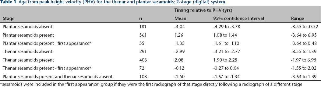
sesamoids were included in the ‘first appearance’ group if they were the first radiograph of that stage directly following a radiograph of a different stage
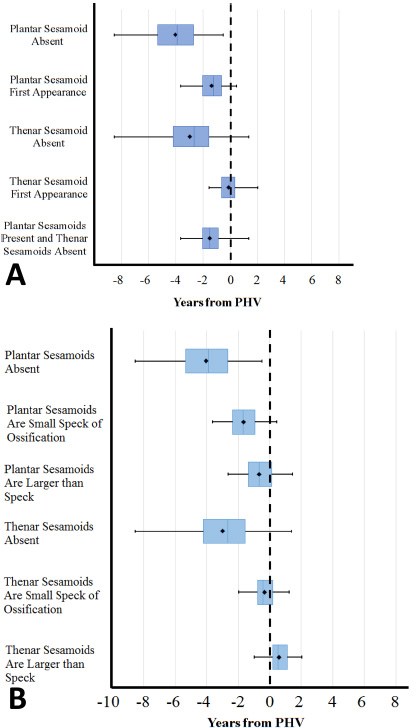
Maturity of the plantar and thenar sesamoids relative to peak height velocity (PHV). A box and whisker plot of the age with respect to PHV for the plantar and thenar sesamoids using three grading schemes. The black lines represent the range for each examined sequence, while the blue box represents the middle 50% of data. The blue line in the middle of each box represents the median, while the black diamond represents the mean: (
The analogue system, consisting of three grades, provides more information regarding PHV than the binary method, as shown in Table 2 and Fig. 5b. The plantar sesamoids are only visible on the lateral radiograph or as a small puff of ossification within a narrow window with respect to PHV, with an mean age of 1.67 years (95% CI 1.90 to 1.43) before PHV. Plantar sesamoids larger than a puff of ossification, first occur at a mean 0.67 years (95% CI 0.90 to 0.45) before PHV. The thenar sesamoids are only visible as a small puff of ossification with a mean age of 0.32 years (95% CI 0.49 to 0.15) before PHV. The thenar sesamoids first become larger than a puff of ossification at a mean 0.58 years (95% CI 0.42 to 0.74) after PHV.
Age from peak height velocity (PHV) for the thenar and plantar sesamoids – 3-stage (analogue) system
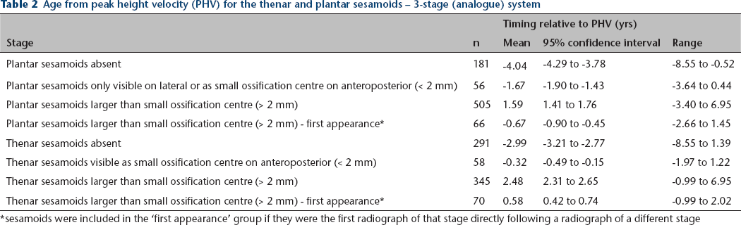
sesamoids were included in the ‘first appearance’ group if they were the first radiograph of that stage directly following a radiograph of a different stage
Most plantar sesamoids appear during calcaneal stage 2, with 64% of calcaneal stage 2 having the plantar sesamoids present. In all, 96% of calcaneal stage 3 have the plantar sesamoids present, and 100% of calcaneal stage 4 have the plantar sesamoids present. The analogue system adds depth to the binary system. For instance, 28.8% of calcaneal stage 2 radiographs have plantar sesamoids that appear more mature than a faint puff of ossification on an anteroposterior radiograph, whereas 84.5% of calcaneal stage 3 radiographs have plantar sesamoids that are more mature than a puff of ossification.
For the binary system, many thenar sesamoids appear during Sanders stage 2, with 40.9% of Sanders stage 2 having the thenar sesamoids present. Only 4.0% of Sanders stage 3 do not have the thenar sesamoids present, indicating that the thenar sesamoids form during Sanders stage 2. The analogue system gives increased detail. For instance, Sanders stage 3 has 67.7% of thenar sesamoids that appear in a more mature form than a puff of ossification, whereas only 14.0% of the thenar sesamoids are more mature than a puff of ossification for Sanders stage 2.
The interrater κ and P, and the intrarater κ and P for the three methods of grading the plantar and thenar sesamoids are shown in Table 3. P is the proportion of measurements that were the same between the two raters.
Interrater reliability and intrarater reliability
Discussion
Many people are uncomfortable with analogue distinctions, especially in the absence of additional data. ‘Capping’ of the finger phalanxes, or a small bend of the epiphysis over the metaphyseal edge, as described by Sanders is easier to appreciate if you have radiographs that immediately precede and follow capping. 13 Many people prefer binary distinctions, which have the advantage of clear distinction between the absent and present forms. Although binary distinctions are simple, they are often not specific enough for exclusive use. They sometimes need to be combined with analogue distinctions like the calcaneal score to add value.
The binary approach, considering the sesamoids as either absent or present, provides useful information regarding maturity, especially when the plantar and thenar sesamoids are considered together. In the absence of serial radiographs it is difficult to assess maturity of the child relative to PHV since there are only two stages of ossification. However, a combination of these two simple binary markers can allow for deduction of maturity relative to PHV, since the plantar sesamoids appear approximately 1.2 years earlier than the thenar sesamoids. As shown in Figure 5a, if the plantar sesamoids are present and the thenar sesamoids are absent, the child is a mean 1.5 years prior to PHV. Of note, there were no instances of any children having the thenar sesamoids present and plantar sesamoids absent.
The three-point system is an analogue scale. Identifying a puff of bone allows maturity to be localized with single radiographs. As shown in Figure 5b, a plantar sesamoid that appears as a puff of ossification or is only visible on the lateral radiograph, indicates that the child is a mean 1.67 years before PHV and a thenar sesamoid visible as a puff of ossification indicates that a child is a mean 0.32 years prior to PHV. Our data is in agreement with the observation by Dimeglio that the onset of puberty coincides with the appearance of the thenar sesamoids, with Dimeglio defining the first two years of puberty as the time during which PHV occurs. 18 We cannot comment directly on the relationship of the thenar and plantar sesamoid appearance to maturity as measured by the Sauvegrain or Dimeglio systems grading of the elbow.
The thenar sesamoids and plantar sesamoids are useful as markers for adding certainty to analogue systems, such as the Sanders and calcaneal scores. The plantar sesamoids can be used to differentiate calcaneal stage 1 from 2, and 2 from 3. Only 5.3% of calcaneal stage 1 radiographs have the plantar sesamoids present, even on a lateral radiograph. In all, 64% of calcaneal stage 2 radiographs have the plantar sesamoids present and 96.4% of calcaneal stage 3 radiographs have the plantar sesamoids present. If the plantar sesamoids are present on a lateral radiograph, the calcaneus is significantly more likely to be calcaneal stage 2 than 1. The three-point scale clarifies the distinction between calcaneal stage 2 and 3. Only 28.8% of calcaneal stage 2 radiographs have plantar sesamoids that appear more mature than a faint puff of ossification on an anteroposterior radiograph. However, 84.5% of calcaneal stage 3 radiographs have plantar sesamoids that are more mature than a puff of ossification on an anteroposterior radiograph. Thus, the presence of clearly defined radiopaque plantar sesamoids on an anteroposterior radiograph indicates that a calcaneus is more likely calcaneal stage 3 than 2.
The thenar sesamoids can be useful to differentiate Sanders stage 2 from Sanders 3, especially if capping of the finger phalanxes is ambiguous. Most Sanders 3 radiographs (96.0%) had the thenar sesamoids present whereas only 40.9% of Sanders 2 radiographs had the thenar sesamoids present. The three-point scale can further clarify the distinction between Sanders stage 2 and Sanders stage 3. Sanders stage 3 has 67.7% of thenar sesamoids that appear in a more mature form than a puff of ossification, whereas only 14.0% of the thenar sesamoids are more mature than a puff of ossification for Sanders stage 2. Thus, if the thenar sesamoids are larger than a puff of ossification, a hand is more likely to be Sanders stage 3 than stage 2. The Sanders hand stages correspond to Greulich and Pyle bone ages, and thus the thenar sesamoids can be used to clarify Greulich and Pyle bone ages as well. Thenar sesamoids larger than a puff of ossification indicates female bone age greater than ten years or male bone age greater than 13 years.
Limitations of this study include the age of the collection used, the fact that the majority of the study participants were white, and that some evidence points to children reaching puberty at earlier ages.19,20 However, the patients in this study are the same as those used for creation of the Greulich and Pyle atlas, which several studies have shown is applicable to modern children with minimal error, and continues to be extensively used today.19,21–23
As binary markers, the thenar and plantar sesamoids can provide limited information relative to timing of PHV by themselves. When the thenar and plantar sesamoids are considered together, the presence of the plantar sesamoid and absence of the thenar sesamoid indicates that a child is a mean 1.5 years before PHV. However, considering the thenar and plantar sesamoids as an analogue three-point scale allows significantly more information to be derived regarding maturity with respect to PHV. By both methods of grading, the sesamoids add certainty to the Sanders and calcaneal skeletal maturity systems.