
Abstract
In our previous studies, WBN/KobSlc was characterized as a rat strain in which only males began to develop pancreatitis, and then presented with diabetic symptoms. In the course of studying their pancreatic inflammation, we detected molar caries in prediabetic males feeding on a standard diet (CRF-1) widely used for experimental animals. The purpose of this study is to confirm whether the WBN/KobSlc strain is caries-susceptible to the diet reported to be non-cariogenic, and to examine the effect of a prediabetic condition on their dental caries. For a morphological study, 25 male WBN/KobSlc rats aged 3.2–7.8 months and 24 females of the same strain aged 3.3–6.6 months were used, along with 10 males and 10 females of 8.2-month-old F344 rats. Marked dental caries were detected in the mandibular molars of male and female WBN/KobSlc rats regardless of pancreatitis, although no similar changes were observed in any teeth of the F344 strain fed the same diet. Soft X-ray examination revealed that the caries began in the crown and progressed horizontally and vertically, and that a severe radiolucent lesion extensively expanded to the entire crown, corresponding to a macroscopically deleted molar. The caries had gradually developed mainly in the second mandibular molar from more than 3.5 months of age, while none were seen in any rats before that time. The WBN/KobSlc rats were caries-susceptible even to the standard laboratory diet, and pancreatitis was not directly associated with the onset of dental caries in this strain.
On the pathogenesis of dental caries, Keyes 1 proposed that its aetiology requires three factors, i.e. pathogenic bacteria, sugar and a predisposing host. In addition, a host time has been considered as a fourth factor. 2 To investigate that aetiology, hamsters and mice have been used as animal models. Up till now, since the onset of naturally occurring dental caries has not been reported in these animal models, researchers have used a diet containing large amounts of sugar and/or an inoculation of infectious cariogenic bacteria into the oral cavity to induce caries. Rats are thought of as one of the most suitable animal species for caries research because of advantages such as a brief lifespan and short experimental period as well as their ease of handling. However, inducing the onset of caries requires experimental manipulations such as an infection with pathogenic bacteria and/or a cariogenic diet high in sucrose. 3
In our previous studies, WBN/KobSlc was characterized as an albino rat strain in which almost all males began to develop pancreatitis as early as three months old, and to subsequently present diabetic symptoms from about 10 months of age, whereas females showed no such changes over their entire lifespan. 4,5 Although there have been no reports of dental caries in the WBN/KobSlc rat, in the course of studying their pancreatic inflammation, we detected molar caries in male prediabetic animals at five months of age fed a standard diet (Charles River Formula 1 [CRF-1]) widely used for experimental rats and never before reported to be cariogenic.
The purpose of this study is to confirm whether WBN/KobSlc rats are caries-susceptible to the standard laboratory diet, which is non-cariogenic to general rat strains, and to examine the effect of a prediabetic condition on dental caries in this strain.
Materials and methods
Experimental animals
The animals were handled according to the principles outlined in the Guide for the Care and Use of Laboratory Animals prepared by our institution (Setsunan University) for all experimental procedures. WBN/KobSlc and F344 rats were used in this study. The F344 rat was selected for the control because it is a popular inbred albino strain in many long-term studies and the body weight change seems to be more similar to that of WBN/KobSlc strain in comparison with other strains. Animals of both strains were born in our laboratory from females supplied by Japan SLC, Inc (Hamamatsu, Japan). All were housed in polycarbonate cages in an air-conditioned animal room at a temperature of 20-26°C and a relative humidity of 40–70% under a 12/12 h light/dark cycle and ventilation with filtered fresh air. They were allowed free access to a widely used pelletized diet for experimental mice and rats (CRF-1; Oriental Yeast, Japan) and tap water ad libitum.
Morphological study and grading for caries evaluation
For a morphological study, 25 male WBN/KobSlc rats aged 3.2–7.8 months and 24 females of the same strain aged 3.3–6.6 months were used, along with 10 males and 10 females of 8.2-month-old F344 rats. Each WBN/KobSlc rat undergoing a morphological study in a blinded evaluation was allocated to one of the following three groups: less than 3.5 months of age (phase I: 10 male and 6 female rats), between 3.5 and 5.5 months (phase II: 9 male and 10 female rats) and more than 5.5 months (phase III: 6 male and 8 female rats). Animals were euthanized by exsanguination under deep anaesthesia, after which the mandible and maxilla were removed and fixed in 10% neutral-buffered formalin (pH 7.4). After a 24 h fixation, the occlusal, buccolingual and proximal surfaces of all teeth were intensively observed under a binocular stereoscope, and any macroscopic defect on the teeth was categorized as belonging to one of the following three types according to the severity of the defect (no defect, partial molar defect and complete molar defect). Following macroscopic examination, soft X-ray and histological examinations were made.
In this study, dental caries was characterized by teeth that were macroscopically defective or had a radiolucent area on soft X-ray film. In addition ‘Ratio of rats developing dental caries’, ‘Ratio of teeth with caries’ or ‘Ratio of caries by molar region’ was calculated as follows.
Ratio of rats developing dental caries = number of rats developing dental caries/total number of rats × 100 (%).
Ratio of teeth with caries = number of molar teeth with caries/total number of molar teeth × 100 (%).
Ratio of caries by molar region = total number of teeth with caries by molar region/total number of all molar teeth with caries × 100 (%).
Soft X-ray observation and grading for caries evaluation
To identify the vertical and horizontal expansion of dental caries, soft X-ray photographs of the mesiodistal plane were taken under conditions of 35 kV, 2 mA for 4 min. By observation and measurement of those photographs, the caries conditions were classified into four groups: no radiolucent change (grade 0), radiolucent area only on the occlusal surface of the crown (grade 1), radiolucent areas on both occlusal surfaces and either of the mesiodistal surfaces of the crown (grade 2) and radiolucent areas over the entire surface of the crown (grade 3).
Histological observation
After soft X-ray observation, the left mandible of each rat was subjected to histological examination. The sample was decalcified in a 5% solution of ethylenediaminetetraacetic acid 4 Na (EDTA 4 Na) for two weeks at 4°C after fixation with 10% neutral-buffered formalin. After decalcification, specimens of the mandible were trimmed, then dehydrated in a sequential ethanol series using an automated processor, and embedded in paraffin wax. Serial 7 μm thick mesiodistal sections were made through the centres of all molars, then stained with haematoxylin and eosin for examination by light microscopy.
Measurement of sugar content in diet
The contents of free sugar (monosaccharide or disaccharide) in the diet were measured by the high-performance liquid chromatographic method.
Bacteriological examination of oral cavity
Rats undergoing a bacteriological examination of the oral cavity, i.e. 4.2-month-old male WBN/KobSlc rats without caries (n = 2), 8.6-month-old male WBN/KobSlc rats with caries (n = 3) and male F344 rats without caries aged 10.5–12.1 months (n = 3) were separated from rats undergoing morphological examination. Samples of oral bacterial flora were collected with sterile cotton swabs along the occlusal surfaces of the mandibular molars. Viable bacterial counts in the flora of mandibular occlusal molar surfaces were made on BCP-added plate count agar (Nissui Pharmaceutical, Japan) after two days of anaerobic incubation at 37°C. The colonies were then classified into genus Streptococcus, Lactobacillus and other genera by their Gram-stain properties, shapes observed microscopically and/or catalase activity. Biochemical identification of the two genera from rats with caries was done using the commercial kits STREPTOGRAM (Wako Pure Chemicals, Japan) or API 50CHL (bioMerieux, Japan). Additional tests were conducted according to Bergey's method. 6,7 Cariogenicity of these bacteria was also assessed by their acid production and adherent ability observed in brain heart infusion (BHI) broth (Difco Laboratory, Japan) containing 5% sucrose at 37°C and a 35° angle for 48 h.
Statistical analysis
We employed the Wilcoxon rank-sum test for grading caries evaluation in addition to the chi-square test for the ratio of rats developing dental caries and the ratio of the teeth with caries by soft X-ray examination; the statistical differences between each group were analysed.
Results
Morphological characteristics of caries in WBN/KobSlc rats
Based on macroscopic and soft X-ray observations, dental caries originated in the crown and became apparent in the mandibular molars of both sexes in WBN/KobSlc rats, although such lesions were not detected in any incisors or molars of maxilla in this strain. On the other hand, in all F344 rats no dental caries were found in any teeth including mandibular molars.
Macroscopic observation of caries in WBN/KobSlc rats
The typical macroscopic appearance of carious molars of the mandible in WBN/KobSlc rats is shown in Figures 1B–D. Macroscopically, the dental caries developed mainly in occlusal fissures, and was initially detected as a partial coronal defect of the mandibular molar (Figures 1B and C). The lesion then expanded horizontally until finally the crown of the carious molar was completely invisible (Figure 1D).
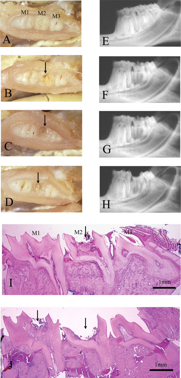
Morphological characteristics of molar caries in mandible of WBN/KobSlc rats. (A–D) Macroscopic appearance of mandibular molars in WBN/KobSlc rats. (A) Normal molars. (B, C) Partial coronal defect (arrows) of second molar (M2). (D) Complete coronal defect (arrow) of M2. (E–H) Soft X-ray photograph of molars of the mandible in WBN/KobSlc rats. (E) Normal molars. (F) Grade 1-type caries is detected as radiolucent lesion only in occlusal surface of the crown (M2). (G) Grade 2-type caries is detected as radiolucent lesion in both occlusal surfaces and either of mesiodistal surfaces of the crown (M2). (H) Grade 3-type radiolucent lesion extensively expands over entire crown (M2). Parts E, F, G and H correspond to A, B, C and D, respectively. (I, J) Histopathological features of caries in WBN/KobSlc rats. (I) Corresponding to grade 2-type caries on soft X-ray. Occlusal caries of M2 (arrow) extends into dental pulp, but first (M1) and third (M3) molars are almost intact. (J) Corresponding to grade 3-type caries on soft X-ray. Occlusal caries of M2 (arrowhead) is more severe than grade 2-type. M1 also had occlusal caries (arrow) similar to grade 2-type caries of M2 (I)
Ratio of caries in the mandible of WBN/KobSlc rats by molar region
Soft X-ray examination of caries in WBN/KobSlc rats
Soft X-ray photographs of the carious molars of the mandible in representative cases of WBN/KobSlc rats of both sexes are shown in Figures 1F–H. By a soft X-ray examination, grade 1-type of caries was detected as a radiolucent lesion only in the occlusal surface of the crown (Figures 1F), which was macroscopically normal, or as a partially defective crown of the molar (Figures 1B). Grade 2-type of caries was detected as a radiolucent lesion in both occlusal surfaces and either of the mesiodistal surfaces of the crown (Figures 1G) corresponding to a macroscopically partially defective crown of the molar (Figures 1C). In severely affected teeth, dental caries progressed both horizontally and vertically, and the grade 3-type of radiolucent lesion extensively expanded covering the entire crown (Figures 1H) and corresponding to a macroscopically complete defect of the crown of the molar (Figures 1D).
Progression of caries by age and caries region
In WBN/KobSlc rats, caries developed mainly in the second mandibular molar after 3.5 months of age, while none were seen in any rats earlier than 3.5 months. Moreover, the number of affected teeth and the severity of the caries increased with age in this strain.
Ratio of rats developing dental caries
The ratio of rats developing caries of the mandibular molars in the WBN/KobSlc strain based on age is shown in Table 1A. No rats with dental caries were seen before 3.5 months of age, whereas beyond that age the ratio of rats developing caries increased, reaching more than 50%.
The caries incidence in mandibular molars of WBN/KobSlc rats
Group I: <3.5 months of age; Group II: 3.5–5.5 months of age; Group III: >5.5 months of age
*Significant difference from group I (P < 0.01)
Ratio of teeth with caries
The ratio of teeth with caries of the mandibular molars in the WBN/KobSlc strain based on age is shown in Table 1B. The respective rates for males and females were 0/60 (0%) and 0/36 (0%) before 3.5 months of age (phase I); 10/54 (18.5%) and 7/60 (11.7%) between 3.5 months and 5.5 months (phase II); 11/36 (30.5%) and 20/48 (41.7%) beyond 5.5 months (phase III). The number of teeth developing dental caries increased with age, with the rate beyond 5.5 months of age (phase III) being significantly higher than that below 3.5 months of age (phase I).
Change of caries scores in WBN/KobSlc rats by soft X-ray examination
Caries scores of individual WBN/KobSlc rats by soft X-ray examination are shown in Table 2. No radiolucent lesion was observed in any molars of male or female WBN/KobSlc rats before 3.5 months of age (phase I). Dental caries of WBN/KobSlc rats were scored as grade 1 or grade 2 in both sexes between the ages of 3.5 and 5.5 months (phase II), with radiolucent lesions mainly observed on the occlusal surfaces or/and either of the mesiodistal surfaces of the crown. A grade 3-type of carious molar was newly detected in both sexes when a radiolucent lesion had expanded throughout the molar and the crown had been completely lost since more than 5.5 months of age (phase III). The scores of caries in the age phases II and III were significantly higher than that in phase I, and the severity of the caries had apparently worsened with age.
Change of caries grades of mandibular molars in individual WBN/KobSlc rats by soft X-ray photography
Group I: <3.5 months of age; Group II: 3.5–5.5 months of age; Group III: >5.5 months of age
Significant difference from group I (*P < 0.05, **P < 0.01)
Caries region
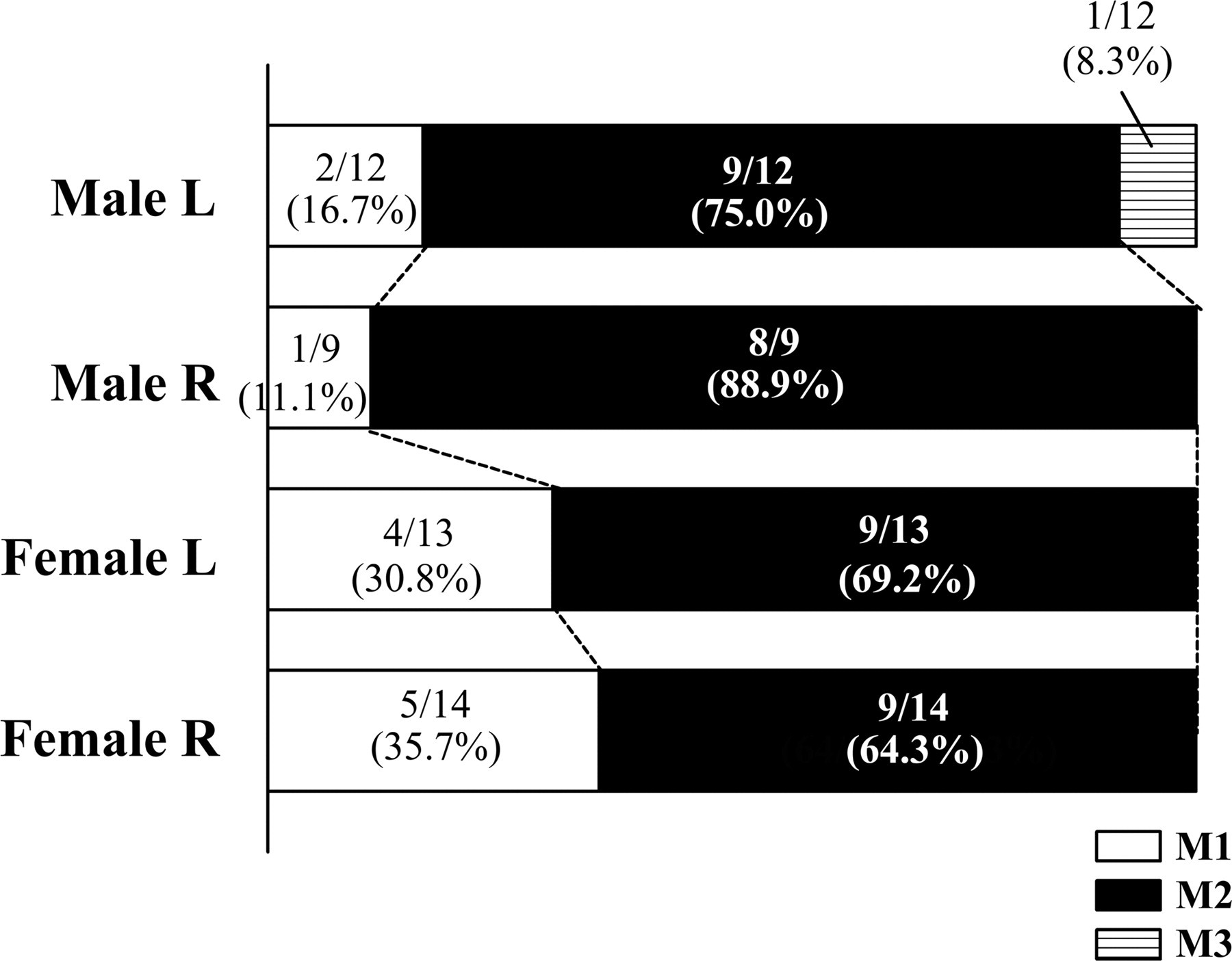
The frequency of caries in the mandibular molar region of WBN/KobSlc rats is shown in Figure 2. It was most frequent in the second molar (M2) at more than 60%, followed by the first (M1) (11.1–35.7%) and third (M3) molar (0.0–8.3%). M2 was affected far more often than other molars. There was no significant difference between M1 and M3.
Histopathological examination of caries in WBN/KobSlc rat
Histopathological lesions corresponding to grade 1-type caries on a soft X-ray were necrotic dentin, dentinal tubules with a bacterial colony, and a beading and coalescence of dental tubules on the crown surface. Progressing to grade 2-type caries on a soft X-ray, a cleft had formed both vertically and horizontally in the crown (Figure 1I). That lesion was accompanied with a second dentin formation and telangiectasia in the dental pulp. The cleft spread throughout almost the entire crown in grade 3-type caries on a soft X-ray (Figure 1J), and an abscess with necrosis was evident in the pulp.
Sugar content in diet
The contents of free sucrose, glucose, fructose, lactose and maltose in the diet (CRF-1) used in the present study were 2.61%, 0.06%, 0.10%, 0.30% and 0.05%, respectively. Free ribose and galactose were not detected.
Bacteriological study
Numbers of Streptococcus and Lactobacillus species as well as the total bacterial count had increased remarkably in male WBN/KobSlc rats with caries compared with those in male WBN/KobSlc and male F344 rats without caries (Figure 3). By biochemical identification, S. mutans (pH < 5.1), S. ferus (pH < 4.5), L. plantarum (pH < 4.5) and L. fermentum (pH < 6.5) were isolated from the oral bacteria of male WBN/KobSlc rats with caries. Adhering materials were clearly observed in the medium of S. mutans and S. ferus.
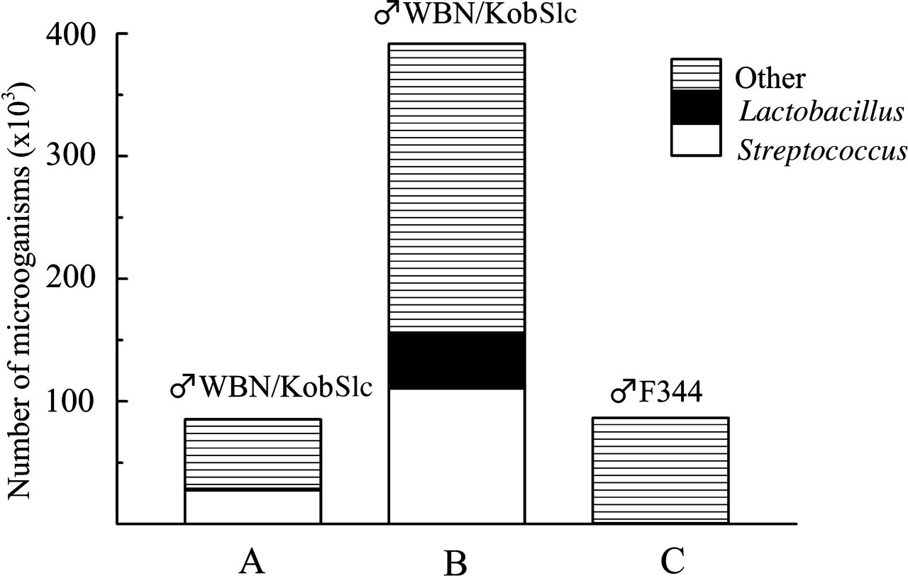
Mean viable bacterial counts in the flora of mandibular occlusal molar surface of WBN/KobSlc rats. (A) WBN/KobSlc male rats of 4.2-month-old without caries (n = 2). (B) WBN/KobSlc male rats of 8.6-month-old with caries (n = 3). (C) Male F344 rats aged 10.5–12.1 months without caries (n = 3)
Discussion
Dental caries were not usually induced without cariogenic diets in any experimental animal models including rats. Sucrose in the diet has strong cariogenicity because it is a specific substrate for synthesis of insoluble glucan and has fermentable capacity for acid formation. Many other fermentable monosaccharides or disaccharides including glucose, fructose, lactose and maltose, oligosaccharides and starches also have caries-promoting potential as substrates to acid formation but their cariogenic risk is far lower than that of sucrose. 8 Therefore, large amounts of sugars or carbohydrate were added to the standard diet to achieve the successful induction of caries in experimental animals. These cariogenic diets facilitated the development of dental caries within three months in various rat strains, including F344. 3 An analysis of the diet in our experiment showed that CRF-1 contained merely 2.6% of free sucrose, and that it provided a far lower level of sugar than the established cariogenic diet containing a high sucrose content. 3 It is highly probable that the CRF-1 diet containing a low sucrose content shows little caries activity and does not usually induce caries in general rat strains.
A CRF-1 diet apparently induced caries from the rather young age of more than 3.5 months in both sexes of WBN/KobSlc rats. Rats susceptible to caries have been reported in several strains such as the Hunt-Hoppert rat, 9 Harvard caries-susceptible rat 10 or Osborne-Mendel rat. 11 Yet, even these susceptible strains require caries-inducing factors such as a diet high in sucrose or carbohydrate, or an infection with pathogenic bacteria to deliberately induce caries. It is not yet clear that there is a definite difference in caries susceptibility to exactly the same diets including CRF-1 between reported caries-susceptible strains and the WBN/KobSlc strain. At any rate, it is apparent that WBN/KobSlc is a caries-susceptible strain, and is of considerable interest that even a standard diet low in sucrose may induce the early onset of dental caries in such a strain.
König 12 studied the effect of particle size of corn and sugar diets on the caries incidence of rats and observed that diets containing finely ground corn were more caries-conducive than those containing coarse ground corn. Meanwhile, some cereals were reported to be conducive to caries when coarsely ground than when finely ground because coarse particles were packed in and retained by the fissure. 13 In addition, Bibby and Sedwick 14 observed that rats which fed a coarse-particle diet exhibited a considerable increase in fractures of cusp tips. In contrast, Will et al. 15 reported that the number and degree of fractures in the teeth of rats fed coarse- and fine-particle diets were not statistically significant. We cannot completely deny the possibility that the particle size or fractures of cusp tip may affect dental caries in WBN/KobSlc rats. However, the CRF-1 diet has never before been reported to be cariogenic while being widely used for experimental rats in many studies including long-term toxicological and carcinogenicity investigations lasting over two years (corresponding to the lifespan of rats). Moreover, in the present study no dental carious tooth was observed in F344 rats fed CRF-1, even under the same conditions as the WBN/KobSlc strain. Therefore, it is highly unlikely that the fractures of cusp tips induced by the particle size of the CRF-1 diet may be confined to the WBN/KobSlc strain. Thus, follow-up studies are necessary to confirm the effect of finely- or coarsely-powdered cariogenic diet on dental enamels of the WBN/KobSlc strain in future.
The oral flora of male WBN/KobSlc rats contained similar ratios of Streptococcus spp. regardless of whether they had dental caries or not, while those bacteria were scarcely visible in F344 rats. S. mutans is a well-known major cariogenic bacterium not only forming adherent insoluble glucan but also producing acids. 16,17 S. ferus, a bacterial species originally isolated from rats, has ‘mutans-like’ cariogenicity. 18 Both of those genera were isolated from the plaque of male WBN/KobSlc rats with caries. Lactobacillus spp. are also known to be cariogenic genera and capable of causing caries in enamel 19 due to decalcification by Lactobacillus-produced lactic acid. In the present study, the numbers of genera were remarkably increased in the oral flora of male WBN/KobSlc rats compared with that of F344 rats, and the pH reading of the culture from Lactobacillus spp. was less than 6.5, which is sufficient to decalcify tooth components. Therefore, the incidence of dental caries correlated well with the proliferation of inherent cariogenic bacteria in the oral flora of WBN/KobSlc rats.
As with F344 rats in the present report, many other strains do not develop any dental caries, even though they have immature enamel on the occlusal surface of their molars, 3 suggesting that most rat strains are likely to be able to eliminate cariogenic bacteria on the enamel of molars. On the other hand, it seems that WBN/KobSlc rats lose this ability at around the postnatal age of 3.5 months. It has been reported that the incidence of dental caries rises under prediabetic or diabetic conditions. 20 WBN/KobSlc is a strain in which almost all males show prediabetic symptoms attributable to pancreatitis, 5 with the onset corresponding to that of dental caries. However, female WBN/KobSlc rats have developed neither pancreatitis nor diabetes, and there was no difference in the incidence or severity of molar caries between the sexes. Thus, it is unlikely that prediabetic or diabetic conditions are directly associated with the onset of dental caries in this strain.
Based on all the data in this report, it is clear that WBN/KobSlc rats may provide a new model for dental caries needing no bacterial infection or addition of sugar.