
Abstract
Diagnostic medical sonography (DMS) is an operator and patient dependent examination where anatomical and hemodynamic analysis settings have to be adjusted in real-time while scanning. Several techniques were developed so far to perform the DMS acquisition and post processing adjustments quickly and to maximize their easiness. A new easyMode Technology was recently implemented in order to drastically simplify the operator system setup to only three macro-parameters related to the desired effect in terms of: resolution/penetration, contrast/soft, smooth/sharp. A multidimensional algorithm varies simultaneously several internal system parameters to modify the image in the direction of the macro-parameter varied by the user. No prior knowledge of ultrasound physics or technology is required to adjust the macro-parameter, the effects of which are directly visible on the sonographic image in real-time (for instance, to optimize the level of smoothness with respect to sonographic image contrast). The aim of the easyMode is to increase productivity, reduce sonographer’s work-related musculoskeletal disorders due to extensive use of the ultrasound system control panel, and increase diagnostic confidence based on the intuitive optimization of the sonographer’s desired effect, instead of an engineering approach to real-time scanning optimization.
Introduction
Diagnostic medical sonography (DMS) is an operator and patient dependent examination where anatomical and hemodynamic analysis settings have to be adjusted in real-time while scanning (Figure 1).
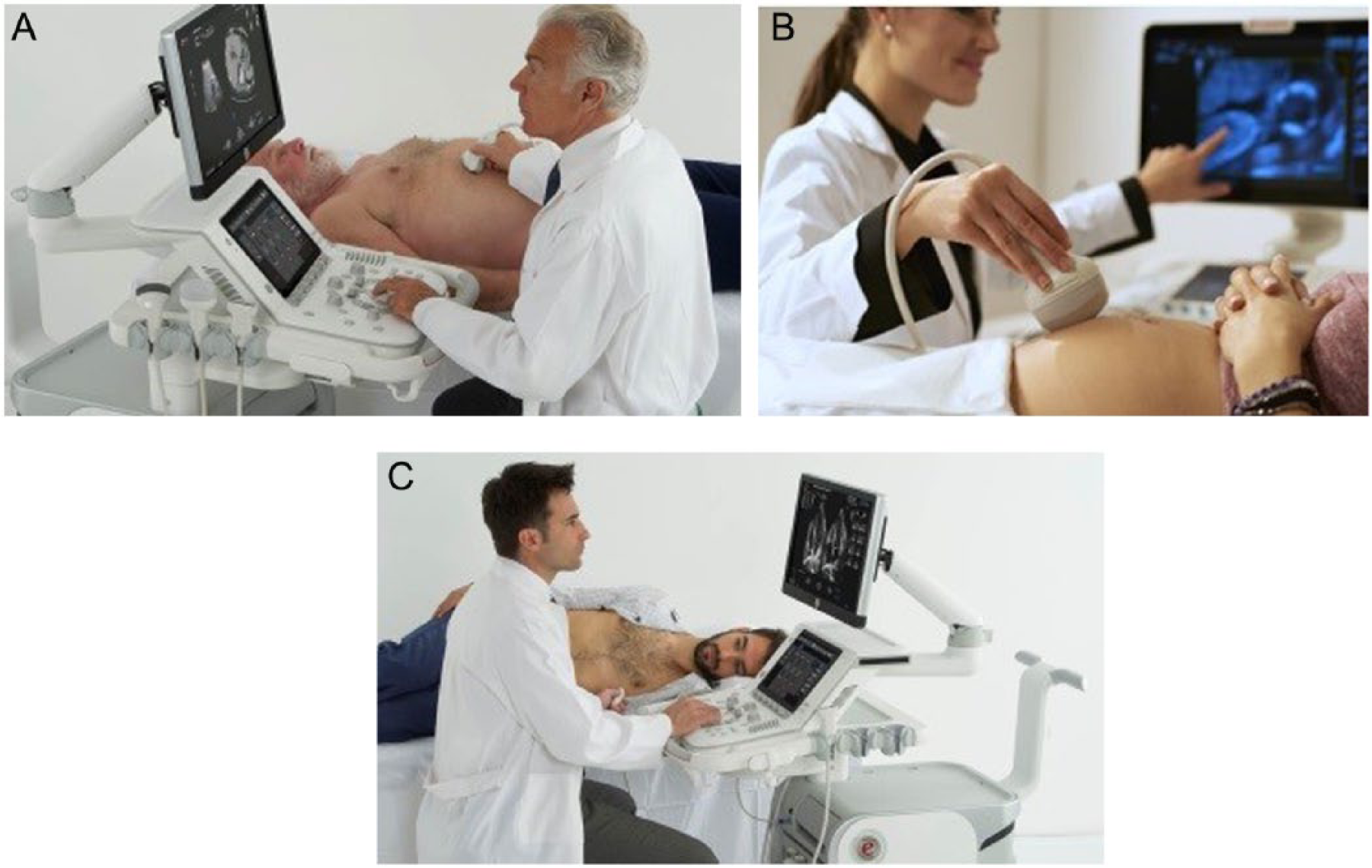
Examples of DMS scanning in different clinical applications. Abdomen (A); Obstetrics (B); Cardiac (C).
More and more DMS investigations are performed per day worldwide, with productivity being one of the main key drivers for users. 1
Ultrasound systems are used for many clinical applications (Adult Cardiology, Pediatric Cardiology, Neonatology, Vascular, Adult Cephalic, Ophthalmic, Abdomen, Small Parts, Thyroid, Urology, Musculoskeletal, Obstetrics, Gynecology, Breast, Lung ultrasound, Intraoperative, Neurosurgery, Fusion Imaging for traditional and non-traditional applications for both diagnostic and interventional purposes, Interventional Radiology, Critical Care, Emergency).
Furthermore, the patient population can be represented by Fetal, Neonatal, Pediatrics, Adult, while the user profile, intended to cover the full range from novices to expert users, is populated by Sonographers, Medical Doctors, Radiologists, Surgeons, Veterinary Practitioners, Midwives, Paramedics. Due to their wide diffusion, ultrasound systems are used also by non-sonographers; therefore, a higher level of usability has been urgently requested in recent years.
These new paths in the use of DMS have completely changed the customers’ approach to the technologies and devices, as well as the market perception of such diagnostic technology, which is the only one, in the field of complex diagnostic imaging, characterized by a real-time nature. 2
The ergonomics and workflow of ultrasound systems is nowadays of primary importance due to the increased use of ultrasound systems in the everyday clinical practice even by “non-sonographers,” as well as the increased attention to the problem of Work-Related Musculoskeletal Disorders (WRMSD) for sonographers.3–6
Methods
Several techniques were developed so far to perform the DMS acquisition and post processing adjustments quickly and to maximize their ease of use. Just to highlight the most important:
Preset—Pre-defined image setting depending on the chosen body district;
Auto Gain—Automatic Gain reset to a pre-defined level (which usually varies per application);
Automatic Adjustment—Real-time parameter adjustment to a pre-defined target type of image;
Continuous Automatic Adjustment—Continuous real-time parameter adjustment, which varies depending on the acquired echo image characteristics.
An easy adjustment technology (easyMode; Esaote S.p.A., Genova, Italy) was recently implemented with the goal to simplify the operator system setup. easyMode offers a real-time parameter modification, which takes into account the final effect the operator wants to obtain on the image (resolution/penetration; contrast/soft; smooth/sharp), instead of being based on the adjustment of the technical parameters of the ultrasound system (direct changes on Dynamic Range, Gray Map, Scanning Lines, Spatial Compound, etc.).
a. Presets
Most ultrasound systems have preset modes that provide better imaging of specific organs of the body. 7
Preset (to set the acquisition parameters to pre-defined values) is the basic technique to group and speed up the optimization of acquisition parameters.
The Preset activation and selection control is one of the most important ones in an ultrasound system. Selecting the right preset for the particular region of the body under examination makes scanning much easier. Built-in software assists not only with dedicated labelling and measurements, but it enables better images and clips to be acquired. Moreover, also the physical parameters are optimized for the desired application. 8
The correct scanning preset has a large impact on the quality of the image. The ultrasound system optimizes the image for each particular scan type, for example, abdominal preset vs. pelvic or gynecological preset. Any sonographic study has to begin by selecting the correct probe and appropriate preset for the type of exam being performed. 9
In any case as ultrasound machines have developed, the amount of practitioner input required to achieve an acceptable image has diminished, possibly resulting in an increased reliance on presets and the ultrasound machine’s optimization button. Whilst under average conditions presets provide an appropriate starting point, the practitioner is still expected to utilize the full range of ultrasound machine functions to make adjustments and manually optimize the image. 10
Additional manual adjustments may be necessary in most of the cases, which forces the operator to know and apply in real-time changes related to physical and technical principles of sonographic technology.
b. Auto Gain
Changing the gain is analogous to amplifying or suppressing the volume of signal in an image. By adjusting the gain up and down, you may find it easier to visualize certain structures. Most ultrasound machines are equipped with an Auto Gain, which is the machine’s interpretation of the optimal level of gain for the body part or structure being scanned. When first starting out with scanning, the use of Auto Gain is likely going to be a useful tool. 11
As the ultrasound beam travels deeper into the patient body, the returning echoes are attenuated, resulting in less resolution. A feature known as Time Gain Compensation (TGC) allows the sonographer to adjust the image brightness at specific depths. The top row of sliders controls near field gain, whereas the bottom row of buttons controls far field gain (Figure 2). The Auto Gain control resets the ultrasound machine back to standard gain presets for the type of scan (Preset selected) being performed. 12
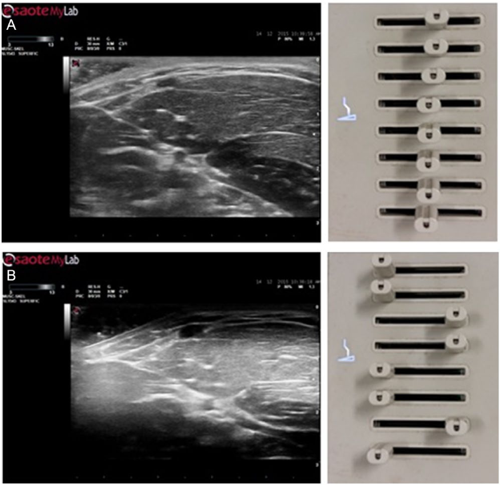
(A) TGC sliders position effect on the sonographic image—homogenous TGC setting; (B) inhomogeneous TGC of the near and distant fields (dark) with respect to the middle field (saturation).
c. Automatic Adjustment
Organ specific imaging (or tissue specific imaging) is an approach applying automatic adjustment of the scanner commands (gain, image optimization algorithms, TGC, etc.) depending on the organ that the operator has selected for viewing (through dedicated Preset selection). This results in shortening the investigation time and also in some degree of standardization of images. 13
Automatic adjustment has been introduced relatively recently in order to automatically optimize the echo image to a defined histogram obtained by the analysis of expected similar acquisitions.
This technology, even if recently improved, cannot consider specific patient and/or examination dependent characteristics, which anyway forces the image appearance to a predefined histogram representation (always related to ultrasound physics and technical knowledge).
d. Continuous Automatic Adjustment
It represents the latest update of the Automatic Adjustment, where it is the operator that recalls the Automatic Adjustment function once the image quality is not satisfactory, instead of having the ultrasound system automatically and virtually continuously re-apply the Automatic Adjustment function once the sonographic image characteristics change with respect to a pre-defined group of parameters.
e. easyMode Technology
A new technology was recently implemented on an ultrasound system prototype with the goal to simplify the operator system setup to only three macro-parameters related to the desired effect in terms of: resolution/penetration, contrast/soft, smooth/sharp.
The goal of such technology was to simplify the tuning process of the image by the operator considering the adjustment based on the desired effect to apply to the image, instead of forcing the ultrasound system user to understand and master the technical and physical sonographic parameters and then to act on them for their optimization.
Results
In all the traditional techniques for image quality adjustment listed above (Preset; Auto Gain; Adjustment; Continuous Automatic Adjustment) some manual adjustments may be needed on the image obtained after the technique re-call (Preset; Auto Gain) or after the output of image processing (Automatic Adjustment; Continuous Automatic Adjustment). These adjustments have to be performed considering the usual technical and physical parameters of the ultrasound system, therefore forcing the sonographic user to master the physics and implemented technology, which is behind the sonographic image acquisition and formation.
These approaches, therefore, assume a deep technical knowledge of the sonographer, therefore possibly limiting the scanning to expert users and, in any case, possibly lengthening the sonographic use learning curve, as it must include a large section related to physics, technology, and the so-called “knobology,” which considers how the technical theory has been implemented within the ultrasound system and how the operator has to deal with (usually such implementation is ultrasound system manufacturer dependent and it may vary also between different systems of the same manufacturer).
easyMode was developed in order to overcome the limits of traditional image set up and adjustment technologies, and it is currently in the implementation phase on an ultrasound prototype system. Tests are in place on phantoms and ex vivo with respect to the above listed technologies.
In order to develop easyMode, the operator approach to ultrasound system parameters adjustment was completely reconsidered: instead of starting from an engineering point of view and then determining how the clinicians have to approach and apply it, the focus of the technology development and implementation was the sonographer’s point of view.
With easyMode, the ultrasound system understands and reacts according to the user’s needs, instead of forcing the sonographer to understand the technology and its internal processes.
In the traditional approach, the ultrasound technology and physics has to be understood to obtain the desired clinical outcome (in terms of sonographic image characteristics needed for a confident diagnosis), while in the easyMode environment it is the ultrasound system which has to re-organize itself in terms of acquisition, processing, and visualization parameters to ensure the outcome desired by the system user.
This outcome was obtained by re-thinking the signal processing and its related controls in a multi-dimensional space, where the standard parameters are varied along one of the easyMode space dimensions, following a pre-defined curve with a particular path and slope depending on its content, variation, and starting/ending levels.
Within the easyMode multidimensional space each single traditional parameter represents a line in a single dimension. While the easyMode space is adjusted, several single dimension parameters are changed according to a multi-dimensional matrix.
For a given easyMode multidimensional space direction (for instance, contrast/smoothness) some single dimension space parameters can be involved (e.g., dynamic range, dynamic compression, gray map, image quality enhancement algorithm parameters, etc.; see Figure 3). To correlate the three points of the multidimensional space to the one-dimensional space of each single parameter, a correspondence matrix with single dimension parameters was defined (for instance, the two range extreme points: max contrast/max smoothness and the factory default setting as multidimensional intermediate point). A parametric curve for each single dimension parameter was computed as the best fit with respect to the above-mentioned three points. Therefore, when the system user adjusts the easyMode along one of the multidimensional lines, all the related ultrasound system acquisition and processing parameters change, each one following the computed curve simultaneously, modifying the image in real-time from one extreme setting to the other. The parameters, which are changed within the easyMode multidimensional space, are available to the system user within the traditional user interface, as well as some internal—not user-accessible—parameters of the system.
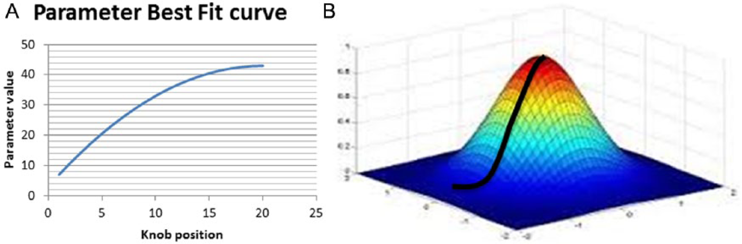
Single dimension space parameter (A); easyMode multidimensional space (B).
The easyMode user interface is constituted by three virtual sliders on the touch screen, which are an integral part of the ultrasound system control panel (see Figure 4).
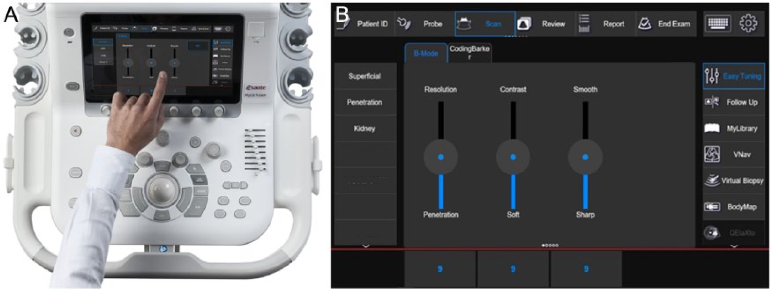
easyMode virtual sliders (A); close up on the easyMode touch screen user interface (B).
In easyMode a multidimensional algorithm simultaneously varies several internal system parameters to modify the image in the direction of macro-parameter varied by the user (resolution/penetration; contrast/soft; smooth/sharp; see Figure 5). No prior knowledge of ultrasound physics or technology is required to adjust the macro-parameter, whose effect is directly visible on the image in real-time (for instance, to optimize the level of smoothness with respect to image contrast).
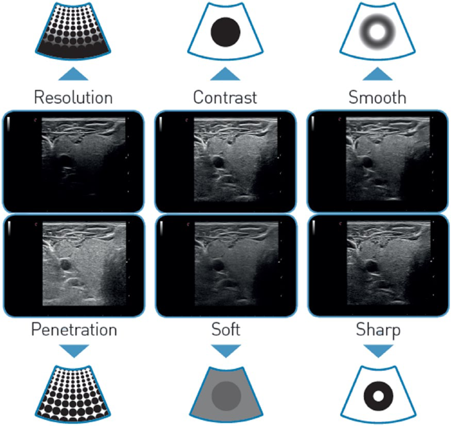
easyMode examples of resolution/penetration (left), contrast/soft (center), smooth/sharp (right) image adjustment.
The traditional system adjustment environment is separate with respect to the easyMode environment, due to the completely opposite approach to the ultrasound system setup and tuning process, and also because the easyMode result may have an outcome in a multidimensional space point that cannot be achieved with any multi-parameter adjustment obtained in the traditional technically/physically-focused approach).
Conclusion
The easyMode ultrasound system optimization approach was carried out by completely re-thinking the system user approach to image optimization: from forcing the sonographer to deeply understand ultrasound physics and technology (the implementation of which varies from one system manufacturer to another), to an intuitive approach that enables the operator to concentrate only on the effects he/she wants to obtain on the final sonographic image in terms of optimization and balancing between resolution and penetration, smoothness and contrast, improved image details of increased frame rate in terms of acquisition and visualization.
easyMode was developed with the goal to:
reduce the number of End User image controls;
obtain faster changes in image settings also for sonographic users with limited knowledge of the system workflow;
reduce the possibility to obtain «bad tuning» configuration for less experienced sonographer.
The aim of the easyMode is to increase productivity, reduce sonographer’s work-related musculoskeletal disorders due to extensive use of ultrasound system control panel, and increase diagnostic confidence based on the intuitive optimization of the sonographer’s desired effect, instead of an engineering approach to real-time scanning optimization.
easyMode advanced optimization technique is suitable for drastically simplifying the sonographer approach to sonography by freeing the operator from the deep knowledge needed to optimize image and hemodynamics acquisition parameters, especially in more complex clinical applications such as radiology, cardiology and ob-gyn.
Footnotes
Declaration of Conflicting Interests
Carlo Biagini, MD, PhD, was involved in the ideation and evaluation of the technology. Giampaolo Borreani, Roberto Pesce, Luca Bombino, MSc, and Leonardo Forzoni, MSc, are employees of Esaote S.p.A., and were the technical developers of the technology discussed in this article. Their contribution for the article was limited to the technical explanation and technological support.
Funding
The authors received no financial support for the research, authorship, and/or publication of this article.
HCI International 2017 – Posters’ Extended Abstracts. HCI 2017. Communications in Computer and Information Science, “Intuitive Real-Time Multidimensional Diagnostic Ultrasound Image Optimization Technology,” vol 713, 2017, pgs. 511–518, Borreani G., Biagini C., Pesce R., Bombino L., Forzoni L., (original copyright notice as given in the publication in which the material was originally published). “With permission of Springer.”