
Abstract
A galactocele is a cyst that forms from the obstruction of a lactiferous duct within the breast. These cysts typically form when a woman is pregnant or lactating and are usually of periareolar location. Mammography and sonography are common imaging modalities used to detect these cysts. This case study presents the sonographic detection of a galactocele with an abnormal location and appearnace in the left axilla.
Introduction
Galactoceles are cysts that typically develop within the breast. 1 These cysts occur when milk in the ducts of the breast fails to travel outward, becoming stagnant. This ultimately produces a fatty or milk-containing lump. 2 These cysts are most commonly detected after a woman ceases breastfeeding but may also develop anytime from the third trimester of pregnancy through the beginning of lactation. 3 This case study presents a variable location and sonographic appearance of a galactocele, which was located in the patient’s left axilla and did not exhibit internal echoes but appeared completely anechoic. This case also shows the use of sonography to identify, image, and diagnose the presented pathology.
Case Report
A two-week postpartum woman presented to the sonography department for a soft tissue sonographic examination because of a palpable lump noted in the left axilla. The lump was not painful, and the patient stated that the lump had been decreasing in size since taking a prescribed antibiotic. A linear array transducer with a frequency of 12 MHz was used with a Philips iU22 (Philips Ultrasound, Bothel, Washington) ultrasound machine.
Sonography showed a single, well-circumscribed, anechoic structure measuring 2.32 × 1.57 × 0.50 cm and displaying posterior enhancement in the left mid axillary region (Figures 1 and 2). Hetereogeneous tissue was visualized surrounding the anechoic area, measuring 3.34 cm in width (Figure 3). Color Doppler evaluation demonstrated little to no vascularity within the mass or surrounding tissue (Figure 4). Based on the sonographic findings, a probable diagnosis of galactocele versus cyst was suggested. Due to the benign nature of the diagnosis, no further imaging was suggested, and the patient was informed to monitor the lump and notify the doctor of changes.
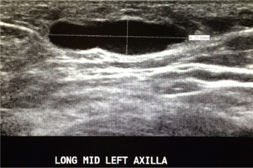
Longitudinal gray-scale sonogram in the mid left axilla showing a single, anechoic structure with posterior enhancement.
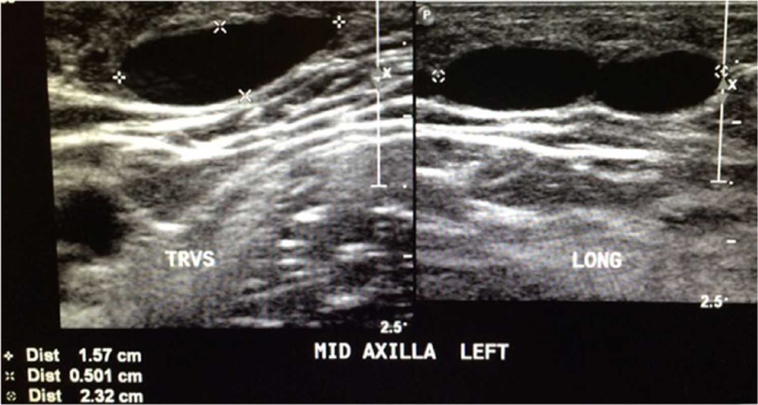
Transverse and sagittal gray-scale sonogram in the mid left axilla showing the anechoic mass measuring 2.32 × 1.57 × 0.50 cm.
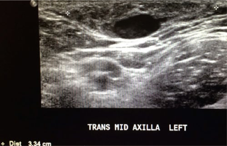
Transverse gray-scale sonogram in the mid left axilla showing the mass surrounded by heterogeneous tissue measuring 3.34 cm in width.
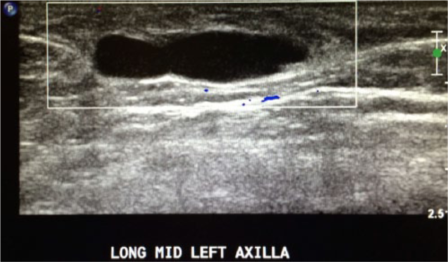
Sagittal color Doppler image of the left axillary mass showing little to no vascularity within the mass or surrounding tissue.
Discussion
The breast is a superficial organ containing the mammary, or milk-producing, glands. The breast itself consists of three different layers: the subcutaneous layer, the mammary layer, and the retromammary layer. The mammary, or middle, layer of the breast houses the parenchymal elements. Parenchymal elements include the lobes, ducts, and acini. 4 Several changes occur in the breast of a pregnant or lactating woman because of the effects of hormones like estrogen, prolactin, and progesterone. Specifically, an increase in estrogen production causes changes in the ducts, while an increase in progesterone causes the lobules of the breast to increase in size. 3 Lactation causes the lobules of the breast to expand and milk secretion to accumulate in the ducts. If the accumulated milk remains in the duct for an extended period of time, a galactocele may begin to form. 3
Galactoceles are considered common, accounting for approximately 4% to 5% of Breast Imaging Reporting and Data System (BI-RADS) category four lesions when core needle biopsies are performed. 5 A galactocele typically contains low-level echoes and is located within the actual breast itself. The breast itself is divided into four quadrants: the upper outer and inner quadrants and the lower outer and inner quadrants. The upper outer quadrant, which contains the superior and lateral regions of the breast, extends to the axilla and houses a higher concentration of lobes compared to any other breast quadrant. 6 Because of this, tumors and other masses may arise in this area. The axilla contains ducts and lobes as well as lymphatics that drain the breast. 6 Although this is not a typical location for galactoceles to occur, it is possible due to the multiple ducts and channels located in this area.
Galactoceles are often periareolar in location. 1 The lactiferous ducts, which secrete milk, lie posterior to the areola and converge before exiting through the nipple. Because of the higher concentration and closer proximity of the ducts in this area, a galactocele is more likely to form. 4 Ductal dilation often accompanies this simple cyst. Mastitis and abscesses are also associated with this abnormality due to milk becoming inert. 1 The patient reported here had no signs of either.
Mammography is typically used as the initial screening tool for breast-related abnormalities; however, the pathology presented in this case was found through sonographic evaluation only. Sonographically, galactoceles typically will appear to be well marginated, having a round or oval shape. Galactoceles may also exhibit internal echoes. The presence of these echoes is caused by the fatty, milky material associated with nursing and breastfeeding. 2 Some galactoceles may appear to have intracystic hypoechoic material and will exhibit some form of posterior acoustic enhancement or echogenic boarders. 5 Color Doppler imaging may show vascularity surrounding the cyst but not within. 5 Galactoceles are considered benign and often resolve to oil cysts after a patient has finished nursing. 1
Complex internal echo patterns and posterior acoustic enhancement are two characteristics often seen with pregnancy-associated breast diseases. 3 Differential considerations for galactoceles include simple breast cysts, oil cysts, and ductal dilation. 1 Sonography is a dependable imaging modality used to diagnose breast diseases that occur during pregnancy and lactation. 3 The more subtle features of fibroglandular tissue become more noticeable, compared to their appearance on mammography, exhibiting increased echogenicity. The vascular characteristics of surrounding tissues also increases. 3 Sonography is also preferred in aiding such a diagnosis because it does not expose the patient to ionizing energy.
Ultrasound guided cyst aspiration can be performed to confirm the contents of the mass, as is true with any breast mass that does not meet the criteria of a simple cyst. 5 Cytopathology may be done in conjunction with cyst aspiration to determine if the contents of the cyst match the diagnosis of galactocele and to determine if the cyst is simple or complex. 5 A core needle biopsy is often performed to aid in the diagnosis of breast pathologies, but such a biopsy poses the risk for the formation of a milk fistula, a tract between a lactiferous duct and the skin, or infection.5,7 However, if a mass found during pregnancy is concerning for malignancy, a biopsy should still be considered. 7
Galactoceles have been widely reported in the literature. In a similar case to the one here, a 36-year-old, one-week postpartum woman was diagnosed with an axillary galactocele. 5 She had presented with a palpable lump in the right axillary region. The lump was tender but had not caused any changes in the skin. The lump appeared to increase in size after the patient began breastfeeding. The sonographic evaluation in that case showed an anechoic cystic structure that also contained low-level echoes and heterogeneous appearing subcutaneous tissue. Color Doppler imaging showed little to no vascularity within the cystic area but increased vascularity in the surrounding heterogeneous tissue. 5 That case also reported that galactoceles appear sonographically as well-circumscribed cystic structures that exhibit posterior enhancement. 5 A “wavy line” may be seen separating a galactocele into compartments of varied echogenicities. The internal echogenicity or fat fluid levels of a galactocele is highly dependent on its content. A galactocele with higher milk or fat content will appear more hypoechoic rather than anechoic. To identify a mass as a galactocele, the cystic structure should exhibit either an anterior or posterior echogenic rim. 5
Conclusion
Mammography is typically done before examining the breast sonographically when a lump is palpated. However, because of the atypical location of the pathology in this report, sonographic evaluation was done as the initial screening tool to image and examine this axillary galactocele. The sonogram characterized a single, anechoic structure located in the patient’s left axillary region. This case depicted a less common variation in a known pathology of nursing women and supports sonography as a beneficial tool in imaging and assisting with diagnosis of similar conditions.
Footnotes
Acknowledgements
The authors would like to thank Amy Gerardot, RDMS, for her support and guidance and Sharlette Anderson, MHS, RDMS, RDCS, RVT.
Declaration of Conflicting Interests
The authors declare no potential conflicts of interest with respect to the research, authorship, and/or publication of this article.
Funding
The authors received no financial support for the research, authorship, and/or publication of this article.