
Abstract
Hepatocellular carcinoma (HCC) is the most common type of primary hepatic malignancy. Most cases of primary hepatic carcinoma are secondary to either viral hepatitis infections (hepatitis C or hepatitis B) or cirrhosis, with chronic alcohol abuse being the most common cause of hepatic cirrhosis. Echocardiography is done primarily to evaluate the overall function of the heart; the condition of the heart muscle, heart valves, and chamber sizes; and the risk for heart disease. It is also used to evaluate the effectiveness of medical or surgical treatment over time. The case presented emphasizes the importance of performing a complete echocardiographic protocol on each patient and being aware of the surrounding structures that might be seen at the periphery of standard images. During an echocardiographic examination, diseases other than heart disease may be found incidentally and should be reported to the referring physician to optimally manage the patient’s care.
Keywords
Echocardiography is used primarily for the diagnosis of heart diseases such as hypertrophic cardiomyopathies (myocardial disease), valvular stenosis or insufficiency (endocardial disease), and pericardial effusion and constrictive pericarditis (pericardial disease). 1 It is also very important in the follow-up of patients after medical or surgical therapy to provide physicians the necessary anatomic and hemodynamic data on which to base management decisions. Although echocardiography is used primarily to diagnose heart disease, the operator must be aware of all the findings of an examination, including any incidental findings unrelated to cardiac disease that might appear on the periphery of images taken during a scan.
Hepatocellular carcinoma (HCC) is the most common type of primary hepatic malignancy and ranks in the top five of malignancies worldwide, although it is seen less commonly in the Western world than in Africa or Asia and parts of the Far East.2,3 The incidence of HCC is increasing in the United States,3,4 and if it can be diagnosed at an early stage, typically with sonography, it can be treated more successfully than in its later stages, where the five-year survival rate is only approximately 5%.5–11 This case report presents a patient with an unsuspected hepatocellular carcinoma found during a routine echocardiographic examination.
Case Report
A healthy asymptomatic white male in his 90s was referred for echocardiography as a follow-up after coronary angioplasty in 2003 and a pacemaker placement in 2005. A complete echocardiographic study protocol was followed using a Vingmed cardiovascular system (GE Vingmed Ultrasound, Horten, Norway) with a 2.5-MHz phased array transducer. During the subcostal view, a 4 × 3.7-cm hyperechoic mass was detected in the left lobe of the liver. This finding was documented (Figures 1–3) and reported to the referring physician, who followed up with a computed tomography (CT) scan. The CT scan confirmed the size and location of the liver mass, and the patient was sent for a liver biopsy. Pathology showed the mass to be a hepatocellular carcinoma.
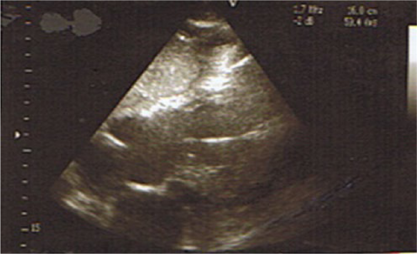
Two-dimensional subcostal four-chamber view showing the pacemaker wire in the right ventricle; a small segment of the left lobe of the liver can be seen along the upper left edge of the image, which shows a part of the hyperechoic mass.
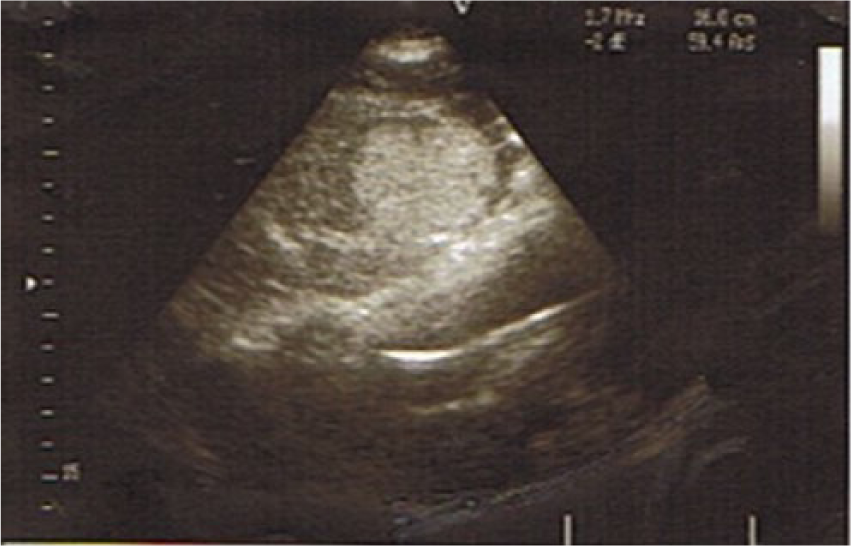
Two-dimensional subcostal four-chamber view showing the pacemaker wire in the right ventricle; the left lobe of the liver is clearly seen showing the extent of the hyperechoic mass.
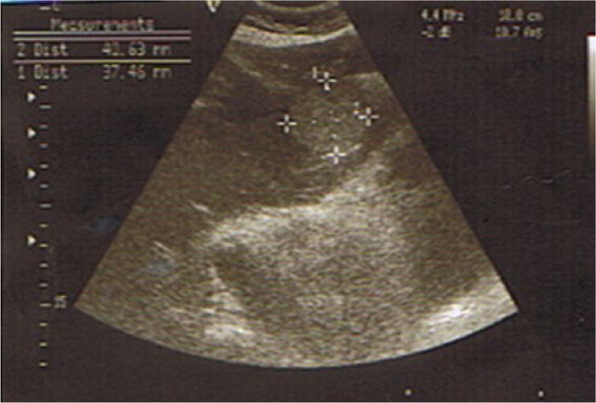
Two-dimensional subcostal four-chamber view focused on the left lobe of the liver showing the measurement of the hyperechoic 4 × 3.7–cm mass.
Review of the patient’s history showed a resection of early stage rectal cancer in 2003. Follow-up CT scans and a magnetic resonance imaging (MRI) study in 2006 showed the patient to be free of any detectable disease. In 2012, serum blood markers used to detect the presence of primary colorectal cancer were negative. The patient had a history of cigarette smoking, although he had quit 15 years ago; the patient is an active drinker and averages two to three alcoholic drinks per day.
Discussion
Hepatocellular carcinoma is the most common primary tumor of the liver; however, the most common tumors found in the liver are not primary but secondary ones that arise from metastases from other sites of the body such as the colon, breast, or upper gastrointestinal system.2,3 Although the prevalence of hepatocellular carcinoma in the Western world is less than in Africa or Asia and parts of the Far East, there has been an increase in the number of cases of hepatocellular carcinoma in the United States over the past two decades. 2 Risk factors for the development of HCC include infection with the hepatitis B (HBV) and/or C (HCV) virus, particular comorbidities or conditions, and certain external sources. 12 Chronic HBV infection is the leading cause of HCC in Asia and Africa, whereas HCV infection is the leading cause of HCC in Europe, Japan, and North America.5,13 The incidence of HCC is increasing in the United States, where currently there are approximately four million patients chronically infected with HCV.4,14 In the United States, HCC is more common in men than in women by a factor of approximately 3:1 and is more common in African Americans than in whites. It is usually found in patients 50 years and older.2,6,7 However, because of the high incidence of HCV in specific patient groups, such as transfusion-related HCV, and with more evidence supporting intravenous drug use as one of the leading risk factors for spread of the virus, the age-specific incidence of this cancer in the United States has progressively shifted toward younger people. 4 Other common risk factors for HCC are alcoholism, cirrhosis, hemochromatosis, Wilson disease, and type 2 diabetes.12,15
Patients with HCC typically present with symptoms of anorexia, an enlarged abdomen, easy bruising or bleeding, jaundice, and abdominal pain or tenderness, especially in the right upper quadrant.3,6 Physical examination may show an enlarged tender liver. In the case presented, however, the finding was truly an incidental one as the patient showed no signs or symptoms.
When patients present with any of the signs or symptoms noted above, the initial examination is usually abdominal sonography, which may be followed up by a CT scan or MRI and laboratory bloodwork such as liver enzyme studies and serum α-fetoprotein (which is elevated in approximately 70% of cases). 3 Any positive findings are then followed by a liver biopsy for tissue diagnosis.3,5 In this case, the initial examination was transthoracic echocardiography, which then led to appropriate patient management.
The sonographic appearance of HCC is generally nonspecific and changes as the lesion becomes larger. Most small lesions initially appear as a solid hypoechoic mass. As the lesion grows larger, it may develop central necrosis, which leads to a heterogeneous, somewhat hyperechoic mass that often has posterior acoustic enhancement.16–23 In the case above, the sonographic appearance was a nearly round solid hyperechoic (relative to the patient’s normal surrounding liver) mass, as shown in Figures 2 and 3. Most cases of HCC present with associated cirrhosis of the liver, causing diffuse hyperechogenicity of the liver, which likely contributes to the relative hypoechoic appearance of HCC.12,16,22 A characteristic of HCC in all stages of development is its hypervascularity, which can be readily identified sonographically using color or power Doppler imaging. 3 Newer technologies in ultrasound such as compound scanning, which can significantly decrease the amount of posterior acoustic enhancement seen, and elastography may affect the sonographic characteristics of HCC, and sonographers should be aware of that possibility.23,24
Treatment options and the prognosis for HCC depend on many factors, particularly tumor size, staging, and tumor grade. A high-grade tumor will have a poor prognosis, while a low-grade tumor may go unnoticed for many years. Aggressive surgery or a liver transplant can successfully treat a small or slowly growing tumor if it is diagnosed early.6–9 If the disease is found at a later stage, it may be unresectable, leaving fewer treatment options. Catheter-directed chemotherapy delivered into the liver may help palliate the disease, but it will not provide a cure. Sorafenib tosylate (Nexaqvar), an oral medicine that blocks tumor growth, is now approved for patients with advanced hepatocellular carcinoma.10,11
Although echocardiography is used primarily for the diagnosis of heart disease and the follow-up of treatment, it also may have incidental findings of extracardiac pathology either in the chest or the abdomen.25–28 Echocardiography has been reported to play a role in the evaluation of patients with systemic amylidosis and bacteremia. Other extracardiac findings do not occur often, and most articles in the literature are case reports of findings such as pleural effusion or pneumoperitoneum (seen with a left parasternal view), mediastinal masses, and liver tumors involving the left lobe of the liver (seen with a subcostal view) such as the one in this case report. More recently, Alkhouli et al 28 published a comprehensive review article of extracardiac incidental findings, which found only 41 articles in the world’s literature reporting incidental liver abnormalities (only one of which was a metastatic cancer). A single case report has been published of an incidental finding of hepatocellular carcinoma during an intraoperative transesophageal echocardiogram. 29
Conclusion
To my knowledge, this is the first reported case of hepatocellular carcinoma that was detected incidentally during a routine transthoracic echocardiogram. This finding emphasizes the importance of the sonographer and his or her ability to recognize an incidental finding outside the scope of a typical echocardiographic examination protocol. It is important that sonographers perform all the views in the complete echocardiographic protocol as indicated by the guidelines of the American Society of Echocardiography30,31 and to make note of the surrounding structures, reporting any abnormal finding even if it is extracardiac. This type of information is important and essential for the referring physician to optimally manage the patient’s care.
Footnotes
Acknowledgements
I thank the referring physician in this report, Mehran Khorsandi, CCDS, FACP, FACC, FSCAI, for his permission to publish the case study.
Declaration of Conflicting Interests
The author declared no potential conflicts of interest with respect to the authorship and/publication of this article.
Funding
The author received no financial support for the research, authorship, and publication of this article.