
Abstract
The purpose of this study was to determine the feasibility and reliability of using elastography and subsequent image analysis to evaluate two rotator cuff muscles. Elastography imaging was conducted on the infraspinatus muscles of four cadavers. Next, elastography imaging was conducted on the supraspinatus muscles of 10 human subjects. Image analysis was conducted retrospectively to determine the relative computed strain value. Pearson correlations and two-tailed t tests for equal variance were used to demonstrate an interclass correlation (ICC) for two researchers performing image analysis. A significance value of P < .05 was set a priori. In the cadaver portion of the study, a Pearson correlation had an r = 0.99, and a two-tailed t test for equal variance showed P = .08. In the human portion of the study, a Pearson correlation had an r = 0.92, and a two-tailed t test for equal variance yielded a P < .05. When all elastography images from both portions of the experiment were combined, the analysis showed an r = 0.97 and a P < .05. This descriptive research study provides lower level scientific evidence that steady-state quasi-static elastography could provide valid and reliable information that will assist in the evaluation of the rotator cuff muscles of the shoulder.
Keywords
Rotator cuff tears commonly involve the supraspinatus tendon and can also be noted to extend posteriorly to the infraspinatus, which can also be a site for degenerative cuff tears. 1 It is theorized that these rotator cuff tears may occur due to advancing age as well as repeated injuries. 2 Because of this, the American College of Radiology has published guidelines for imaging to determine how to diagnosis an acutely painful shoulder. This scale is a reference for physicians that ranks the appropriateness of a variety of imaging modalities for different presenting pathologies and situations. This scale ranks each imaging modality with a score of 1 to 10, with 10 being the most appropriate. For the patient presenting with acute and persistent shoulder pain, with noncontributory radiographs, and with a physical examination and history that is nonspecific, the Appropriateness Guidelines suggest further imaging. 3 Sonography of the shoulder is given a rating of 5 (may be appropriate) based on the existing literature. The literature that contributed to this rating was the gray-scale evaluation of the shoulder, but the use of elastography applied to evaluate the integrity of the supraspinatus and infraspinatus tendon/muscle systems could elevate the diagnostic yield when evaluating the painful shoulder. By examining the elastosonographic characteristics of the muscles surrounding the shoulder, another dimension of information is added to the sonographic examination. This presumably would increase the examination’s sensitivity and specificity, ultimately yielding improved outcomes.
Patients can be evaluated for a suspected rotator cuff tear with a manual examination and radiographs, and this often leads to a need for further diagnostic evaluation. Elastography could play a role in helping to determine the stiffness or elasticity of the rotator cuff tissue. This is based on the principle that as soft tissue hardens or becomes inelastic, more force is needed to displace the tissue. Therefore, the measurement of stress, or shear wave force required to displace soft tissue, will increase with decreasing tissue elasticity. In two studies that were conducted using shear wave force on soft tissue, the resulting data on stiffness added to the palpation information.4,5 Shear wave evaluation of muscle has been accomplished with magnetic resonance imaging (MRI), and in a case-control study of the tibialis anterior and the gastrocnemius muscles, the difference in stiffness was detectible between those with and without neuromuscular disease. 6 An early study of the quadriceps muscles of 10 volunteers with the use of elastography was conducted to determine differences in muscle stiffness when volunteers underwent a series of contractions. 7 Further evidence is needed on the use of elastography to evaluate muscles as it relates to detecting tissue injury. To advance the potential clinical use of elastography to assist in the evaluation of the acutely painful shoulder, it is important to gather preclinical evidence on the validity of the technique as well as some cohort evidence as to the reliability of the technique. In the United States, this sonographic technique is restricted to the use of steady-state quasi-static elastosonography. This article is organized based on the Guidelines for Reporting Reliability and Agreement Studies (GRRAS) format set forth by Kottner et al, 8 which provides a standardized method for reporting outcomes. The objective of this study was to determine the feasibility and reliability of evaluating the rotator cuff muscles with elastography. This was done through creating a reproducible scanning protocol to test feasibility, ensuring that standardized images by multiple sonographers could be obtained. Furthermore, an interclass correlation (ICC) was completed to determine reliability of image analysis between two sonographers.
Materials and Methods
This study was granted exemption under category 4 of The Ohio State University Institutional Review Board (IRB) exemption categories, for the collection of data on existing pathological specimens that were de-identified to the investigator.
Cadaver Study
Preclinical evidence was gathered using four fresh cadavers that were scanned using a refined protocol involving both gray-scale and elastography imaging. The cadavers were placed in an upright, sitting position to allow for a posterior scanning approach. A Twice Ultrasound unit (Esaote, Indianapolis, Indiana) was equipped with a 13-MHz linear array transducer to obtain an optimized gray-scale image of the infraspinatus muscle. This was confirmed by imaging the bony landmark of the scapula at the edge of the humerus. Once this image was obtained, the elastography function was activated, producing an elastography region-of-interest (ROI) box on the instrument’s screen. The ROI box was placed over the infraspinatus to allow for image acquisition (Figure 1). The same size ROI was used throughout the study to ensure standardization.
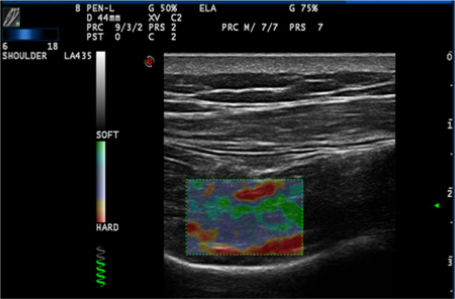
An elastography image of the infraspinatus muscle in a cadaveric shoulder.
Elastography images were acquired using the steady-state quasi-static elastosonographic technique. Steady-state quasi-static elastography is characterized by the manual deformation of the shoulder muscles using free-hand compression facilitated by the transducer. The deformation of the infraspinatus muscle tissue being examined allows the ultrasound unit to calculate a relative computed strain value. Two experienced musculoskeletal sonographers independently obtained elastography images of the infraspinatus muscle bilaterally on all four cadavers. After three shoulders were excluded because of evidence of previous shoulder surgery, a total of 10 elastography images were available for analysis.
Human Study
A purposive cohort sample of 10 human subjects (ages 20–29 years) was used in the second portion of the study to translate the preclinical technique arrived at from the cadaver portion of the study. The subjects were placed in an upright sitting position to allow for a posterior scanning approach. Again, a Twice Ultrasound unit equipped with a 13-MHz transducer was used. In this portion of the study, the supraspinatus muscle was evaluated bilaterally. A gray-scale image of the supraspinatus muscle was obtained and confirmed using the bony landmarks of the head of the humerus and the greater tuberosity. Once this image was obtained, the elastography function was activated, producing an elastography ROI box on the ultrasound screen (Figure 2). This ROI box was kept the same size for all subjects to ensure standardization. The ROI was then placed over the supraspinatus muscle for analysis.
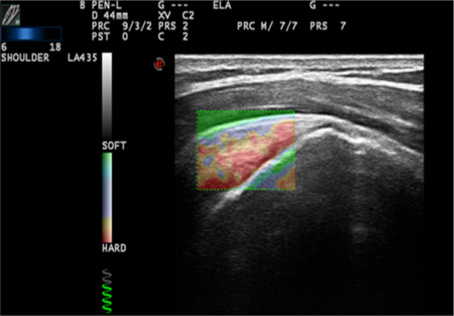
An elastography image of the supraspinatus muscle in a human shoulder.
As in the cadaver portion of the study, a steady-state quasi-static elastosonographic technique was used to acquire the elastography images. Manual deformation of the shoulder muscles was completed using free-hand compression facilitated by the transducer. This deformation of the supraspinatus muscle allowed the ultrasound unit to calculate the relative computed strain value. Two experienced musculoskeletal sonographers independently obtained elastography images of the supraspinatus muscle bilaterally on all 10 subjects, yielding a total of 40 elastography images per investigator for analysis.
Image Analysis
Elastography images were retrospectively analyzed using the system’s MyLabDesk Software (Esaote). Two researchers independently traced the outline of the targeted muscle within the elastography ROI (Figures 3 and 4). Once the trace was completed, the computer software generated a computed relative strain value, representative of the amount of muscle tissue deformation within the ROI. These data were then recorded and given to a third researcher for database entry to ensure that all researchers were blinded to eliminate the possibility of bias.
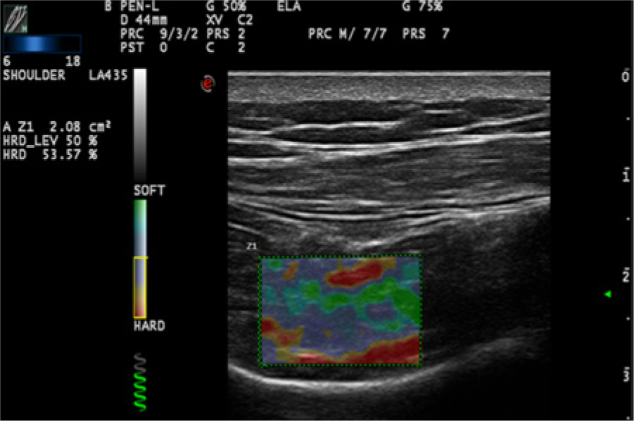
Image analysis of the infraspinatus muscle in a cadaveric shoulder. A traced region of interest (ROI) can be seen within the elastography ROI box, ensuring only the strain of the muscle tissue is being analyzed.
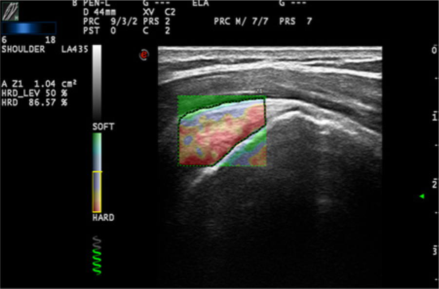
Image analysis of the supraspinatus muscle in a human shoulder. A traced region of interest (ROI) can be seen within the elastography ROI box, ensuring only the strain of the muscle tissue is being analyzed.
A two-tailed t test for equal variance was calculated to determine the significance of the statistics. A Pearson correlation was used to determine the interclass correlation between the two researchers performing the analysis. This was done to determine if reliable data regarding rotator cuff elasticity could be obtained between the two investigators who performed the scan. These statistical measures were completed for both data sets, cadaver and human, as well as the two data sets combined, to determine the intrarater reliability of the elastography image analysis.
Results
In the cadaver portion of the study, a Pearson correlation revealed a strong positive correlation (r = 0.99), but a two-tailed t test for equal variance revealed a P = .08, deeming the results not statistically significant. In the second portion of the study, a Pearson correlation again showed a strong positive, statistically significant correlation (r = 0.92), with a P < .05.
The computed relative strain values from both portions of the studies were then combined and put through the same statistical measures as previously described. This showed a strong positive, statistically significant correlation (r = 0.97, P < .05) (Table 1). All data were then visually represented in the form of scatter plots in order for subjective analysis to be performed. The scatter plots are segregated into the cadaver data points (Figure 5), human data points (Figure 6), and the combined data points, representing both the cadaver and human portions of the study (Figure 7).
The r Values Calculated from a Pearson Correlation Representing Intrarater Reliability and Their Associated Significance Values.
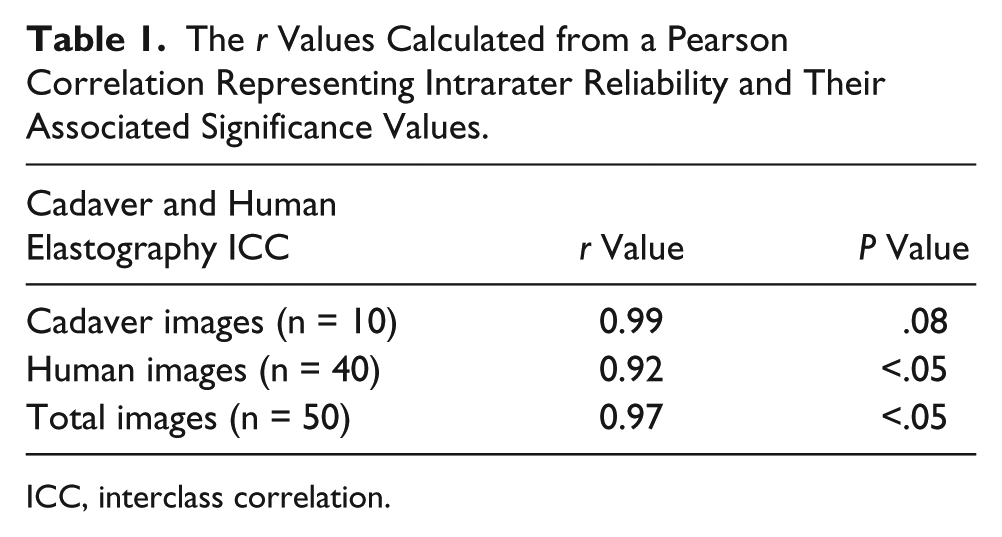
ICC, interclass correlation.
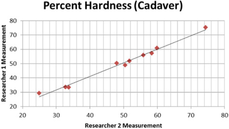
Scatter plot portraying a visual representation of researcher correlation of rotator cuff muscle percent hardness in the cadaver portion of the study.
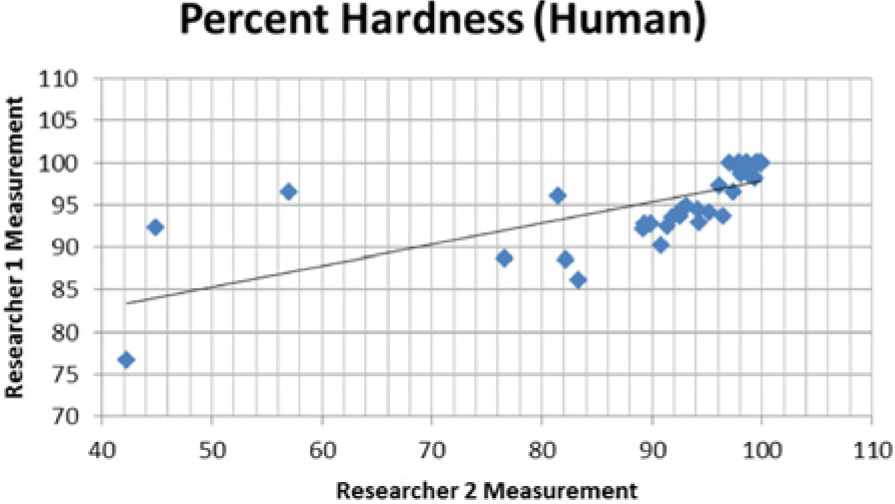
Scatter plot portraying a visual representation of researcher correlation of rotator cuff muscle percent hardness in the human portion of the study.
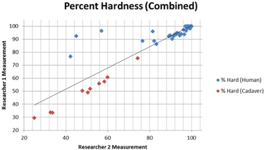
Scatter plot portraying a visual representation of researcher correlation for both the cadaver (red) and human (blue) portions of the study.
Discussion
The purpose of this study was to investigate the feasibility and reliability of an elastographic evaluation of the rotator cuff muscles. This is the first step in a line of research in which the ultimate goal is to raise the level of evidence in the emerging imaging modality of elastography. Multiple studies have been carried out investigating the use of musculoskeletal ultrasound as a substitute for MRI while imaging the rotator cuff muscles.9,10 Minimal preliminary reports have been documented that use elastography for early diagnosis and evaluation of the rotator cuff muscles; there are currently no published case-controlled studies.11–14 The ultimate goal of our research in the area of elastography imaging is to gather enough evidence to demonstrate validity and reliability, which could lead to the eventual raising of the grade for sonography in the American College of Radiology (ACR) Appropriateness Guidelines, making the use of elastographic imaging in the acutely painful shoulder “usually appropriate.” As mentioned previously, for patients presenting with acute shoulder pain, in which the radiographs are noncontributory and the patient has persistent pain with a physical examination and history that is nonspecific, the Appropriateness Guidelines require further imaging, and sonography is given a ranking of 5 out of 10 (may be appropriate). 3 Therefore, preclinical research must be done to ensure feasibility and translatability if the use of elastography is to be encouraged.
To begin at the lowest level of evidence, elastography images were obtained and analyzed of the infraspinatus muscle in a cohort of cadavers. This was done to ensure feasibility as well as provide initial preclinical data points that would allow reliability statistics to be calculated. An attempt was also made to image the supraspinatus as well, but due to the age of the cadavers and the degenerative nature of the muscle, as well as the inability to image around the acromion process, much of this muscle was unable to be visualized. A Pearson correlation was used to determine intrarater reliability and an r value of 0.99 was found, indicating a very strong positive correlation. However, a two-tailed t test for equal variance indicated no statistical significance, with a P value of .08. Because a significance level of .05 was set a priori to the study, these results required further scientific investigation. Our hypothesis for the nonsignificance of these results was based on having only 10 elastography images for analysis, raising the possibility of a type II statistical error, that is, concluding there is no significance when in fact it may exist. Therefore, since feasibility was demonstrated in the cadaver model, the next logical step was to translate the research into a human sample. This human sample would also provide a larger sample size, which, if our hypothesis was correct, could render our reliability results significant.
A purposive sample of 10 human subjects between ages 20 and 29 years was scanned using a steady-state quasi-static elastosonographic technique to obtain elastographic images of their supraspinatus muscle bilaterally. The supraspinatus was chosen in this population of subjects due to it being the most common rotator cuff muscle involved in degenerative rotator cuff tears. 1 This yielded a total of 40 sets of elastography images from which measurements and statistical calculations could be made. For these subjects, a Pearson correlation r value of 0.92 was found, demonstrating a strong positive correlation. Again using a two-tailed t test for equal variance, a P < .05 was calculated, deeming the results statistically significant as well.
This finding supported our hypothesis that the low number of images being analyzed was the reason our statistics were not significant in the cadaver portion of the study. However, this does not fully prove this, given the statistical measures were done on two different data sets. The next step was to combine the two data sets (cadaver and human) and perform a third Pearson correlation to demonstrate intrarater reliability. It is important to note that the basis of this experiment was examining reliability of elastography image analysis. For this reason, we believe that this study did not violate assumptions, by combining data sets that include data derived from both preclinical and clinical measures, as well as elastography investigations of the supraspinatus and infraspinatus muscles. Once all data points were combined, a total of 50 sets of elastography images yielded a Pearson correlation r value of 0.97, which represents a very strong positive correlation. The significance of these results was confirmed by again using a two-tailed t test, which yielded a P < .05.
This study demonstrates the feasibility and high reliability of elastography image acquisition and analysis between two researchers in a preclinical and purposive human sample. This sonographic technique has great potential to be used clinically for the evaluation of rotator cuff muscles, although at present there is limited existing evidence regarding the strain of healthy and diseased skeletal muscle tissue. 11 Clinically, it is known that multiple forms of pain and discomfort originate from improper muscle contraction. 15 For this reason, physical therapists already employ the assessment of muscle stiffness through palpation.15,16 This assessment, however, is subjective and provides no standardization among therapists. By employing elastography, quantitative values representing muscle stiffness can be obtained, eliminating subjectivity and standardizing skeletal muscle tissue assessment.
To eliminate subjectivity and error in the manual assessment of muscle stiffness through palpation, it is important that the elastographic assessment of skeletal muscles is done using a standardized method, to ensure reproducibility and accurate results. Feasibility studies have been conducted, demonstrating that it is possible to produce stiffness maps using elastography coupled with biomechanical pressure measurements acting as the gold standard.11,15 This information provides a foundation for using elastography in evaluating muscle strain, and continued research needs to be conducted before this can confidently be implemented clinically. Another question that still remains is whether muscle strain variance is dependent on the muscle group being evaluated. 11 The current study represents the first step in this line of scientific inquiry.
Limitations
This two-part study is limited in its generalizability due to the bias that is inherent with preexperimentally designed studies. Although low-level evidence is provided, the aim was to gather enough data to move forward to staging translational experiments that generate valid and reliable data on humans. In addition, there are various methods and techniques for image acquisition as well as processing algorithms for the display of elastography images, making translation difficult. 11 For example, the current study used a steady-state quasi-static technique that required manual deformation using the transducer to acquire tissue elasticity information. However, while using shear wave force technology, in which manual compression is not done, our developed protocol and technical settings would not be appropriate. At its current status in development, elastography also seems to be highly user dependent due to the need for manual compression to deform the tissue. This introduces variance in levels of pressure, probe alignment, and the introduction of artifacts. 11
Conclusion
The goal of this study was to adhere to the GRRAS template for a reliability study, as well as attempt an elastography imaging experiment and subsequent image analysis on two of the four rotator cuff muscles of the shoulder. Once the elastography images were obtained, a method of image analysis was determined and tested to ensure it was able to produce valid and reliable results. This descriptive research study provides lower level scientific evidence that steady-state quasi-static elastography can provide valid and reliable information that will assist in the evaluation of the shoulder muscles. This method of shoulder evaluation also has the potential and should be considered a means of augmenting the manual shoulder examination.
By gathering evidence demonstrating a reliable method of evaluating elastography images, it should be considered that this imaging specialty not be limited to the evaluation of shoulder muscles. With more extensive research, the methods used in this study have the potential to be translated to other anatomical areas and tissues within the body, in which the amount of deformation could be an important diagnostic tool.
Future Directions
Although statistically significant results were obtained from these data, this study needs to be replicated in a larger human study to strengthen the results. Without a larger sample size, these results cannot be generalized and no conclusive results can be made. Furthermore, this study would be enhanced with the clinical release of acoustic force elastography. This emerging technology would provide a standard quantitative value of sheer force being used to deform the targeted muscle tissue. This would eliminate a potentially significant source of operator error and allow for a more standardized method of data collection. Once validity of this method is achieved, elastography data could then be coupled with laboratory values and used as a diagnostic tool to prevent the progression of muscle pathologies, as a correlation has been found between quantitative elastographic strain values and elevated serum markers while monitoring inflammatory myopathies.11,17
Footnotes
Acknowledgements
We thank Mark and Michelle Whitmer for their assistance in preparing the cadavers for this data collection. We also thank our data technician, Christopher Kanner, for his support.
Declaration of Conflicting Interests
The authors declared no potential conflicts of interest with respect to the research, authorship, and/or publication of this article.
Funding
The authors received no financial support for the research, authorship, and/or publication of this article.