
Abstract
Iniencephaly is a rare defect caused by improper closure of the neural tube. The incidence is 0.1-10 per 10,000 live births. The head is disproportionately large as compared to the body and displays excessive retroflexion. The neck is absent with the face directed upward. Prenatal sonography is an accurate means of identifying this anomaly.
Introduction
Iniencephaly, a constituent of the group of the neural tube defects, is rare and severe as compared to others. No exact etiological factor has been found for this defect and prognosis is extremely poor. Most infants with iniencephaly have additional birth defects such as anencephaly or cephalocele. Other body systems may also be affected and there can be cardiovascular and gastro-intestinal tract malformations. Sonography is the imaging modality of choice for detection of iniencephaly and other associated anomalies.
Case Report
A woman in her early 30s, third gravida with 30 weeks amenorrhea, diagnosed clinically as having polyhydroamnios, was referred for obstetric sonography. There was a history of polyhydroamnios and mid-trimester abortion during the second pregnancy two years earlier, though no clinical/sonographic details were available with the patient. Her first child, a male seven years old, was alive and healthy.
Sonography (GE Logiq 200, 3.5MHz curvilinear transducer) confirmed polyhydramnios. The scan revealed a single live fetus with biparietal diameter (BPD) measuring 7.05 cm and femur length (FL) of 5.21 cm, corresponding to 28 weeks of gestation. The fetal head was hyperextended, the neck region could not be seen, and the trunk was noted to be in close proximity to the fetal head. The trunk also was noted to be disproportionately small compared to the fetal head (Figures 1 and 2). Varus deformity of the left foot was seen; the right foot appeared normal. Sonography also showed a short and rotated spine (Figure 3). A diagnosis of iniencephaly was made and the patient was referred back to her obstetrician. No further follow-up was available.
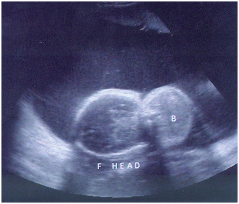
Body (B) in proximity to the fetal head and disproportionately smaller as compared to the fetal head. Note the excessive amniotic fluid.
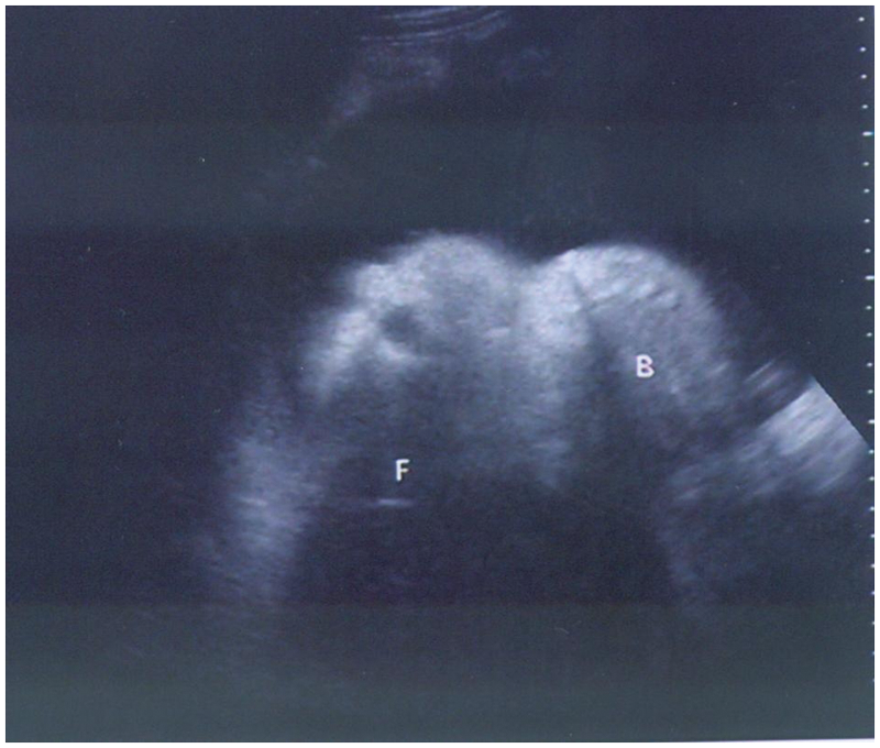
The head is so retro-flexed that the face looks upwards. (F, fetal head; B, body)
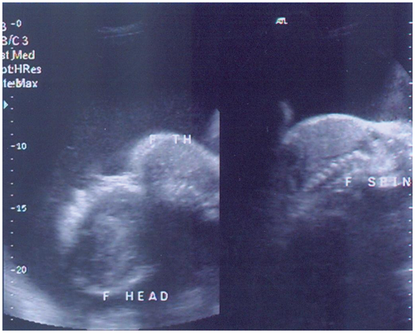
Retroflexion of head and absence of neck is seen in the left image. A short and rotated spine is seen in the right sided scan. (F TH, fetal thorax)
Discussion
Iniencephaly is a rare and fatal neural tube defect developing after the cephalic neural tube has closed, characterized by fusion between posterior occipital bone and the cervical spine. Several other anomalies have been associated with iniencephaly, including anencephaly, cephalocele, holoprosencephaly, agnathia-microstomia-synmelia, spina bifida, omphalocele, gastroschisis, diaphragmatic hernia or agenesis, pulmonary hypoplasia or hyperplasia, cardiac malformations, renal anomalies, overgrowth of the arms compared to the legs, genu recurvatum, arthrogryposis, club-foot, and gastrointestinal atresia.1,2
The mother in most cases presents prenatally with high alpha-fetoproteins or clinical polyhydramnios as was seen in the case presented. Sonographically, the diagnosis is made based on the extreme dorsal flexion seen of the head caused by its fusion with the spine, with the head so retro-flexed that the face looks upward, leading to the term “star gazing” anomaly (Figure 4). There is significant shortening of the spine due to marked lordosis and there is overall shortening of the fetus. Also there is polyhydramnios detected sonographically.3–5
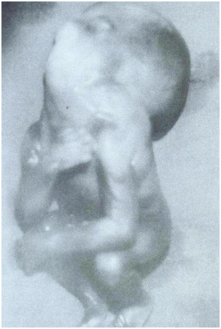
Fetal specimen showing characteristic features of iniencephaly.
This entity was first described by Saint-Hilaire in the mid 19th century. 3 Historically, the incidence has varied from 0.1-10:10,000; however, the incidence reported has increased as more cases are being recognized by sonography. The disorder is more common among females, with a female:male ratio of 9:1. Risk factors include low parity and low socioeconomic status. Maternal obesity, diabetes, and intake of diuretics, antiepileptic drugs, antihistamines, and sulfa drugs have all been shown to be associated with an increased risk of neural tube defects, but there is no definitive evidence of their association with iniencephaly. Iniencephaly may be secondary to a mix of genetic and environmental factors as it is a common malformation in families with a history neural tube defects, and the recurrence risk is 1% to 4%.1–3 The patient presented here gave the history of polyhydramnios and mid-trimester abortion during an earlier pregnancy two years prior. No clinical/ultrasound details were available, but a similar anomaly at that time could not be ruled out.
The prognosis for this anomaly is extremely poor and it is very nearly uniformly fatal. Babies are either stillborn or die a few hours after birth. The deformation of the fetal body may also pose a danger to the mother’s life during delivery. Radiologists as well as clinicians appear to be less aware of iniencephaly as compared to other neural defects like anencephaly. Specific sonographic characteristics can differentiate the two, though both can coexist.5–10 Antenatal sonography remains the imaging modality of choice for detection of these neural tube defects, and effective ways of patient management can then be implemented.
Footnotes
Declaration of Conflicting Interests
The authors declared no potential conflicts of interest with respect to the research, authorship, and/or publication of this article.
Funding
The authors received no financial support for the research, authorship, and/or publication of this article.