




Abstract
Background
Fanconi anemia (FA) is an uncommon inherited disorder characterized by bone marrow failure, congenital anomalies, and increased susceptibility to malignancy. Early signs may appear in the oral cavity, making dental evaluation important for prompt detection in pediatric patients. This case highlights the importance of oral findings in early recognition of FA and the role of dentists in prompt referral and interdisciplinary management.
Case report
A 9-year-old girl presented with gingival bleeding for 1 month. Examination revealed severe gingival inflammation with intraoral petechiae. Routine blood tests revealed pancytopenia. Bone marrow biopsy revealed a hypocellular marrow, and FA was confirmed by cytogenetic analysis. Tooth #55 was extracted under hospital admission, following platelet transfusion and antibiotic prophylaxis. The patient subsequently underwent bone marrow transplantation (BMT) with immunosuppressive therapy and radiotherapy, resulting in complete resolution of gingival inflammation. She continues to be under regular follow-up with hematology and pediatric dental care.
Conclusion
Early identification of oral signs can aid dental professionals in suspecting systemic disorders such as FA, allowing timely referral, diagnosis, and multidisciplinary management.
Introduction
Fanconi anemia (FA) is an inherited disorder of bone marrow failure characterized by genomic alterations, congenital abnormalities, and a predisposition to malignancy. It was first described by the Swiss pediatrician Guido Fanconi in 1927, who reported a familial occurrence of pancytopenia associated with physical malformations. 1 The condition is exceptionally rare, with an estimated prevalence of about 1 in 350,000 births, and is typically identified at a mean age of 7 years. It follows an autosomal recessive pattern predominantly, though X-linked inheritance has also been identified. 2
Mutations in any of over 15 FA-associated genes disrupt the repair of deoxyribonucleic acid (DNA) interstrand crosslinks, producing genomic instability and hematologic suppression. Clinically, affected individuals may present with growth retardation, skeletal anomalies, skin hyperpigmentation, renal malformations, and progressive bone marrow failure, typically manifesting during childhood. 3
Oral findings may include mucosal pallor, petechiae, gingival bleeding, ulcerations, delayed tooth eruption, microdontia, hypoplastic enamel, and generalized growth delay of the craniofacial complex. Recognition of these oral manifestations is essential for timely diagnosis, particularly when hematological symptoms are yet to develop or are nonspecific. 4
Early identification of FA through oral and dental features can prompt timely hematologic evaluation and genetic testing, allowing for appropriate management and prevention of severe complications. This case report highlights the role of oral findings in the initial recognition of FA in a pediatric patient and underscores the importance of interdisciplinary collaboration between dental and medical professionals. Written informed consent was obtained from the patient’s parent/legal guardian for publication of the clinical details and accompanying images.
The unique learning aspect of this case lies in the early recognition of FA based on oral manifestations in the absence of overt congenital anomalies. Persistent gingival bleeding and mucosal petechiae represented the initial clinical indicators that prompted hematologic evaluation and subsequent diagnosis. This case underscores the critical role of pediatric dentists in identifying subtle oral signs of underlying systemic disease and emphasizes the importance of timely referral and interdisciplinary collaboration to facilitate early diagnosis and optimize patient outcomes.
Case Report
A 9-year-old female patient presented to the Department of Pedodontics with a chief complaint of bleeding gums persisting for approximately 1 month. According to the parents, the bleeding episodes were preceded by a mild, short-lasting febrile illness. The patient also reported noticing blood-stained stools over the preceding 2 weeks, suggesting possible systemic involvement. A detailed review of the prenatal, perinatal, and past medical histories revealed no significant findings, and there was no history of similar illness or hematologic disorders in the family.
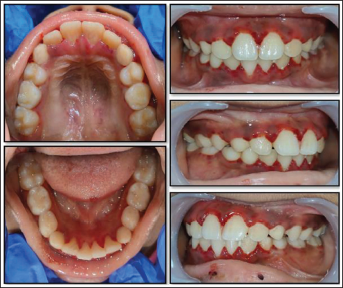
On general examination, the child was alert, conscious, and cooperative. Her height and weight were appropriate for her chronological age. The skin appeared pale with scattered hyperpigmented macules over the trunk and extremities, raising suspicion of a systemic or hematologic abnormality. No overt congenital malformations were detected. Intraoral examination revealed severe gingival inflammation with spontaneous bleeding and petechial spots on the labial and buccal mucosa (Figure 1). The presence of mucocutaneous pallor, petechiae, and persistent gingival bleeding prompted consideration of an underlying bleeding or bone marrow disorder. The child was in the mixed dentition stage, with no dental developmental anomalies.
Pre-operative Intraoral Images Showing Gingival Inflammation.
Routine hematological investigations were advised to exclude systemic causes of bleeding. The reports revealed pancytopenia, indicating suppression of all major blood cell lineages (Table 1). Given the history of blood-stained stools, the patient underwent gastrointestinal evaluation, including stool examination and upper gastrointestinal endoscopy, which revealed no active mucosal lesions or identifiable source of bleeding. The bleeding was therefore attributed to severe thrombocytopenia. Considering the hematologic findings, no invasive dental procedures were initiated during the initial visit. Supra-gingival scaling was performed, and the patient and parents were counseled regarding oral hygiene maintenance and were referred to a hematologist for further evaluation. The child was subsequently admitted to a government hospital, where whole blood and platelet transfusions were administered to stabilize the hematologic parameters. Additional investigations were conducted to exclude infectious or systemic etiologies. As the hematologic profile showed minimal improvement, a bone marrow biopsy was performed, revealing a hypocellular marrow, suggestive of bone marrow failure. The diagnosis of FA was confirmed through a cytogenetic assay, which demonstrated characteristic chromosomal breakage (>30%) upon exposure to mitomycin C.
Blood Investigations at the First Visit.

During the course of hospitalization and ongoing diagnostic evaluation, the patient was referred back to the Department of Pedodontics with pain associated with tooth #55. Clinical examination revealed that the tooth exhibited pre-shedding mobility and was interfering with occlusion and mastication. Considering the discomfort and potential for traumatic injury, the extraction of tooth #55 was planned. The procedure was performed under hospital admission, following platelet transfusion and antibiotic prophylaxis to minimize the risk of bleeding and postoperative infection. Post-transfusion hematological evaluation revealed a platelet count of approximately 52,000 cells/µL, which met the recommended threshold for invasive dental procedures. Prior to extraction, the patient rinsed with 0.2% chlorhexidine for 2 min. Hemostasis was achieved by local compression using gauze soaked in tranexamic acid, and the postoperative course was uneventful (Figure 2).
Post-extraction Intraoral View of Tooth #55 Showing an Uneventful Surgical Site with Achieved Hemostasis.

Three months after the confirmed diagnosis, the patient underwent bone marrow transplantation (BMT) along with ionizing radiation and immunosuppressive therapy. Post-transplant follow-up revealed complete resolution of gingival inflammation and normalization of oral health status (Figure 3). The patient continues to remain under regular multidisciplinary follow-up with the hematologist and pediatric dentist to monitor hematologic stability and maintain optimal oral health.
Intraoral Images After Bone Marrow Transplantation.

Discussion
FA is a rare hereditary disorder characterized by congenital anomalies, cellular hypersensitivity to DNA crosslinking agents, and a marked predisposition to malignancies. It exhibits genetic heterogeneity, with mutations identified in at least 15 distinct genes, whose protein products interact within a common DNA repair pathway to maintain genomic stability and regulate cellular resistance to DNA damage. Clinically, FA is recognized among the disorders of bone marrow failure, manifesting with progressive pancytopenia, developmental abnormalities, and increased cancer susceptibility.2, 3
Clinically, individuals with FA may present with a wide spectrum of anomalies, including bifid thumbs, upper limb malformations, skin pigmentation abnormalities, gastrointestinal defects, and genital anomalies. 2 However, a subset of patients may lack overt physical or cutaneous manifestations. Data from the International FA Registry (IFAR) indicate that approximately one-third of patients with FA exhibit no congenital malformations. 5 Similarly, in the present case, the patient did not demonstrate any physical anomalies, underscoring the variability of phenotypic expression in FA.
Hematologic abnormalities arising from bone marrow failure represent the most significant clinical manifestation of FA. The most common hematologic complications include pancytopenia, anemia, thrombocytopenia, and leukopenia. 4 Consequently, affected patients often present with bleeding, pallor, and susceptibility to repeated infections, highlighting the importance of early recognition and multidisciplinary management in pediatric cases. At the time of presentation, our patient exhibited pancytopenia with marked thrombocytopenia, which likely contributed to the gingival bleeding observed.
Gingivitis and periodontitis are the most commonly reported oral manifestations in individuals with FA. 2 In the present case, the patient exhibited severe gingivitis, likely resulting from a combination of suboptimal oral hygiene and thrombocytopenia, the latter contributing to exacerbated gingival bleeding.
Various dental anomalies have been reported in patients with FA, including microdontia, supernumerary teeth, tooth agenesis, discoloration, abnormal tooth morphology, rotation, transposition, delayed eruption or shedding, and micrognathia. Certain dental anomalies, such as supernumerary teeth and tooth agenesis, may arise due to alterations in calcium metabolism occurring during the process of odontogenesis. Other dental irregularities can be explained by craniofacial anomalies, including micrognathia and microcephaly, which influence tooth development and eruption patterns.2, 6 In the present case, no dental anomalies were observed.
Individuals affected by FA are significantly more prone to developing squamous cell carcinoma (SCC). Following BMT, chromosomal instability is further exacerbated by ionizing radiation, graft-versus-host disease, and immunosuppressive therapy, collectively increasing the risk of SCC by approximately 4.4-fold in this population. 7 Approximately two-thirds of SCC cases in FA patients arise in the oral cavity, most commonly affecting the lateral borders of the tongue and the gingival regions. 2
Close collaboration between the hematologist and dentist is essential to minimize the risk of excessive bleeding during dental procedures. It is recommended that the platelet count be raised to at least 50,000/µL prior to such procedures. 3 Consequently, dental management for such patients should ideally be conducted in a hospital setting. In the present case, the patient was hospitalized during the extraction, and platelet transfusion was administered beforehand to ensure adequate hemostasis. Local hemostatic measures such as sutures, topical agents (oxidized cellulose, gelatin, fibrin glue, cyanoacrylate), and antifibrinolytic therapy with tranexamic acid are recommended to minimize postoperative bleeding.
In patients with FA and an absolute neutrophil count below 500/µL, antibiotic prophylaxis—commonly with amoxicillin or clindamycin—should be administered 1 h before invasive dental procedures and continued until mucosal healing to prevent serious infections. 3 Additionally, a chlorhexidine mouth rinse 30 min prior can help reduce the risk of postoperative infection.
Dental treatment in patients with FA should be focused on infection control. Non-invasive procedures typically do not require prophylactic platelet transfusions, whereas invasive procedures may necessitate desmopressin or antifibrinolytic agents in mild cases and platelet transfusions in severe cases. Whenever possible, completing treatment in a single hospital visit with platelet support is preferred over multiple sessions. 8 Given the increased risk of oral cancer, early and regular screening with careful mucosal examination is recommended, with dental evaluations every 3–6 months. 9
Patients with FA require close follow-up by a multidisciplinary team, including a hematologist, a pediatrician, an endocrinologist, an ear, nose, and throat (ENT) specialist, a cardiologist, and an oncologist. A pediatric dentist should also be integrated into the team to monitor oral health, provide preventive care, and counsel the family. A carefully planned preventive dental program is essential to minimize the need for extensive interventions later and can be effectively managed by a pediatric dentist. Acute dental infections should be treated promptly, and all potential oral sources of infection should be eliminated to safeguard the patient’s overall systemic health.2, 10
Conclusion
This case highlights the importance of early recognition of FA through oral manifestations, even in the absence of overt congenital anomalies. Prompt identification of unexplained gingival bleeding by the pediatric dentist facilitated timely hematologic referral and definitive diagnosis. Effective management of such patients relies on close interdisciplinary collaboration, with the dentist playing a crucial role in early detection, preventive care, and long-term oral surveillance.
Footnotes
Author Contributions
This manuscript has never been presented. This manuscript has not been submitted or published in any other journal. The manuscript has been read and approved by all the authors.
Declaration of Conflicting Interests
The authors declared no potential conflicts of interest with respect to the research, authorship, and/or publication of this article.
Funding
The authors received no financial support for the research, authorship, and/or publication of this article.
Patient Consent
Patients’ written consent has been obtained for publication.
