
Abstract
Despite extensive clinical research and management protocols applied in the field of coronary artery diseases (CAD), it still holds the number 1 position in mortality worldwide. This indicates that we need to work on precision medicine to discover the diagnostic, therapeutic, and prognostic targets to improve the outcome of CAD. In precision medicine, epigenetic changes play a vital role in disease onset and progression. Epigenetics is the study of heritable changes that do not affect the alterations of DNA sequence in the genome. It comprises various covalent modifications that occur in DNA or histone proteins affecting the spatial arrangement of the DNA and histones. These multiple modifications include DNA/histone methylation, acetylation, phosphorylation, and SUMOylation. Besides these covalent modifications, non-coding RNAs—viz. miRNA, lncRNA, and circRNA are also involved in epigenetics. Smoking, alcohol, diet, environmental pollutants, obesity, and lifestyle are some of the prime factors affecting epigenetic alterations. Novel molecular techniques such as next-generation sequencing, chromatin immunoprecipitation, and mass spectrometry have been developed to identify important cross points in the epigenetic web in relation to various diseases. The studies regarding exploration of epigenetics, have led researchers to identify multiple diagnostic markers and therapeutic targets that are being used in different disease diagnosis and management. Here in this review, we will discuss various ground-breaking contributions of past and recent studies in the epigenetic field in concert with coronary artery diseases. Future prospects of epigenetics and its implication in CAD personalized medicine will also be discussed in brief.
Keywords
Introduction
Epigenetics is a term coined from Greek prefix, “epi,” meaning “over,” “external,” or “around,” and the traditional word “genetics” that is inheritance. Hence, epigenetics can be described as the changes affecting heritable phenotype variations without altering the DNA sequence of a genome. Epigenetic change is associated with the covalent modifications in the structural components of chromatin, nitrogenous bases of DNA, and expression of various non-coding RNA species, which decide the expression pattern of genes. They ultimately help in keeping the cell identity, which is censoriously important for normal development and causation of various diseases. During recent 2 decades, epigenetics has seen an extensive surge in the field of precision medicine, and epigenetic changes have been associated with almost all disease states, including malignant and non-malignant, especially lifestyle disorders. They mostly occur due to changes in the microenvironment of cells, which in turn is decided by various modifiable and nonmodifiable factors. 1 Based on these facts, epigenetics plays an inevitable role in disease pathogenesis of coronary artery disease (CAD). Further, exploration of epigenetics underlying various diseases has paved way for the development of novel therapeutic agents. Several drugs targeted to epigenetic factors are under trial. 1
Acute Myocardial Infarction (AMI) is the foremost cause of death worldwide. 2 The role of epigenetic changes in patients with CAD is comparatively novel concept with interesting findings. Studies have shown that epigenetic alterations significantly contribute to the development of acute myocardial infarction. Endothelial dysfunction, imbalance in cholesterol metabolism, and atherosclerotic plaques are the primary pathophysiological modification leading to CAD. These changes, along with the risk factors of CAD-like obesity, diabetes mellitus (DM), hypertension, and insulin resistance, are interrelated to environmental risk factors associated to increased rate of CAD. 3 The development of atherosclerotic plaque follows epigenetic changes of vascular smooth muscle cells (VSMCs), endothelial cells (ECs), and macrophages. Inflammation, cholesterol metabolism, and homocysteine homeostasis are associated with the development of atherosclerotic plaque, the main pathologic change resulting in CAD. 4
A number of reports regarding influence of epigenetics on CAD are being added to the literature on an exponential rate, paving the way to explore the underlying epigenetic mechanisms of the disease. In this review, we will discuss the latest discoveries related to epigenetic modifications in fetal and adult stages that contribute to the CAD development and its progression. Also, how the environment coordinates with epigenetics of an individual towards the initiation and development of CAD, as well as the role of epigenetics in personalized medicine will also be discussed to provide a comprehensive view to readers.
Pathophysiology of CAD
Atherosclerosis is a chronic inflammatory condition which is characterized by thickening of vascular wall leading to luminal obstruction. 5 It is the basic pathogenic lesion in vaso-occlusive conditions of different organ systems like coronary artery disease and peripheral artery disease. Atherosclerotic plaque is composed of an accumulation of lipids, T lymphocytes, macrophages and fibrous tissues in arterial walls. This theory is known as the “response to injury” hypothesis, implicating endothelial disorder as the primary step in the cascade of different pathological stages of this chronic inflammatory condition, which precedes any structural alterations in the arterial wall occur. 6 The result of the process is the endorsed expression of intercellular and vascular cell adhesion molecules due to low-density lipoprotein (LDL) cholesterol oxidation in subendothelial space, which ultimately leads to monocyte adhesion/ migration to the subendothelial area. In the intima monocytes differentiate into macrophage which engulf oxidized LDL via scavenger receptor promoting foam cell formation and exhibit proinflammatory functions by secreting cytokines—viz. interleukins and tumor necrosis factor.
The end step of this process is the formation of atherosclerotic lesion I (fatty streak). Atherosclerotic build up results in plaque formation, vascular remodeling, acute and/or chronic luminal obstruction, abnormalities of blood flow and diminished oxygen/ nutrients supply to myocardium. By impairing or obstructing normal coronary blood flow, atherosclerotic build up causes myocardial ischemia. 7 In this review we will discuss the epigenetics modifications played a significant role in the pathogenesis of CAD.
Risk factors for CAD have been classified into demographic, biochemical parameters, lifestyle and genetic factors. Demographic factors include advancing age and male sex, biochemical parameters include variables such as raised cholesterol levels, lifestyle factors include sedentary working habits, high levels of stress and depression, unbalanced dietary habits, socioeconomic status and alcohol or smoking/tobacco abuse and genetic factors include positive family history for CAD and inherited disorders of lipid metabolism. 5
In demographic factor, age play a pivotal role in development of CAD.8,9 A study by Ravi et al demonstrated that increasing age is an independent risk factor for CAD. 10 Considering gender, men of all ages have higher mortality from CAD than women before menopause. 11 Several studies marked that; the incidence of CAD is similar in post-menopausal woman in age matched men.12-14 Several studies have reported high risk lifestyle choices are associated to epigenetic alterations such as histone modifications and DNA methylations are strongly associated with obesity leading to coronary artery disease. 15 A recent study indicated that different candidate gene methylations are associated to lipid metabolism and inflammation in subjects with obesity. 16 Also, a recently published study demonstrates that tobacco smoking is associated with DNA methylation at several loci of atherosclerotic carotid lesions. 17 Moreover, a study by Rebecca et al. reveals that specific DNA methylation patterns have been found in tobacco smoking CAD patients. 18 Those who smoke may have a vulnerability to illness with a heritable (genetic) component for CAD, T2DM, or hypertension, yet once the patient quits smoking, the likelihood of having/developing the disease phenotype starts to remit. 19 Also, Lohoff et al revealed that inducement of chronic alcohol modifies PCSK9 methylation leads to impairment of lipid metabolism and CAD. 20 Epidemiological studies propose that early life, mainly prenatal, environmental exposure can induce metabolic and physiological changes in fetus by modifying epigenetic profiles leading to predisposition of multiple chronic diseases such as diabetes, obesity, cardiovascular event, and cancer. 21 Accumulating reports from numerous studies suggest that epigenetics might be one of the unique molecular mechanisms with different contributing factors like fetal programming, environmental stimulus, and childhood phenotype. Owing to the reversible nature of these factors, epigenetic modifications are becoming an attractive therapeutic agent. 22
Inception of epigenetics in response to environmental variances
Intrauterine exposure
External stress modulates cellular and tissue level maturity. Severity varies depending time of exposure within narrow window period of genetic variation or specific organogenesis. Intrauterine development is highly sensitive for modifications in response to environmental stress in order to fine tune genotypic and phonotypic traits. This high level of developmental plasticity is not only adaptive but also may result in maladaptive change paving way for pathological processes.21,23 Epigenetic modulations can be inheritable through subsequent generations resulting in transgenerational epigenetic inheritance. Available micro milieu imparts most drastic effect on epigenetic status and modulation of transcriptome in early few weeks of pregnancy. 22 Diet, pathological conditions like hypertension, DM, smoking, chemical pollutants, and psychological stress have been proven to have important role in determining epigenetic landscape for future disease processes (Figure 1)24,25.

Increased risk of developing CAD with the effect of environmental inducements through intrauterine and after birth exposure.
Exposure after birth
Exposure to environmental pollution has been associated with cardiovascular risk factors. It is an established fact that smoking is the strongest independent risk predictor for premature coronary heart disease; moreover, second-hand smoke (SHS) has been characterized as an environmental pollutant contributing approximately to 80% death due to CAD and only <5% death from lung cancer. 26 This suggests coronary atherosclerotic tissues are susceptible to environmental chemical pollutants. A meta-analysis of genome-wide DNA methylation conducted by Roby Joehanes et al in 2017 revealed that changes in DNA methylation sites are associated with cigarette smoking highlighting thousands of differentially methylated CpGs. 27 Dietary influence on CAD has been studied widely suggesting both Mediterranean and DASH (Dietary Approaches to Stop Hypertension) diet, that is filled with vegetables, fruit, and nuts are associated with decreased risk of AMI and improved effects on blood pressure, lipid profile, inflammation, endothelial function, and thrombosis. 28 Specific research to elucidate the effect of dietary determinants on CAD risk modifying epigenetic modifications is lacking, although literature suggests diet may influence factors such as DNA hyper and hypomethylation. 29 Future research strategies might prove to be a worthwhile investment of resources to describe diet—epigenetics interactions leading to atherosclerotic disorders.
Another important factor resulting in epigenetic aberrations includes a sedentary lifestyle, which possesses a significant role in deciding the pattern of expression of different genes. This is achieved through changes in the microenvironment of chromatin affecting its spatial states; hetero/euchromatin ultimately changes the ability of transcription machinery to DNA accessibility despite the absence of DNA alterations. 30 The epigenetic changes lead to gain and loss of function, as observed in animal models.31-35 In this article, we aimed to review Epigenetic factors, including DNA methylation, histone alteration, and noncoding RNA control, as a risk factor of CAD by altering the relationship between genes and the environment.
Epigenetic changes can present as:
(1)
(2)

Environmental factors affecting epigenetic upbringing such as covalent modifications and different expression of non-coding RNAs leading to the development of Coronary Artery Disease over the years.
Covalent Modifications
Methylation of DNA
Amongst the covalent modifications, DNA methylation, which is a pre-transcriptional modification, has been extensively studied. It is the major epigenetic modification in mammals. DNA methylation shows a covalent transfer at CpG dinucleotide regions where methyl group (–CH3) attached to the fifth carbon of cytosine base. This process is catalyzed by particular enzymes called DNA methyltransferases (DNMTs). 36 DNA methylation is involved in multiple factors such as basic biological activities of the cell, gene expression, aging, malignancies and non-malignancies such as CAD.36,37 It is estimated that a total of 70% to 80% of CpG dinucleotides exist in a state of methylation in the human genome’s dinucleotides may cluster as CpG islands and serve as functional units to regulate gene expression by deciding the binding of transcription factors. 38 The frequency of CpG dinucleotide is higher in promoter regions as compared to intronic and exonic region. 39 Hypermethylation of promoter regions in somatic cells inhibits their activity. On the other hand, promoters with strong activation are largely unmethylated. 40 There are 3 angles that are being used to study the association between DNA methylation and CAD, as shown in Figure 3.
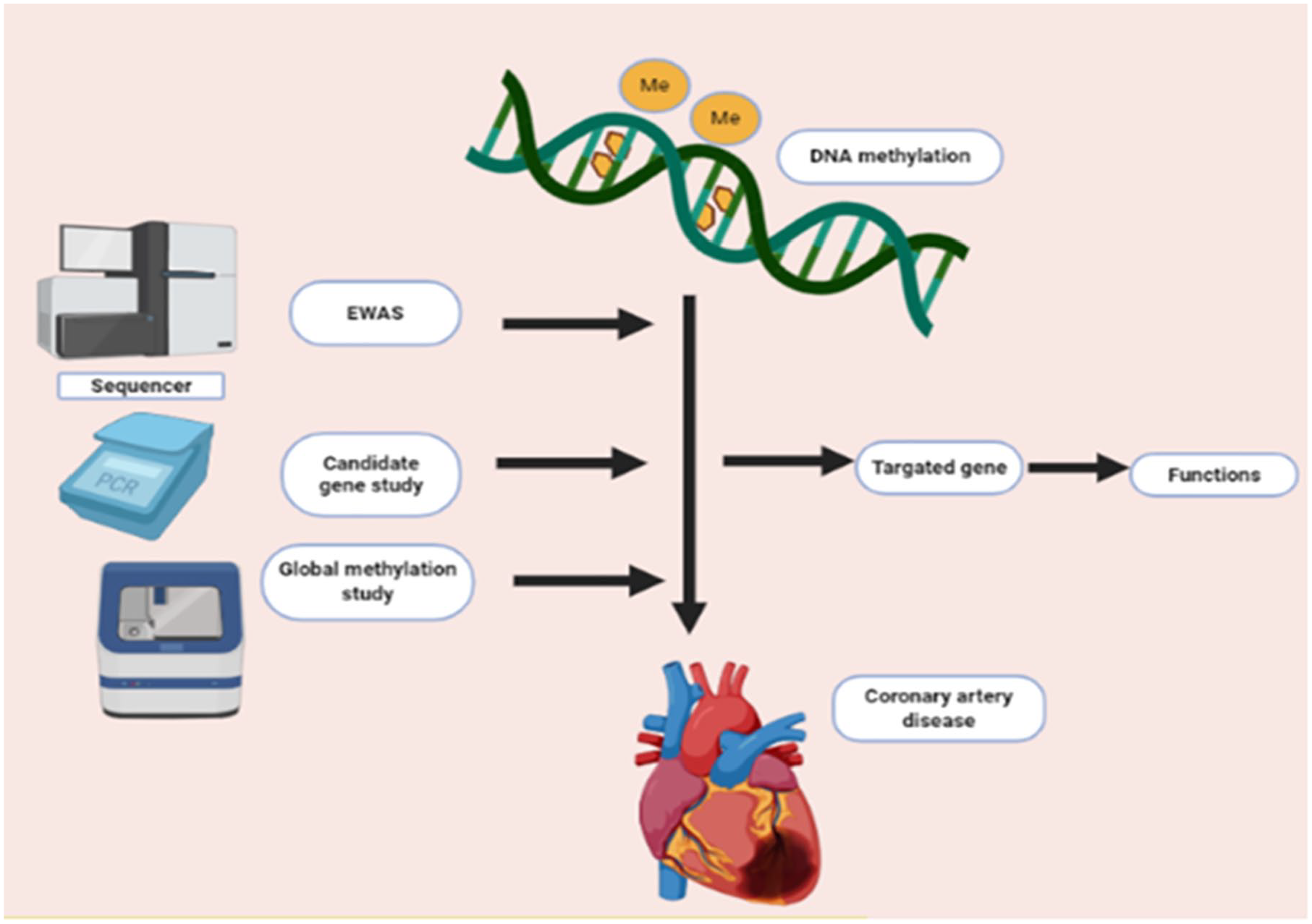
Different approaches to study the association between DNA methylation and CAD.
Role of DNA methylation in CAD
Lipid metabolism plays a pivotal role in atherosclerosis. It is affected by genetic and environmental factors both. Hypermethylation of certain genes like insulin like growth factor-2 (IGF2) and leptin genes is associated with risk factors of CAD, namely obesity and insulin receptor (IR).41,42 Hypermethylation of ATP Binding Cassette Subfamily G Member (ABCG1) 43 and hypomethylation of Carnitine palmitoyltransferase 1A (CPTIA) 44 has been shown to be associated with postprandial lipemia. Differential patterns of methylation are observed in genes involved in the pathogenesis of CAD and lipid homeostasis. 45
Global DNA methylation
The environmental and lifestyle factors which contribute to the development of CAD involve the epigenetic changes in the genome. Epigenetic modifications start in utero itself, which is a vulnerable period that can program the individual for risk of disease in adulthood. 46 Later the early and midlife environmental exposures to smoke, environmental contaminants and pollution lead to methylation of DNA, laying the foundation of non-genetic mechanisms by which CAD might develop and manifest later in life. Also, a protein-restricted diet and diet deficient in vitamin B12, folate, methionine in pregnant animals, and a low-fat flavonoid-rich diet affects the methylation patterns in specific genes contributing to atherogenesis. Smoking, environmental toxins, and pollution contribute to atherogenesis and have been observed to alter the methylation in specific genes.47-49
The various reports have elucidated that modifications in DNA methylation sites contribute to the regulation of different biological processes, including coronary atherosclerosis, hypertension, and inflammation, that play a significant role in the development of CAD.50-52 For instance, mice with hypomethylated DNA had overexpression of inflammatory markers, which leads to the formation of aortic fatty streaks. 53 In CAD prone mice lacking ApoE, modified DNA methylation sites are found in both blood leukocytes and aorta before the formation of vascular lesions. 54 These modifications contribute to inflammation and atherosclerotic plaque.
Different studies have reported that both hypomethylation and hypermethylation status in CAD patients. Although global hypomethylation has been reported in DNA from atherosclerotic tissues,25,55,56 CAD associated genes have been found to be hypermethylated in promoter regions more commonly 51 (Table 1). These differences could either be due to the presence of epigenetically/genetically non-identical cells in atherosclerotic tissue. Moreover, different methylation patterns may be associated with heterogeneous cell types of an individual’s tissue. While hypermethylation looks to be a greater contributing factor for CAD development, compared to hypomethylation, emerging single-cell sequencing technologies could overcome these discrepancies in the future.
Global DNA methylation pattern in CAD.

Candidate gene DNA methylation
An alternative way to investigate the methylation patterns is to analyze methylation at CpG dinucleotides of the targeted genes. Some reports from current studies associated to the pathophysiology of atherogenesis are compiled and depicted in Table 2. Modifications in the methylation status of a specific gene of interest can affect the functional pathways involved in atherogenesis such as LDL metabolism, lipid homeostasis, cholesterol biosynthesis, homocysteine metabolism, endothelial dysfunction and, inflammation.
Candidate gene DNA methylation status in CAD.

Therefore, exploring candidate gene methylation pattern in CAD using novel cutting-edge techniques help us to confirm the contribution of new genes after global methylation studies. This will open up a new therapeutic window for targeted therapies and prognostic markers after validation.
Epigenome-wide association studies
Different epigenome-wide association studies (EWAS) have been conducted in coronary atherosclerosis linked to DNA methylation at the CpG site to elucidate its role at the molecular level.69,70 The results of recent studies are shown in Table 3. EWAS study reveals that patients with CAD have a different pattern of DNA methylation sites at several genomic loci.
Epigenome-wide association studies.

Some investigators have found different methylated CpG sites in a specific gene; while others have observed a CpG island with increased methylation sites. 71 Correspondingly, EWAS scrutinize gene-specific DNA methylation. Gradually, EWAS has substituted the candidate gene method as it is useful to find novel CpG sites linked to disease phenotype. 72 Although a large proportion of previous EWAS confirmed their results on replication, the smaller sample size in these studies asks for further validation of the obtained results. A total of 84 translating loci with varied amounts of methylation were identified in at least more than 1 EWAS which suggests an investigation of epigenetic regulation of the identified genes in coronary atherosclerotic statuses. The results revealed a group of genes highlighting the significance of various conditions including carbohydrate and lipid metabolism along with obesity, inflammation and many others in CAD. 71 Still, these patterns are mostly inconclusive, warranting future research with a larger sample cohort.
Histone modifications
Histones are alkaline proteins around which DNA is wrapped to form bead-like structures called nucleosomes. Histones are characterized by 5 families: H1/H5, H2A, H2B, H3, and H4. 77 Histones are a fundamental unit of chromatin. 78 These proteins are extremely unstable. They modify rapidly in response to any external signal. Histone molecules undergo several post-translational modifications due to different molecular mechanism at chromatin and amino acid levels. 79 Amongst the multiple changes, histone acetylation, methylation, and phosphorylation are found to be associated with transcription regulation and gene expression. Post-translational modifications of histones can interfere with chromatin structure, organization, and function, which in turn alter mechanisms such as gene expression, DNA repair, DNA damage, and ultimately affecting the proliferative capacity of the cells. 80 Moreover, histone methylation and demethylation have been studied in coronary atherosclerosis widely. 81
Histone methylation and demethylation
Histone methylation is directly linked to activation or inactivation of a specific gene. Owing to the modified segment of histone a gene decides whether to activate/inactivate. 82 In 1960s, methylation pattern on histone was first identified. The methylation enzyme was unknown until the event of the lysine methyltransferase (KMT) family was found. 83 Histone methylation was considered to be the ultimate stone post-translational modification until the detection of first histone lysine de-methylase (KDM).84-86 Methylation of histone can occur both in arginine and lysine residues. At histone H3 and H4, the following methylation residues are documented: K4, K9, K27, K36, K79, and K20. Methylation of lysine residues can be found in multiple degrees, such as mono(me1)-, di (me2), or trimethyl (me3) groups.
Histone-lysine methyltransferase acts as a catalyst. It transfers a methyl group from S-adenosylmethionine (SAM) to amino groups on lysine residues to histone tails. 87
Discovery of lysine demethylating enzymes altered the primitive concept of irreversible and stable nature of histone methylation. Now, things are changed, and 2 critical classes of histone-lysine dimethyl enzymes are known. It is reported that flavin adenosine dinucleotide is being used as a cofactor of lysine-specific de-methylases (LSDs). The most studied LSD includeLSD1, which interacts with protein complexes like CoREST (resting corepressor) and other protein complexes catalyzing reversible methylation of H3K4 and H3K9.85,88 A recent study has shown that JmjC domain-containing proteins are the largest class of histone de-methylases. They act as a catalyst and take part in histone demethylation by using Fe (II) and alpha-ketoglutarate as a cofactor. 86 Though arginine demethylating enzymes have been studied extensively, still histone lysine demethylation is found to possess a predominant role in the process of gene transcription. 87 A number of studies have been widely demonstrated the association between histone methylation and atherosclerotic plaque.88-90 In 2015, Greibel et al. established that H3K9me2 and H3K27me2 were significantly reduced in atherosclerotic plaque. They also found the H3K4me2 level in the atherosclerotic and healthy carotid artery. Simultaneously, immunohistochemistry results showed elevation of H3K4me2 in VSMCs, while H3K9me2 showed reduction. Likewise, H3K9me2 and H3k27me2 both the levels are reduced in inflammatory cells. Interestingly, the expression of the corresponding histone methyltransferases MLL2 and G9a was augmented in an advanced stage of atherosclerosis as compared to its early stage. 88 Further, in 2015, Wierda et al observed that the global H3K27me3 level was less expressed in vessels with advanced atherosclerotic plaques. Though, this result does not affect the equivalent histone methyltransferase (EZH2) or de-methylase JMJD3, suggesting H3K9 and H3K27 demethylation were significantly associated with atherosclerotic plaque formation. 89
Histone methylation of H3K4 is linked with the development and progression of coronary atherosclerosis. 91 However, increased demethylation of H3K27 has been detected in the advanced stage of CAD. 89
The significant impact of histone proteins is due to their intimate association with DNA, which makes them vital in a variety of regulatory processes. Hence histones are the main components that are exceptionally important in the onset and progression of CAD phenotype. However, their exact role is substantially diverse, and any specific pattern is yet to be established.
Acetylation of histone tails
Histone acetylation at lysine residues was discovered approximately 50 years ago.93,94 After the discovery of histone acetylation, researchers have identified some enzymes that catalyze reversible acetylation. The protein lysine acetyltransferase (KATs), initially known as histone acetyltransferase (HATs) and histone deacetylases (HDACs), also act on many non-histone substrates. Most acetylation substrates are localized in the nucleus and act as transcription factors and coregulators.95,96 Various studies have established a significant role of acetylation in gene expression and regulation of chromatin structure.97,98 Furthermore, acetylation and deacetylation are the fundamental natures of histone modifications that play a major role in many biological processes. 99 Overall, transcriptional activation induced by HATs results from an addition of acetyl group. In contrast, the transcription inhibition induced by HDACs results from removal of the acetyl group from H3 and H4 in the N-terminal tails at lysine residues. 60 In 2016, Greibel et al. revealed that H3K4 methylation and H3K9 acetylation were significantly correlated with the severity of coronary atherosclerosis. 92 Different expression patterns of class II histone de-acetylase releases acetyl group from histone molecule and found to be associated with coronary atherosclerosis. Also, HDAC2 is less expressed due to oxidized low-density lipoprotein (ex-LDL), which shows increased oxidative stress. Overexpression HDAC2 cDNA in human aortic endothelial cell downregulate Arg2 expression, which in turn block the oxidized low-density lipoprotein mediated vascular dysfunction. 100
HATs and HDACs are the primary enzymes for the regulation of lysine acetylation levels, suggesting these can be used as a therapeutic target in the management of coronary atherosclerosis.
Phosphorylation
Phosphorylation of histone proteins occurs in the hydroxyl groups of serine, threonine, and tyrosine residues. Kinases act as a catalyst and take part in the addition of phosphate group from ATP, whereas phosphatases catalyze the removal of the phosphate group. The phosphate group converses a negative charge in phosphorylation, which is associated with the chromatin model and leads to gene transcription. 101 However, the calcium/calmodulin-dependent protein kinase II δ (CAMII δ) exhibit a significant role in the pathogenesis of cardiac hypertrophy. 99 It is reported that chromatin remodeling is facilitated by nuclear CAMII δ through phosphorylation of H3Ser10, which endorse gene transcription and leads to coronary artery diseases.102,103
SUMOylation and ubiquitination
SUMOylation and ubiquitination are important mechanisms responsible for histone post-translational modification that comprises of addition or removal of large bulky groups. 104 These 2 mechanisms show their resemblances in terms of 3D structures of SUMO and ubiquitin proteins. 105 SUMOylation regulates miscellaneous cellular processes with cell cycle, transcription, protein stability, DNA replication/repair, signal transduction, apoptosis. 106 SUMOylation is essential for cardiac function under pathological and physiological condition. 107 It has been shown that failing heart encourages SUMO2/3 conjugation. Additionally, an elevated level of SUMO2/3-dependent modification has been observed to effect in congestive heart disease (CHD) such as cardiac hypertrophy by promoting cardiac cell death. 108 Similarly, the role of SUMOylation in cardiac protein degradation has also been established. 109 In contradiction, over-expression of SENP5, SUMO2/3-specific deconjugation enzyme has been detected to result in dilated cardiomyopathy or cardiac failure. 110 The role of SUMOylation and ubiquitination has not been specifically explored in atherosclerotic conditions, in spite of the fact that these may prove to be attractive candidates to elucidate the molecular mechanisms of post-AMI cardiac remodeling. This could create a future area of research.
Non-coding RNAs
NIH conducted “ENCODE” project reported that only 3% of the human genome is translated to proteins, where 86% is transcribed to non-coding RNAs (nc RNAs).111,112 In the year 1950, worldwide published data demonstrated that there was no association between the genome and the developmental biology of organisms, and the non-coding genome was considered as a garbage gene.113-115
However, recent studies revealed the significant role of non-coding genome in the mechanism of gene expression, regulation followed by protein expression, and also in the pathogenesis of many diseases, both malignant and non-malignant, including coronary atherosclerosis. 116 nc RNAs are transcribed from DNA and perform important regulatory and structural functions. 117 Mainly, nc RNAs are divided into 2 classes (a) small ncRNAs containing <200 nucleotides and (b) long nc RNAs with a length more than 200 nucleotides. Nowadays, non-coding RNAs are being implicated in the pathogenesis of coronary atherosclerosis and hence act as important biomarkers of cardiovascular injury. 118
miRNAs
Discovery of micro RNAs (miRNAs) is an innovative domain in translational research. These miRNAs can instantly regulate expression of different genes through various molecular mechanism. The nucleotide sequence of miRNAs is extremely preserved across, act as a modulator of gene expression and gene regulation. 119 Till date, approximately 2500 miRNAs have been discovered in the human genome and these are responsible for the regulation of 62% of human genes. 120 The role of miRNAs in coronary artery disease has been widely studied over the years. Thus, miRNAs are found to be major regulator in cellular gene expression for the initiation and progression of CAD. 121 List of reported studies where miRNA played a substantial role in CAD are depicted in Table 4. Also, literature supports that miRNAs such as miRNA1 and miRNA133a can be used as better diagnostic and prognostic biomarkers for AMI in comparison with cardiac troponins. 138
List of miRNA associated with Coronary artery disease.

Long non-coding RNA (lncRNA)
In 2002, Okazaki and his team first identified the long non-coding RNAs (>200 bp) while performing a wide range sequencing in a mouse model. 139 Subsequent studies found that long non-coding RNA (lncRNA) can be expressed in the cytoplasm or nucleus. The high stability of lncRNAs makes them interesting entities and an essential candidate for research. These long non coding RNAs may exist in extracellular fluids (ECF) like plasma with ultra-stability exhibiting a complex behavior on different disease states.140-142 These RNAs are packed in exosomes/extracellular vesicles and can reach to the bloodstream when it released from the dying or apoptotic cells.143,144 Packaging of lncRNAs into exosomes increases their stability and half-life. In addition, these large RNAs are found to be more stable when they linked to RNA binding proteins keeping them resistant against RNases. Due to the longer half-life and tremendously increased stability of lncRNAs make them easily detectible entities and hence may act as suitable and novel non-invasive diagnostic and prognostic biomarkers.
Like miRNAs, lncRNAs also do not encode protein though they exhibit similar structures like mRNA. 145 The ncRNAs play a crucial role in gene regulation processes and epigenetic changes.146,147 The Long intergenic non-coding RNAs (lincRNAs) have been reported to play significant roles in a number of diseases, including coronary atherosclerosis, 148 immune disease, 149 and different type of malignancies. 150 Over the years, many experimental findings have demonstrated that lincRNAs have significant roles in the pathogenesis of cardiovascular disease.151-153 Subsequently, the alterations in specific lncRNA are being proposed to be used as novel diagnostic and prognostic biomarkers of coronary atherosclerosis.154-156 A large-scale single nucleotide polymorphism (SNP) association study conducted in the Japanese population found a long non-coding myocardial infarction association transcript (MIAT). They elucidated 6 SNPs in the MIAT that were statistically increased in patients of AMI. 157 In 2017 Qui and colleagues performed in vivo experiment and described MIAT as pro-fibrotic lncRNA in AMI. 158 Recent studies conducted in different in vitro and vivo model demonstrated that, the long nc RNA named super enhancer associated RNA called Wisper (Wisp2) has been widely expressed in cardiac fibroblast (CF).They also observed that Whisper was significantly overexpressed in mice with AMI. 159 Subsequent reports have shown that lncRNAs play a significant role in apoptotic cell death in patients with AMI, viz lncRNAs cardiac apoptosis-related lncRNA (Carl), and mitochondrial dynamic related lncRNA (Mdrl) were down-regulated AMI.160,161
The lncRNA called long intergenic non-coding RNA predicting cardiac remodeling (LIPCAR) is significantly associated with the remodeling of left ventricular (LV) in patients of AMI, which leads to heart failure. 162 They demonstrated that circulating LIPCAR levels were higher in AMI patients with repeated heart failure. Moreover, the LIPCAR was also found to be significantly associated with chronic heart failure patients with a severe coronary atherosclerosis condition. The plasma levels of LIPCAR was recognized as an important risk predictor for diastolic dysfunction in patients with type 2 diabetes. 163 It is also reported that circulating lncRNA, LIPCAR, and H19 were significantly associated with CAD and increased risk of chronic heart failure.164,165 In 2017, another study showed that raised levels of circulating ANRIL was significantly associated with stent restenosis. 164 Therefore, detecting lncRNA in extracellular body fluids can be used as a prognostic and diagnostic biomarker repository for the pathology of coronary atherosclerosis and other diseases.142,167
Circular RNA (circRNA)
Circular RNAs (circRNAs) are an addition to the advanced group of non-coding RNAs with an exciting finding in molecular research. 168 These circRNA are produced by the event of back-splicing method with a ring structure lacking 5′ cap and 3′ tail, which makes them more stable against RNAse degradation compared to linear nc RNA such as mRNA, miRNA, lncRNA etc. In the year of 1970s, scientists have first discovered circRNA in RNA viruses by the help of electron microscope. 169 Till the development of high throughput RNA sequencing circRNAs were considered as junk RNAs for last 2 decade. 170
Circular RNAs have added the worth of non-coding RNAs as a biomarker. 171 Though the research about circRNAs is in its infancy, it has a role in various diseases. Studies have shown that circRNAs are significantly associated with Coronary Artery Disease. They are found ubiquitously in the eukaryotic world, most predominantly in the cytoplasm. Circular RNAs are found in different kind of species, comprising fungi, archaea, plants, insects, fish, and mammals.170,172
Circular RNAs have been designated to have multiple roles such as regulator of transcription and translation, splicing participants, protein binding, miRNA sponges, etc. 170 Also, some circRNAs have been reported in protein synthesis using the cap-independent translational mechanism. 169 They exhibit a tissue-specific and developmental stage-specific pattern and serve as gene expression regulators in mammalian cells. Moreover, circRNAs have been reported to play diverse roles in the initiation and progression of neurological disorders, atherosclerotic disease risk, and a variety of cancers. 173
List of circRNA associated with coronary artery disease.
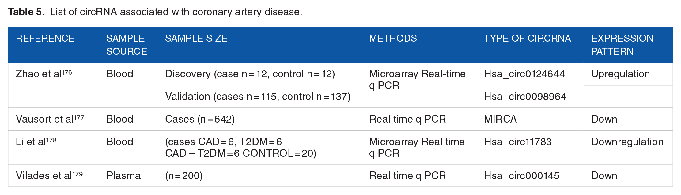
The research reports that have implicated the potential role of circRNAs in the initiation, development, and progression of Coronary artery disease are shown in Table 5. Most importantly, circRNAs have been found to be indispensable participants in the occurrence and progression of CAD.
Here we summarized the recent studies related to the discovery and effect of circRNAs in CAD pathogenesis (Table 5) and discussed some of research articles commenting on the feasibility of circRNAs to serve as biomarkers for cardiovascular diseases.
circRNAs as miRNA Sponge in Coronary Artery Disease
Studies on circRNAs have evidence that some of these RNA species exert their control in the expression of a specific gene through miRNA sponging effect. Due to the interaction with RNA binding proteins, circRNAs are involved in disease progression in CAD and are summarized in Table 6.
circRNA with targeted miRNA in CAD.

Epigenetic Biomarkers of CAD
Epigenetic biomarker defines any epigenetic mechanism which exposes the information about pathology of disease, detection of disease, predicts the risk of the disease development in future, predicts the significance of the disease, monitors to medication and therapy. 182 Many epigenetics biomarkers have been reported for CAD and its progression.183,184 Epigenetic modifications can present in both the form of covalent modifications in DNA/ Histone and pattern nc RNA expressions. Evidence suggests that candidate gene methylation patterns can serve as a potential epigenetic biomarker for diagnosis and prognosis of CAD (Table 2). The role of miRNAs in coronary artery disease has been widely studied over the years. Therefore, miRNAs are found to be major regulators in cellular gene expression for the initiation and progression of CAD. As discussed above altered levels of different circulating miRNAs could be used as diagnostic and prognostic markers of CAD (Table 4). Moreover, lncRNAs (sec b) and circRNAs have been reported to be potential biomarkers of CAD (Table 5).
Epigenetic Diversity Among CAD Patients
In contrast to the conventional principle, 1 individual’s genome does not differ from another. Scientists have established by their extensive research work that abundant differences do exist in nature for an individual’s epigenome. Thus, epigenetic modifications come from the diversity of nature comprising environmental exposure and pollutants, which lead to the development of deleterious diseases like CAD. Keeping this in consideration, scientists and physicians are trying to evolve novel therapeutic strategies based on these differences. 185 Furnishing the Human Genome project is the keystone of the inherent blueprint and backbone of genomics.186,187 Within a decade of declaring the human genome project, researchers have reached to next-generation technology to understand the mechanism behind structural DNA to crucial activities in genome biology. With the advent of next-generation sequencing, scientists have been able to understand the role played by candidate genes and alterations in their nucleotide sequence towards the development of different diseases. Despite these approaches, the epigenetic world remains unexplored about precision medicine in CAD as of now. Worldwide researchers have already initiated efforts to comprehend epigenomics related to coronary artery disease.188-190
Nowadays, several epigenome-based techniques have come up to explore the epigenetics related to CAD (Figure 4).

Epigenetic modifications and techniques involved with different kind of sample sources: WGBS, whole-genome bisulfite sequencing; MSRE, methylation-sensitive restriction enzymes; HPLC, high-performance liquid chromatography; RRBS, reduced representation bisulfite sequencing; ONT, Oxford nanopore technologies; Pyrosequencing—attractive alternative to the conventional bisulfite sequencing PCR (BSP); HRM assay; RT-PCR based high resolution melting curve analysis; ChIP, chromatin immunoprecipitation; MS, mass spectrometry; microarray; RNA seq, RNA sequencing.
Epigenetic Therapeutic Targets in CAD
The veracity of altered environmental exposure could be an epigenetic marker for upcoming treatment. According to researchers, these modifications can be inverted using pharmaceutical agents and nutraceuticals. For example, DNA methyltransferase inhibitors (DNMTis), histone acetyltransferase inhibitors, histone deacetylase inhibitors, histone methylase inhibitors, and bromodomain and extra terminal motif (BET) inhibitors have been extensively studied as conceivable therapeutics in inflammation and CAD. 191 Although DNMTs are present in various food items naturally, many synthetic man-made ones are being used widely as well. 192
Epigenetics Toward Personalized Medicine
Since the remote past, our health care professionals have been striving to offer the best care and effective treatment to their patients, as already discussed above. They have been doing this by experimenting with different treatment strategies, selecting the better successful ones based on their observational analysis, sharing their inferences with an expectation of improving the strategy of their predecessors. The ultimate goal of the efforts was to provide more accurate, precise, and effective treatment for each individual no matter how necessary the tools were at their disposal. However, nowadays, the advent of modern innovative techniques of genetic testing, data analysis, supercomputing devices, and electronic data storage has enabled physicians and scientists to take the area of precision medicine to a level which their historical predecessors could not even think of.
Precision medicine can take the management of a disease to its ultimate goal, i.e., complete cure of the disease. For example, Alexis and Noah, the famous twins who were misdiagnosed and mistreated, for about 14 years, for a condition called palsy. They received the treatment for palsy until human genome sequencing, and DNA sequencing made it possible to correctly diagnose their actual disease condition, that is, dystonia. 193 The sequencing of the human genome took an international collaboration of 13 years and amounted to a financial costing of US$3 billion. However, the advancement in techniques involved, human genome sequencing, can be completed in 24 hours for just US$1000. This has made us capable of tailoring the prevention to an individual patient or group of patients using their genomic information in combination with their environment and lifestyles.
Although human genome sequencing has opened up the window for the interesting discipline of genetic medicine, which then leads the path to develop personalized medicine, it has become clear now, after 17 years, that our genetic material (genes) alone is not able to thoroughly explain the fundamental aspects of development and aging. Moreover, our genes are not enough to predict our susceptibility to different types of diseases. Epigenetics has already answered a lot of curiosity questions that bothered physicians and scientists for a large amount of time in the past. Epigenetics and factors affecting epigenetics mechanisms are attracting the attention of researchers and are being used to elucidate the underlying causes of the onset and progression of numerous life-threatening diseases. Innovative techniques that are harnessed to study epigenetics and epigenetic mechanisms are revolutionizing the approaches to personalized medicine (Figure 4). Innovative technologies to study epigenetics, such as bisulfite sequencing, have the ability to accurately quantify and map epigenetic marks underlying the causation of a particular disease phenotype, including CAD. This was not possible using traditionally used genetic research techniques. Epigenetic modifications determine the cell identity and make a skin cell different from a liver cell or heart cell. In many disease phenotypes like cardiac diseases, neurological diseases, or cancer, epigenetic changes are much more frequent than genetic variants, therefore, providing more vital insights into understanding the development and progression of these disease phenotypes.
Accurate mapping and quantitation of epigenetic markers help us in improving clinical practices and approaches by improving the diagnosis of diseases at the early stages of disease development, even prior to the happening of genetic changes. 194 Research reports are demonstrating that epigenetic variations precede frequent genetic alterations in disease development. 195 Therefore, detection of early disease signals by exploiting epigenetic based diagnostics offers opportunities for clinical intervention even before the appearance or progression of symptoms impact life quality when the patient is relatively fit. This favors our treatment process and success to treat the condition even before it appears. In this way, management of disease and prognosis is performed in more accurate manner.196-198
The concept of personalization of medicine in the context of coronary atherosclerosis could take the management of CAD to even more refinement than other diseases. Environmental factors are potential influencers of CADs. These environmental factors induce epigenetic changes, which in turn affect the genetic expression of an individual in the course of disease development. Therefore, taken together, the characterization of epigenetic aberrations in CAD precision medicine requires careful consideration.
Conclusion
This review summarized recent findings linked to epigenetic study in CAD and illustrated the impact of the epigenetic mechanism in CAD. Epigenetics has significantly influenced our understanding of gene regulation in CAD, although consensus regarding the magnitude of effect size is lacking. Epigenome intensely responds to variations in the environment during an individual’s lifetime contributions in the regulation of critical biological mechanisms and provides a significant association among life experiences, phenotypic expression, and risk of disease development. Since environmental inducements associated with epigenetics are inconsistent with the ethnicity of the population, results are difficult to generalize, and regional studies are imperative. Epigenetic aspects of in-utero exposure and after birth exposure, like dietary factors, have been inadequately explored. Future strategy for further research must incorporate elucidation of diagnostic biomarker potential and therapeutic targets for atherosclerotic disorders in order to translate emerging epigenetic data in bedside care. Moreover, with the help of epigenetics and techniques to study epigenetic factors, the present-day physician and researchers could take personalized medicine to a level unreachable to their ancestors.
Footnotes
Acknowledgements
Facilities provided by Govind Ballabh Pant Postgraduate Institute of Medical Education and Research (GIPMER), New Delhi, are acknowledged. Authors are grateful to the Department of Biotechnology Government of India for financial support provided under the head of “Establishment of Central Molecular Lab in GIPMER to study the diagnosis for precision medicine in order to understand the disease process and utilize it as Clinical Research Facility” with Grant No. BT/INF/22/SP33063/2019.
Funding:
The author(s) disclosed receipt of the following financial support for the research, authorship, and/or publication of this article: This work is funded by the Department of Biotechnology (DBT), Govt. of India, India (BT/INF/22/SP33063/2019).
Declaration of conflicting interests:
The author(s) declared no potential conflicts of interest with respect to the research, authorship, and/or publication of this article.
Author Contributions
MPS and BM were involved in planning, designing the manuscript. MPS was involved in the writing of the manuscript. RSAS, N, A, AK, AKS, EA, and AA were involved in reviewing the manuscript. Finally, the manuscript was approved by BM and SSS.
List of Abbreviation