
Abstract

Unilateral mydriasis in the context of recurrent headache episodes might suggest increased intracranial pressure with transtentorial herniation and is frequently considered an ominous sign. In this report, we present a patient with this particular combination of features but with an entirely different, relatively innocent etiology.
A 28-year-old woman presented with episodes of right-sided pupillary dilation lasting several minutes which were accompanied by transient unilateral visual blurring and impaired focusing (Figure 1). Her medical history was significant for an atrial septal defect (ASD) that had been closed 6 years earlier. The episodes of mydriasis commenced 7 years prior and disappeared after closure of the ASD but recurred a year ago. Their frequency ranges from 10 times a day to once in a few months. The patient did not have any medication, could not identify any precipitating factors, and reported no simultaneous headache, orbital pain, nausea, photophobia, ptosis, diplopia, redness of the eye, or other facial autonomic symptoms. Aside from these episodes, she had attacks of right-sided pulsating headache lasting 1–3 days without nausea, photophobia, phonophobia, or obvious interference with activities of daily life. These attacks also started a year earlier and occurred on average once a month. We were not able to examine the patient during an episode of mydriasis. Interictal neurological examination was unremarkable, and brain magnetic resonance imaging including angiography excluded structural intracranial pathology. She was diagnosed with benign episodic unilateral mydriasis.
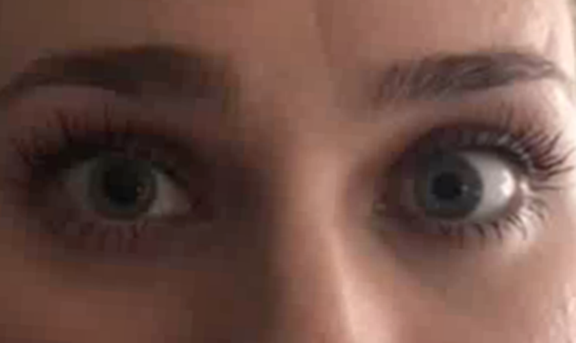
Episode of right-sided pupillary dilation.
Benign episodic unilateral mydriasis is a rare condition most often described in young women with a history of migraine or intermittent headache displaying features of migraine but not strictly fulfilling diagnostic criteria. The duration of episodes, which usually do not have precipitating factors, varies from seconds to weeks but most typically they last several minutes to hours. The most common associated symptoms in a series of 24 patients included monocular visual blurring (62%), headache (37%), orbital pain (21%), unilateral photophobia (17%), and redness of the eye (17%). 1 By definition, there are no structural brain abnormalities, and it is a diagnosis of exclusion. 2
Some authors have previously suggested it to be a limited form of “ophthalmoplegic migraine” (nowadays known as “recurrent painful ophthalmoplegic neuropathy”), while others consider this unlikely. 1 Alternatively, the subsequent development of migraine with typical visual aura after 3 years of recurrent transient mydriasis episodes in one case led Maggioni et al. to regard the latter as possible or atypical auras. 3 Although not completely elucidated, the underlying pathophysiological mechanism has been postulated to involve a temporarily disturbed parasympathetic innervation of the iris sphincter muscle rather than excessive sympathetic outflow to the iris dilator muscle. 4
Of interest, our patient experienced an initial disappearance of mydriasis episodes after closure of an ASD. To our knowledge, no previous report on benign episodic unilateral mydriasis addressed such a temporal association. A recent systematic review that examined the change in migraine headache following percutaneous closure of an ASD showed that preexisting migraine improved in 18 of 126 (14%) patients and resolved in 47 of 126 (37%) patients within a median follow-up period of 12 months. 5 It is possible that the initially favorable course of our patient’s episodes following the ASD closure rests on mere coincidence. Alternatively, this similar response might provide a pathophysiological link between benign episodic unilateral mydriasis and migraine.
Other disorders or phenomena affecting pupillary diameter such as Adie’s pupil (tonic dilation accompanied by light-near dissociation), hippus (physiological rhythmic fluctuation between dilation and constriction), pharmacological influences (sympathomimetic or parasympatholytic eye drops, aerosols, or herbs), orbit trauma, transtentorial herniation, posterior communicating artery aneurysm, and acute angle-closure glaucoma are usually easily excluded following a thorough medical history and careful neurological examination. Recognition of the typical features of episodic unilateral mydriasis is important as subsequent reassurance of its benign course suffices.
Footnotes
Informed consent
The patient was offered to read the manuscript and has given informed consent to the publication of this report.