
Abstract
Overproduction of free radicals in excess of antioxidants leads to oxidative stress which can cause harm to the body. Conventional antioxidants have drawbacks and are believed to be carcinogenic. The present study seeked to confirm folklore use and validate the antioxidant potentials of Grewia tembensis and Xerophyta spekei which have been widely used in the Mbeere community as medicinal plants. Antioxidant properties were determined through scavenging effects of diphenyl-1-picrylhydrazyl (DPPH) and hydrogen peroxide radicals as well as iron chelating effects. The data obtained was assayed in comparison to the standards (Ascorbic acid and EDTA). Ascorbic acid had a significantly greater DPPH radical scavenging property with an inhibitory concentration (IC50) value of 20.54 ± 2.24 µg/mL in comparison to the plant extracts, which had IC50 values of 33.00 ± 1.47 µg/mL, 69.66 ± 1.01 µg/mL and 86.88 ± 2.64 µg/mL for X. spekei, G. tembensis leaf and G. tembensis stem bark extracts, respectively. EDTA demonstrated a significantly greater iron chelating effect having a significantly lesser IC50 value of 25.05 ± 0.79 µg/mL as opposed to 43.56 ± 0.46 µg/mL, 89.78 ± 0.55 µg/mL, and 120.70 ± 0.71 µg/mL for X. spekei, G. tembensis leaf, and G. tembensis stem bark extracts respectively. Additionally, ascorbic acid also exhibited stronger hydrogen peroxide radical scavenging effect than the studied extracts. Generally, X. spekei extract had higher antioxidant activities as compared to both the leaf and stem bark extracts of G. tembensis. The phytochemical screening demonstrated the presence of secondary metabolites associated with antioxidant properties. The present study therefore, recommends ethno medicinal and therapeutic use of G. tembensis and X. spekei in the treatment and management of oxidative stress related infections.
Keywords
Introduction
During energy generation in body cells, free radicals are generated, which, at high concentrations, results in oxidative stress, but at low or moderate levels, they are effective in cellular and immune responses. 1 The development of chronic infections like arthritis, cancer, cardiovascular and other neurodegenerative diseases are known to be mediated by oxidative stress. 1 Excessive production of free radicals above available antioxidants in the body results in oxidative stress (OS), which can potentiate apoptosis, tumorigenesis, and immune response, 2 thereby causing harm to the body. 3
The effects of OS are preventable by the use of antioxidants which protect cells and tissues from the destructive effects of these unstable oxidants. 4 During oxidative stress, the body produces antioxidants which include enzymes and vitamins, to counteract the effects caused by the oxidants. However, these antioxidants can get overwhelmed by some factors like environmental factors that prevent the antioxidants from detoxifying the oxidants hence making the oxidants remain in the body as they cannot be detoxified and eliminated from the body. 5 Synthetic antioxidants such as tertiary butyl hydroquinone, butylated hydroxytoluene as well as propyl gallate, which are used in the preservation of food for human consumption, have been thought to cause serious harmful health effects on the sensitive organs of humans like the liver and the brain and are also proved to be carcinogenic. 6 In addition, these synthetic antioxidants are not readily available to patients due to factors such as cost and inaccessibility. 7
Conventional drugs used to manage and treat illnesses caused by oxidative stress are costly, unaffordable, and inaccessible, especially in rural and remote regions. Therefore, new therapeutic agents with antioxidant activities are more desirable. The challenges linked to conventional management and treatment of OS have led to scientists shifting their attention to finding alternative therapies such as using medicinal plants as they are valued not only for their dietary use but also for their medicinal use. 8 There is a greater need for the use of plant-derived antioxidants due to the challenges of conventional antioxidants and their high cost. Since time immemorial, medicinal plants have been relied upon as therapeutic agents to manage or heal diseases as they are linked to fewer side effects and are affordable. 9
Plants are important sources of molecules, which can be utilized in developing new antioxidant agents, and it is estimated that 44% of new drugs being developed are of plant origin. 10 Some plants like Senna singueana have previously exhibited antioxidant properties. 11 Plant antioxidants like polyphenols, carotenoids, glutathione have exhibited antioxidant effects by interrupting free radicals’ chain reactions. 12 Due to these properties, plant antioxidants are being considered as a source of alternative and complementary therapy against oxidative stress. Natural antioxidants have been discovered in medicinal plants. 13 In the past, traditional herbalists have used herbal plants to cure various diseases, including oxidative stress related illnesses. 14 However, only a few have been scientifically evaluated and validated. 15 The two plants used in the current study; Xerophyta spekei and Grewia tembensis are used by herbalists as medicinal herbs in the Mbeere community in Embu County as remedy to cure a variety of diseases. 16 However, their antioxidant activities have yet to be shown scientifically. Therefore, this experiment was executed to explore, establish and confirm the two medicinal plant extracts’ antioxidant capabilities in vitro. The current study also determined the phytocompound compositions of ethyl acetate extracts of G. tembensis and X. spekei to establish the basis of their medicinal potentials.
Xerophyta spekei (Baker) commonly known as Kianduri by the Embu community, 16 is a member of the Velloziaceae family and is also called Vellozia spekei. It's common in Kenya, Zambia, Tanzania, Zimbabwe, and Ethiopia. In the Embu community, it is used to treat dog bites and diabetes. 16 It is also used as an antivenom where the ashes of the whole plant are applied at the site of the snake bite. 17 The Mbeere community uses it to treat dog bites and diabetes. 16 Traditional herbalists in South Africa use it as an analgesic and an anti-inflammatory. 18
Grewia tembensis (Fresen) is a medicinal plant species commonly known as Muruba by the Mbeere community from Embu County. 19 It's classified under the genus Grewia belonging to the family Tiliaceae which are known for their various biological effects like anti-oxidant, anti-bacterial, hepatoprotective, anti-inflammatory, anti-emetic, anti-malarial, analgesic, and anti-pyretic activities. 20 It grows in moderately dry areas in Eastern Africa. 21 The Kipsigis call it Chesarebut, 19 while the Maasai in Kajiado county calls it Eirri. 21 G. tembensis is used in Djibouti to treat microbial infections like abscesses and furuncle, 22 while the fruits are eaten by the Kamba from the Ithanga division. 23 The Turkana community uses G. tembensis to treat coughs and eat its fruits as food. 24 The Kamba community calls it Muvindaviti or Mutuva and uses its roots to treat typhoid, 19 heartburn, hypochondriasis, 25 reduced appetite, and swollen diaphragm. 24 In Tanzania, the Pare community calls it Mkokoro and uses it to treat breast cancer. 26
Materials and Methods
Plant Materials Collection and Preparation
Fresh X. spekei (whole plant without roots) and G. tembensis (leaf and stem bark) parts were gathered from Gikuyari village, Thura Sub Location, Thura Location, Siakago Division, Mbeere North Sub County in Embu County, Kenya in May 2021 with the help of a local practicing traditional herbalist. GPS coordinates for X. spekei were 0°36′34″ N, 37°37′15″ E while for G. tembensis were 0°35′38.55552″ S, 37°38′12.3252″ E. The plants were transported to the University, where they were identified by a recognized taxonomist and a specimen for each plant was preserved at Kenya National Museum's herbarium for future reference. Voucher numbers were allocated as PN/002/27698/2018 and PN/003/27698/2018 for X. spekei and G. tembensis respectively. The plants were thoroughly washed in running tap water (H2O), rinsed using distilled water (DH2O), and then chopped into small parts. Thereafter, shade dried for 28 days then ground into fine powder. The powders were then stored in separate airtight containers at room temperature awaiting extraction process.
Extraction Process
Four hundred grams (400 grams) of fine dry powder of G. tembensis stem bark were separately soaked in 1.2 L of ethyl acetate for 72 h. In addition, 800 grams of X. spekei whole plant dry powder and 300 grams of G. tembensis dry leaves powder were separately soaked in 2.4 L and 0.9 L of ethyl acetate, respectively, for 72 h. To achieve complete dissolution, the solutions were occasionally swirled. After 72 h, the solutions were decanted and vacuum filtered using a Buchner funnel and No. 1 Whatman's filter paper. Following that, the filtrates were separately concentrated using a rotary evaporator to evaporate the solvent at 90 rpm at 60 °C under vacuum. The following equation was used to determine the extract yields of the plants:

The resultant extracts were stored at 4 °C in clean, sterile glasses ready for use in the bioassay studies.
In Vitro Antioxidant Properties Determination
In Vitro DPPH Radical Scavenging Capacity
The plant extracts’ ability to quench 2, 2-diphenyl-1-picrylhydrazyl (DPPH) radicals was done in triplicates as described by Oyedemi et al,
28
with little modifications. Plant extracts and ascorbic acid (reference) were prepared in a range of concentrations ranging from 15.625 µg/mL to 500 µg/mL. DPPH (1 mM) solution was prepared in methanol. In clean test tubes, one (1) mL of each dilution of the test extract and the standard were placed, followed by DPPH (0.5 mL) and methanol (3 mL) solutions. Following that, the blend was thoroughly vortexed for 5 min before being placed in a dark cupboard for 30 min at room temperature. A blank solution containing 3 mL of methanol and 0.5 mL of DPPH was also prepared. The absorbances of the solutions were measured at 517 nm against a blank using a spectrophotometer. The plant extracts’ % DPPH free radical scavenging effects were computed as;

The % radical scavenging capacity was then plotted versus various concentrations. Therafter, half maximal inhibitory concentration (IC50), representing the concentration at which 50% of the DPPH radicals were scavenged, was graphically determined.
Iron (Fe2+) Chelating Activity Assay
Iron (Fe2+) Chelating assay was demonstrated following protocols of Arika et al, 30 with slight adjustments. Various dilutions (15.625 µg/mL to 500 µg/mL) of test extracts and EDTA (control) at a volume of 1 mL of each concentration were separately blended with 1 mL of ferrous sulfate (0.125 mM) thereafter, ferrozine (0.3125 mM) 1000 µL was added to commence the reaction. The blend was vortexed, incubated at ambient room temperature for 10 mins before reading the absorbance of the plant extracts/reference at 562 nm against blank solution, which had ferrous sulfate (0.125 mM) and ferrozine (0.3125 mM) reagents without the extracts and the standard. The iron chelating experiment was conducted in triplicates.
The iron chelating capabilities of the plant extracts as well as the standard were calculated as follows;

Where; AB = Blank Absorbance, AS = Sample/standard Absorbance.
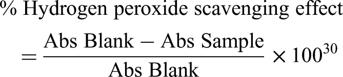
Determination of Hydrogen Peroxide (H2O2) Radical Scavenging Effect
Hydrogen peroxide (H2O2) free radical scavenging capacity of studied plant extracts was done using the protocol of Arika et al
30
Six hundred (600) µL of H2O2 (40 mM) prepared using buffered phosphate solution (1.0 M, pH 7.4) was separately blended with 400 µL of various dilutions (15.625 µg/mL to 500 µg/mL) of the extracts and ascorbic acid (reference). The blend was then allowed to stand for 10 mins at standard room temperature. Optical density was read at 230 nm against a blank that only had phosphate buffer solution.
32
Hydrogen peroxide radical scavenging effect experiment was performed in triplicates. H2O2 free radical scavenging capability was computed as follows;
Gas Chromatography-Mass Spectrometry Analysis
Phytochemical screening to detect and quantify phytochemicals available in the ethyl acetate extracts of X. spekei and G. tembensis was done by GC-MS using 7890A Gas-Chromatograph linked to an Agilent Technologies 5975C mass selective detector, which is made up of an HP-5 MS low bleed capillary column (30 m long, 0.25 mm wide, and 0.25 m film thick). The mass spectrometer's operating parameters included; relative detector gain mode, 70 eV of ionization energy, 3.3 min of filament delay time, 1666 μ/sec of scan speed, 40-550 m/z of scan range, 230 °C ion source temp, and 180 °C quadrupole temp. At a constant flow rate of 1.25 mL per minute, helium gas (99.9% purity) was used as a carrier gas. With 1 μL injection volume, the mass transfer temperature was set at 200 °C while injector line transfer temperature at 250 °C. The oven temperature was set at 35 °C for 5 min then increased at a rate of 10 °C/min to 280 °C for 24.5 min and then increased at a rate of 50 °C per minute to 285 °C for 20.5 min for a total run time of 50 min.
Data Management and Statistical Analysis
Data from the current study were tabulated and organized in a Microsoft Excel spreadsheet prior to being imported into Minitab software version 17.00 for analysis where the values were presented as mean ± STD (standard) error of mean (SEM). For inferential statistics, one (1) way ANOVA (Analysis of Variance) was used, as well as Tukey's post hoc test for pairwise comparison and separation of means. A P value of <.05 was considered statistically significant. The findings were presented in bar graphs and tables. For the identification of phytochemicals found in each extract, a comparison of the data obtained was matched with mass-spectra library search report from the National Institute of Standards and Technology 08 and 11, where each unique peak represented a particular chemical substance.
Results
Yields of the Plant Extracts
Among the tested extracts, G. tembensis leaf (2.99%), which was a dark green semi-solid extract had the highest yield followed by X. spekei (2.01%), a dark brown sticky extract, and G. tembensis stem bark (1.87%), which produced a brown solid extract (Table 1).
Plant Extracts’ Percentage Yield and Description.

In Vitro Antioxidant Properties
In Vitro DPPH Radical Scavenging Properties
The tested plant extracts/standard exhibited dose-dependent DPPH radical scavenging properties across 15.625 µg/mL to 500.00 µg/mL concentrations. As plant extracts/standards’ concentrations decreased, the DPPH radical scavenging capability also decreased. Both stem bark and leaf extracts of G. tembensis demonstrated statistically different DPPH activities in 15.625 µg/mL to 500.00 µg/mL concentratons (P < .05). Additionally, X. spekei extract demonstrated significantly distinct DPPH radical scavenging activity in 15.625 µg/mL to 500.00 µg/mL concentrations (P < .05) except at 125.00 µg/mL and 250.00 µg/mL concentrations where the extract showed statistical similarity in DPPH radical scavenging activity (P > .05). Ascorbic acid, exhibited a significantly higher DPPH radicals scavenging capability having 20.54 ± 2.24 µg/mL of IC50 value as opposed to the plant extracts, which had IC50 values of 33.00 ± 1.47 µg/mL, 69.66 ± 1.01 µg/mL and 86.88 ± 2.64 µg/mL for X. spekei, G. tembensis leaf and G. tembensis stem bark extracts, respectively P < .05).
Findings further showed that the reference had a significantly higher DPPH free radical quenching effect than X. spekei, G. tembensis leaf, and G. tembensis stem bark extracts at concentrations ranges of 15.625 µg/mL to 62.500 µg/mL (P < .05; Figure 1). At 125.00 µg/mL to 500.00 µg/mL concentration ranges, ascorbic acid and X. spekei extract had statistically similar DPPH radical scavenging activities. X. spekei extract showed significantly greater DPPH free radical scavenging capacity than both stem bark and leaf extracts of G. tembensis at 15.625 µg/mL to 500.00 µg/mL concentrations (P < .05; Figure 1). Both stem bark as well as leaf extracts of G. tembensis demonstrated statistically similar DPPH radical scavenging activities in 15.625 µg/mL to 500.00 µg/mL concentrations (P > .05; Figure 1) except at 15.625 µg/mL, 125.00 µg/mL and 250.00 µg/mL concentrations, where the leaf extract showed significantly greater DPPH radical scavenging effect than the stem bark extract (P < .05; Figure 1).

Comparison of DPPH radical scavenging properties of X. spekei, and G. tembensis extracts. Bar graphs having distinct letters within a given concentration are significantly different (P < .05). (1-way Analysis of Variance and Tukey's Post hoc tests).
Iron Chelating Properties
The tested plant extracts revealed a strong efficacy of iron chelating activity across all the plant extracts concentrations, which occurred in a concentration dependent manner. Plant extracts’ ability to chelate irons decreased with a decrease in extracts’ concentration. There were no statistical similarities in iron chelating activities of plant extracts and the standard EDTA across 15.625 µg/mL to 500.00 µg/mL concentrations (P < .05). EDTA exhibited the highest iron chelating effect having a significantly lower IC50 value of 25.05 ± 0.79 µg/mL as opposed to the plant extracts, which had IC50 values of 43.56 ± 0.46 µg/mL, 89.78 ± 0.55 µg/mL, and 120.70 ± 0.71 µg/mL for X. spekei, G. tembensis leaf, and G. tembensis stem bark extracts respectively. Similarly, X. spekei, G. tembensis leaf, and G. tembensis stem bark extracts demonstrated significantly different IC50 values (P < .05).
In comparison, EDTA had a significantly greater iron chelating potency than X. spekei, G. tembensis leaf, and G. tembensis stem bark ethyl acetate extracts from 15.625 µg/mL to 500.00 µg/mL concentrations (P < .05; Figure 2). Additionally, X. spekei extract had significantly higher iron chelating activity than the other extracts across 15.625 µg/mL to 500.00 µg/mL concentrations (P < .05; Figure 2). Both leaf and stem bark extracts of G. tembensis demonstrated statistically similar iron chelating activities across 15.625 µg/mL to 500.00 µg/mL concentrations (P > .05; Figure 2), aside from 62.50 µg/mL and 125.00 µg/mL concentrations, where G. tembensis leaf extract had significantly higher iron chelating activity than G. tembensis stem bark extract (P < .05; Figure 2).

Comparison of iron chelating properties of studied extracts. Bar graphs having distinct letters within a particular concentration are significantly different (P < .05). (1-way Analysis of Variance and Tukey's Post hoc tests).
In Vitro H2O2 Radical Quenching Properties
X. spekei, G. tembensis leaf, and G. tembensis stem bark extracts displayed a potent concentration dependent efficacy of hydrogen peroxide (H2O2) radical scavenging ability. As the concentrations increased among all the tested extracts, the H2O2 radical scavenging capacity also increased. Concentrations 15.625 µg/mL to 125.00 µg/mL for G. tembensis stem bark and leaf extracts exhibited significantly different H2O2 scavenging effects from one another (P < .05) while at concentrations 250.00 µg/mL and 500.00 µg/mL, they both demonstrated statistical similarity in hydrogen peroxide scavenging activities (P > 005). X. spekei extract demonstrated statistical similarity in its hydrogen peroxide scavenging activity between concentrations 500.00 µg/mL and 250.00 µg/mL as well as concentrations 125.00 µg/mL and 250.00 µg/mL (P > .05).
Ascorbic acid's IC50 value was 19.14 ± 0.61 µg/mL, which was significantly lower compared to the plant extracts, which had IC50 values of 29.65 ± 0.69 µg/mL, 42.92 ± 0.50 µg/mL, and 55.78 ± 1.91 µg/mL for X. spekei, G. tembensis leaf and G. tembensis stem bark extracts, respectively indicating that ascorbic acid had the greatest hydrogen peroxide scavenging effect followed by X. spekei extract, G. tembensis leaf extract and finally G. tembensis stem bark extract.
In comparison, X. spekei extract demonstrated statistically similar H2O2 radical scavenging activity with ascorbic acid at concentrations ranging from 62.500 µg/mL to 500.00 µg/mL concentration (P > .05; Figure 3), whereas at 31.25 µg/mL and 15.625 µg/mL concentrations, ascorbic acid demonstrated significantly greater H2O2 radical scavenging capability than X. spekei extract (P < .05; Figure 3).

Comparison of in vitro H2O2 radical quenching properties of X. spekei and G. tembensis extracts. Bar graphs having distinct letters within a given concentration are significantly different (P < .05). (One (1) way Analysis of Variance and Tukey's Post hoc tests).
X. spekei extract demonstrated a significantly higher hydrogen peroxide radical scavenging effect than both the leaf as well as stem bark extracts of G. tembensis at 15.625 µg/mL to 500.00 µg/mL concentrations (P < .05; Figure 3). Similarly, G. tembensis leaf extract demonstrated significantly higher hydrogen peroxide radical scavenging activity than G. tembensis stem bark extract at concentration ranges of 15.625 µg/mL to 62.500 µg/mL (P < .05; Figure 3). However, at concentrations ranging from 125.00 µg/mL to 500.00 µg/mL, both the leaf and stem bark extracts of G. tembensis demonstrated statistically similar hydrogen peroxide scavenging effects (P > .05; Figure 3).
Quantitative Phytochemical Composition
Phytochemical screening results for stem bark extract of G. tembensis as presented in Figure 4 and Table 2, showed n-Hexadecanoic acid, a fatty acid derivative, as a major compound with a percentage abundance of 16.2% while the compound 2-Pentanone, a ketone had the lowest percentage abundance of 0.1%. As Table 3 shows, the extract comprised 38.1% fatty acids, 30.6% terpenoids, 11.6% hydrocarbons, 10.2% steroids, 4.0% phenolic compounds, 2.3% aldehyde compounds, 1.4% ketones, 0.6% tocopherol, 0.6% benzene derivatives, 0.4% methoxybenzoic acid compounds and 0.2% aromatic amine compounds.

GC-MS chromatogram of G. tembensis Ethyl Acetate stem bark extract.
Quantitative Phytochemical Compound Analysis in the Tested Ethyl Acetate Extracts.

Abbreviations: NP, Not Present; RT, Retention time in minutes; Conc, Concentration; % Ab, Percentage Abundance; GTB, Grewia tembensis stem bark extract; GTL, Grewia tembensis leaf extract; XS, Xerophyta spekei extract.
Percentage Total Identified Compounds per Studied Plant Extract.

Abbreviations: GTB, Grewia tembensis stem bark extract; GTL, Grewia tembensis leaf extract; XS, Xerophyta spekei extract.
The compounds present in the leaf extract of G. tembensis were identified by GC-MS analysis as seen in Figure 5. Further, phytochemical investigations of this plant extract as presented in Table 2, showed the presence of 40 compounds with trans-3-Penten-2-ol, an alkenol compound, having the lowest percentage abundance of 0.02%, while Squalene, a triterpenoid, having the highest percentage abundance of 18.78%. The classes of secondary metabolites identified in the leaf extract of G. tembensis were 59.33% of terpenoids, 16.17% fatty acids, 15.08% hydrocarbons, 6.58% phenolic compounds, 1.79% ester compounds, 0.79% steroids, 0.19% naphthalene compounds, 0.04% aldehyde, 0.03% toloudine derivative (Table 3).

GC-MS chromatogram of G. tembensis Ethyl Acetate leaf extract.
Phytochemical compounds of X. spekei extract, showed several secondary metabolites with different peak areas (Figure 6). Ursa-9(11), 12-dien-3-one, a triterpenoid, had the highest percentage abundance of 14.14%, whereas 1,16- Cyclocorynan- 17-oic acid, 19, 20- didehydro-, methyl ester, (16S, 19E)-, an alkaloid, had the lowest percentage abundance of 0.02% (Table 2). Additionally, phytochemical screening of ethyl acetate of X. spekei extract, revealed 33.34% terpenoids, 27.90% fatty acids, 23.93% steroids, 9.76% hydrocarbons, 1.92% phenolic compounds, 1.02% tocopherols, 0.83% tetracarboxylic acids, 0.97% benzene derivatives, 0.20% ketones, 0.11% dialkylaminodiphenylbutanol ester and 0.02% alkaloid (Table 3).

GC-MS chromatogram of X. spekei Ethyl Acetate extract.
Discussion
The accumulation of free radicals in excess of available antioxidants results in OS 33 and OS associated illnesses. 34 Antioxidants are therefore produced to counteract the effects of OS in the cells by neutralizing the unstable free radicals through donation of electrons to stabilize them. 30 Synthetic antioxidants, some of which are used as food preservatives and are commercially available, are said to be toxic as well as carcinogenic; therefore, antioxidants derived from plants provide a better and safer alternative. 35 Herbal plants have anciently been used to manage and treat oxidative stress related diseases.
The antioxidant capabilities of plant extracts and standards in all assays were dose dependent in the current study. The antioxidant effect decreased as the concentration of the extracts/standards decreased. This trend is consistent with our previous study that found dose dependant antioxidant properties of ethyl acetate leaf extract of Senna singueana. 36 Our current findings are also in agreement with other previous research that found dose-dependent antioxidant potentials in the extracts studied.37,38
The reference, Ascorbic acid, exhibited a higher dose dependant DPPH radical scavenging potential in the current study, as compared to the studied plant extracts. This is comparable to the findings of Narendhirakannan et al, 39 who discovered that the ability of garlic extracts to scavenge DPPH radicals increased with extract concentration. They also agreed with Shang et al, 40 who found that the DPPH radical scavenging potentials of inulin and Vitamin C followed a similar pattern. The researchers also discovered that vitamin C outperformed inulin in terms of DPPH radical scavenging activity. In another study by Amudha and Rani, 41 DPPH free radical scavenging capacity of the ethanol and ethyl acetate extract of Cordia retusa increased in a dose dependant trend and that the extracts exhibited a lower DPPH radical scavenging activity as compared to the standard.
Accumulation of heavy metals in the body caused by free radicals can be eliminated through chelation therapy. 42 Therefore, metal chelating antioxidant property was done to detect the capability of the ethyl acetate extracts of X. spekei, G. tembensis leaf, and G. tembensis stem bark to chelate irons. This assay relies on the principle that when antioxidants form a complex with metal irons, they can inhibit the electron transfer, which eventually blocks the formation of free radicals. In a solution containing ferrozine, ferrous ions, and chelating agents, the chelating agent competes with ferrozine to chelate ferrous ions, a process that results in a red-colored iron (II)-ferrozine complex leading to a reduction in absorbance at 562 nm. 43
X. spekei extract exhibited significantly the greatest iron chelating impact among the studied extracts trailed by G. tembensis leaf extract and finally G. tembensis stem bark extract. Although, the metal chelating impact of EDTA was significantly greater than the extracts. These findings agree with Loizzo et al, 44 who previously observed that EDTA had higher chelating activity than the peel and the pulp extracts of Annona cherimola Mill. (Cherimoya). Similarly, Wong et al 45 demonstrated that Vernonia amygdalina, Pereskia bleo, Clinacanthus nutans, Hedyotis diffusa, Callicarpa formosana, and Leonurus cardiaca showed strong chelating activities but lower than the standard. Our observations also concur with Amudha and Rani, 41 who observed that the standard EDTA had a higher iron chelating activity than the ethanol and ethyl acetate extracts of Cordia retusa. This was also similar to a report by Al-Trad et al, 46 which also determined that butanol extract from Ephedra alte had good iron chelating activity.
The in vitro hydrogen peroxide (H2O2) free radical scavenging method is built on the concept that when H2O2 radicals are reduced to two molecules of water (H2O) by an antioxidant substance, absorbance of H2O2 decreases. 30 Hydrogen peroxide can rapidly cross the cell membrane, reacting with both Fe2+ and Cu2+ leading to the formation of hydroxyl free radicals, making it toxic to the body thus, it is important to have antioxidants that can prevent the accumulation of hydrogen peroxide radicals in the body. 39 Ideally, hydrogen peroxide occurs naturally in the environment though at low concentrations. Hydrogen peroxide radicals are also known to occur in leafy crops and can also get through to the human body through food intake.43,47 Once in the body, it decomposes into hypochlorous acid which may result in the generation of hydroxyl (•OH) radicals which are believed to cause DNA damage. 47
In the current study, each studied extract demonstrated good hydrogen peroxide radical scavenging activity. However, our findings also showed that ascorbic acid had a significantly higher hydrogen peroxide radical scavenging capacity than the studied extracts. This was consistent with a previous report by Roghini and Vijayalakshmi, 48 who showed that ascorbic acid, had a higher hydrogen peroxide scavenging capability in comparison to the ethanol extracts of Citrus paradisi and Naringin. Another study by Bhatti et al 47 confirmed that Ranunculus arvensis L aqueous, chloroform, methanol together with acetone extracts displayed potent H2O2 radical scavenging potential in a similar trend as the studied extracts in the current experiment. Our current observations also agree with those of Narendhirakannan and Rajeswari, 39 who found that Allium sativum L. extracts had potent hydrogen peroxide scavenging activity though their activities were significantly lesser than those of the reference.
Antioxidant activities exhibited by ethyl acetate extracts of G. tembensis leaf, G. tembensis stem bark, and X. spekei can be due to the various phytochemicals which work synergistically to combat free radicals. 49 Different compounds were identified, including alkaloids, phenolic compounds, phytosterols, fatty acids, terpenoids, and hydrocarbons such as alkanes and alkenes. Terpenoids are plant compounds considered the most common type of plant secondary metabolites. Their known medicinal functions include antibacterial and antioxidant properties. 50 They exert their antioxidant properties by donating hydrogen atoms to free radicals making them stable thus diminishing the lipid oxidation progression. 51 Alpha.-Amyrin, a triterpenoid, present in G. tembensis stem bark extract, has known antioxidant properties, 52 whereas Limonene, a monoterpenoid, which was present in G. tembensis leaf extract, exhibited antioxidant potentials.53,54 Additionally, Ursa-9(11), 12-dien-3-one, a triterpenoid, present in X. spekei extract, has also been shown to possess antioxidant activities. 55
Hexadecane, a hydrocarbon found in X. spekei extract, has been shown to possess antioxidant effects. 56 Octadecane, another hydrocarbon, present in G. tembensis leaf extract, has antioxidant effects. 57 Similarly, Tritriacontane, another hydrocarbon present in G. tembensis stem bark extract, has also been established to have good antioxidant activities. 58
Fatty acids are phytochemicals that have carboxylic acids with either straight or branched aliphatic chains and can be saturated or unsaturated. 59 Fatty acids exert their antioxidant effects by scavenging free radicals. 60 Ethyl hexadecanoate acid ethyl ester, a fatty acid, found in all the extracts, has been shown in past studies to possess antioxidant effects. 61 Linoleic acid, another fatty acid found in X. spekei extract, has been shown to exhibit antioxidant activities. 62 Another fatty acid, 9,12, 15- Octadecatrienoic acid, ethyl ester, (Z,Z,Z)-, present in G. tembensis leaf extract, has also been proved to have antioxidant properties. 63 Lauric acid, a fatty acid found in G. tembensis stem bark extract, possesses good antioxidants effects. 64
Phenols represent the largest portion of plant phytochemicals and are composed of hydroxycinnamic and hydroxybenzoic acids. 13 They protect against free radicals 50 by chelating metal cations, 65 scavenging free unstable radicals through donation of hydrogen atoms or electrons to unstable radicals. 66 Phenol, 3,5-bis (1,1- dimethylethyl)-, a phenolic phytocompound, found in X. spekei extract has been demonstrated to have good antioxidant potentials. 67
Phytosterols are phytochemicals that play important roles in the growth of plant cells. Phytosterols prevent the effects of unstable free radicals by donating electrons to make them stable 68 thus directly scavenging free radicals. 69 Their biological properties include; antioxidant and anti-inflammatory potentials. 70 Campesterol, a phytosterol present in G. tembensis stem bark extract, is a known antioxidant agent.71,72 Additionally, 17-(1, 5-dimethylhexyl)-10,13-dimethyl-2,3,4,7,8,9,10,11,12,13,14,15,16,17-Tetradecahydro-1h-cyclopenta [a] phenanthren -3-ol, a phytosterol present in X. spekei extract, has also been shown to have antioxidant effects. 73
Tocopherol's antioxidant action is primarily accomplished through the donation of phenolic hydrogen to lipid-free radicals, thereby preventing oxidation. 74 Vitamin E, present in G. tembensis stem bark has been confirmed to have antioxidant properties. 75
Conclusions
From the obtained results, the current study demonstrated that, the ethyl acetate extracts of X. spekei demonstrated significantly higher in vitro DPPH and H2O2 radical scavenging activities, in addition to iron chelating activities followed by G. tembensis leaf extract while G. tembensis stem bark extract exhibited the lowest in vitro DPPH and H2O2 radical scavenging activities and iron chelating activities. X. spekei, G. tembensis leaf, and G. tembensis stem bark extracts were also confirmed to possess bioactive compounds with antioxidant properties at varying concentrations. It is also recommended from the study that the ethyl acetate extracts of X. spekei and G. tembensis may be utilized as alternative antioxidant agents. However, future studies need to be done to isolate individual phytocompounds from the studied plant extracts so as to determine their precise antioxidant modes of action.
Footnotes
Acknowledgements
The authors wish to acknowledge the Department of Biochemistry, Microbiology and Biotechnology (DBMB) of Kenyatta University (KU) for allowing them to conduct the study in the laboratories. We also acknowledge Mr Kimani James, Mr Daniel Gitonga, Ms. Sporah Mavanza and Mr Ibrahim Waweru of the same department for their technical support.
Authors’ Contributions
Mathew Ngugi Piero and George Isanda Omwenga supervised Paul Ochieng Nyalo as he conducted the study. Prior to submission of this manuscript, all the authors read and approved its final draft.
Declaration of Conflicting Interests
The authors declared no potential conflicts of interest with respect to the research, authorship, and/or publication of this article.
Funding
The authors received no financial support for the research, authorship, and/or publication of this article.
Ethical Approval
The experimental protocols and procedures in this study were approved by Kenyatta University Graduate School approval committee.