
Abstract
Toxicities due to exposure to arsenic-contaminated water and the occurrence of diabetes mellitus are major health concerns. Treatment of these concerns using therapeutic measures have recorded limited success. Traditionally, Laportea aestuans (LA) has been used in managing various diseases. Hence, we investigated the reno-hepatoprotective/antidiabetic potentials of methanol leaf extract of LA (MeLELA) in male Wistar rats. Thirty rats (100-150 g) were equally distributed into 6 groups: Group I (vehicle-treated); group II received 2.5 mg/kg sodium arsenite (SA) thrice a week for 2 weeks; group III received streptozotocin (STZ, 50 mg/kg once); group IV received 200 mg/kg LA daily for 14 days; group V received SA and LA; group VI received STZ and LA. Sodium arsenite and STZ induced reno-hepatotoxicity and diabetes, respectively. Phytochemical screening, biomarkers/enzyme activities, blood glucose levels, micronucleus assay, kidney, liver and pancreas histologies were determined according to standard procedures. Alkaloids, carotenoids and flavonoids were present in abundance. Both SA-and STZ-treated groups recorded significant (p < 0.05) reductions in serum protein concentrations, while co-treatment with LA significantly restored the levels. The SA-induced significant increase in creatinine/urea levels were significantly reduced by LA. Co-treatment of each of SA-and STZ-treated groups, respectively, with LA significantly decreased the elevated serum alanine and aspartate aminotransferases’ activities. Increased blood glucose level in diabetic group was remarkably lowered by LA. Also, the SA-induced frequency of micronucleated polychromatic erythrocytes was significantly ameliorated by LA. Conclusively, LA is protective against SA-induced toxicity and STZ-induced diabetes in Wistar rats.
Introduction
Long-term arsenic toxicities, due to drinking of arsenic-contaminated water, have been reported to be one of the worst global health concerns since the early 1980s. 1 Arsenic is found in many foods including sea foods, bread, poultry, cereal products, grains, etc. 2 Despite its serious safety concerns, arsenic is still often incorporated as a component of remedies applicable to treat disorders associated with the digestive system, depression, insomnia, allergies, anxiety, food poisoning, etc. 3 On the one hand, a chronic exposure to drinking water with elevated arsenic concentrations has been reported to cause the development of arsenicosis which is associated with several kinds of skin lesions and cancer types, such as skin cancer, lung cancer, etc. 4 On the other hand, diabetes mellitus is a group of diseases associated with high blood sugar levels, which may result from defects in the production of insulin or its function or a combination of both. 4,5 The high prevalence of diabetes mellitus is, no doubt, not only limited to the developing part of the world, but also the developed worlds with the population of the world affected being estimated as 25%. 4 There are several limitations to the use of synthetic drugs in the management of diabetes mellitus. There is therefore the drive to explore the therapeutic efficacies of traditional herbal plants that possess antidiabetic activities. 6 Plants have been generally used in traditional medicine as remedy for various diseases. Many herbal plants have been reported to possess phytochemical constituents that are essentially responsible for such therapeutic activities as: antifungal, antibacterial, anti-inflammatory, antioxidant, cytotoxicity and wound healing properties. 7,8 Reports on the reno-hepatic and antidiabetic properties of medicinal plants have been recorded by analyzing biochemical and histological parameters of the kidney, liver and pancreas. 9,10 Studies have suggested medicinal plants can be useful in controlling blood glucose level, 11 improve renal biomarkers and restore hepatic functions. 12 Previous studies have indicated that plants play major roles in ameliorating toxicities induced by arsenic. Several studies have also shown that some plants possess many bioactive substances that act on these disorders. 13,14 One of such plants is the tropical nettle weed Laportea aestuans (LA). 15 LA is common in the raining season and is found in a cool environment. It is widely distributed in tropical regions and has various Nigerian names, such as: fiyafiya and ofuefue (Yoruba), bulsum fage (Hausa), and ile-nkita (Igbo). LA is a possible host reservoir for cassava (a major Nigerian food crop) food pest, the mosaic virus. 16 Leaves obtained from the plant have been of applications in folkloric medicine in the management of ulcers, diabetes, and swellings due to its medicinal components as documented by Essiet et al. 17 There are no scientific reports on the influence of methanol leaf extract of Laportea aestuans (MeLELA) on toxicities induced by the administration of sodium arsenite and diabetes caused by treatment of male Wistar rats with streptozotocin. Therefore, this study aimed to scientifically assess the possible antitoxic and antidiabetic properties of Laportea aestuans.
Materials and Methods
Preparation of Test Substances
Fresh leaves of L. aestuans were collected from within the Department of Agricultural Extension and Rural Development, University of Ibadan. The specimen was identified and subsequently authenticated at the herbarium of the Department of Botany, University of Ibadan, Ibadan, Nigeria, with the herbarium number, UIH 22940. The leaves were air-dried for 7 days after which they were ground to obtain 800 g which was dissolved in 5 L of absolute methanol and allowed to soak for 72 hours with intermittent shaking and stirring. The leaf suspension was sieved and the solution of extract was concentrated using a rotary evaporator set at 40 oC at the Multi-disciplinary Central Research Laboratory, University of Ibadan, Ibadan, Nigeria. Sodium arsenite (SA) was dissolved in distilled water and administered at a dose of 2.5 mg/kg body weight of animals which corresponds to 1/10th of the oral LD50 of the salt.
Phytochemical Screening
Qualitative analysis
Phytochemical analyses for major constituents were carried out based on the procedures of Harborne, 18 Fadeyi et al 19 and Tiwari. 20 The phytochemicals assessed were: Alkaloids, flavonoids, glycosides, tannins, saponins, triterpenoids compounds, phenols and steroids, and oxalate.
Quantitative analysis
Estimation of total flavonoid level
The total flavonoid level was determined using aluminum chloride colorimetric method. 21 A calibration curve was generated based on quercetin as the standard compound.
Estimation of total phenolic level
The determination of the total phenolic level of the extract was based on the method reported by Singleton, and Rossi 22 using gallic acid as the reference compound.
Estimation of alkaloid content
With the absorbance readings taken at 565 nm and using atropine as the reference compound, the alkaloid content of the extract was determine as earlier indicated. 23
Estimation of tannin content
In determining the tannin content of the extract, gallic acid was used as a reference compound based on the method described by Harborne. 18
Determination of steroid content
Steroid content of the extract was determined using the methods of Evans. 24 The absorbance of both sample and blank were measured spectrophotometrically at 420 nm. The amount of steroid was calculated in reference to vitamin D.
Determination of terpenoid content
Sample (1 g) was weighed into 10 mL petroleum ether, and soaked for 15 mins. The sample was filtered and the absorbance read at a wavelength of 420 nm. Camphor was used as a reference compound. 19
Determination of cardiac glycoside content
The cardiac glycoside content of the extract was determined based on the method described earlier, 19 with quercitrin used as the standard compound.
Determination of carotenoid content
In determining the carotenoid content of the extract, beta carotene was used as a reference compound and the method described by Fadeyi et al, 19 was adopted.
Antioxidant assay
DPPH-free radical scavenging activity
The capability of the extract to scavenge DPPH free radical as described by Blois 25 was used. The FRSA (Free Radical Scavenging Activity), computed as % antiradical activity, was calculated as follows:
Antiradical activity (%) = (Acont. – Asample)/Acont. X 100
Acont and Asample are the absorbance values for the control and sample, respectively.
Experimental Animals
Thirty (30) male albino Wistar rats within the weight range 100 to 150 g were purchased from the Central Animal House, College of Medicine, University of Ibadan. The animals were distributed into six (6) groups of five (5) animals each and were housed in the Experimental Animal House of the Department of Biochemistry. They were housed under conditions of 29 ± 2 oC temperature and 12-hr light-dark cycle, and they were allowed to acclimatize for fourteen (14) days. All animals used for the study were handled in accordance with the University of Ibadan guidelines for the use of experimental animals in research. The rats were fed with commercial pellets and water was given without restrictions.
Treatment of experimental animals
Group 1: Negative control; Corn oil (2 mL/kg body weight) only for 2 weeks.
Group 2: SA (2.5 mg/kg body weight) thrice a week for 2 weeks.
Group 3: Streptozotocin (STZ) (a single effective dose of 50 mg/kg body weight).
Group 4: Methanol leaf extract of LA (200 mg/kg body weight) was administered only for 2 weeks on a daily basis.
Group 5: SA (2.5 mg/kg) and methanol leaf extract of LA (200 mg/kg) for 2 weeks.
Group 6: STZ administration followed by methanol leaf extract of LA by oral gavage (200 mg/kg) for 2 weeks.
On completion of the treatment period, the animals were sacrificed.
Blood Collection and Serum Preparation
Blood samples were collected by ocular puncture using capillary tubes and transferred into non-heparinized sample bottles, and preserved (1 to 2 hours) to allow blood clotting to take place. In order to obtain serum, the blood samples thus obtained were then centrifuged in Falcon tubes at 3000 x g for 30 minutes. Subsequently, to retain biochemical activities, the serum samples were stored in a freezer until required for use.
Organ Function Tests
Kidney function test
Creatinine and urea levels were determined based on the method earlier described by Folin and Wu, 26 and Beale and Croft, 27 respectively.
Liver function test
The serum activities of liver damage biomarkers, namely: Aspartate aminotransferase (AST), alanine aminotransferase (ALT), alkaline phosphatase (ALP), and gamma-glutamyl transferase (GGT) were determined along with the levels of serum albumin (ALB) and total protein (TP). The liver function tests were carried out spectrophotometrically with the aid of QCA kits (QCA, Spain) for AST, ALT ALP, and GGT; Randox kits (Randox, U.K.) for ALB and TP.
Determination of Blood Glucose Level
Animals were fasted for twelve (12) hours to measure the fasting blood glucose using a glucometer (Accu Chek), based on the glucose oxidase method. 28 Blood samples of diabetic rats was collected from the tip of the rat tail using Accu Chek glucose test strip
Preparation of Bone Marrow Smears for Micronucleus Assay
The micronucleus assay involving aspiration of bone marrow cells with the use of needle and syringe was carried out based on the method already described by Heddle and Salmone. 29
Histological Examination
The kidney, liver and pancreas were harvested from experimental animals, and weighed. The sections collected from these organs were processed, fixed in neutral formalin buffer, prior to histological analysis. Photomicrographs of processed sections were taken using a Nikon E200 microscope at a magnification of X400.
Data Analysis
The values were expressed as mean ± standard error of the mean (SEM) (n = 5). Differences between the groups were analyzed by one-way analysis of variance (ANOVA) with the aid of Statistical Package for Social Sciences (SPSS) software. P-values < 0.05 were considered statistically significant for differences in the means.
Results
Alkaloid, carotenoid and oxalate were found in higher quantity, while flavonoid was moderately high, and tannins, anthroquinnones, terpenoids, steroids, phytate, phlobatannins, were found in low quantity, while cyanide and cardiac glycosides were absent (Table 1). The antioxidant activity of Laportea aestuans was monitored by the rate of bleaching of DPPH free radical at the characteristic wavelength of 517 nm with an indication that the extract possesses hydrogen donating ability or can scavenge free radicals (Table 1).
Phytochemical Components (Quantitative and Qualitative Assay) and Antioxidant Assay Experimental Results of Methanol Extract of Laportea Aestuans Leaf.

Abbreviations: ND, not detected; SEM, standard error of mean; (+), means present in small quantity; (++),means present in large quantity.
* Each value in the quantitative analysis is a mean of 3 determination ± standard error of the mean.
Effect of MeLELA on Body, Kidney, Liver, and Pancreas Weights of Male Wistar Rats
The percentage body weight change before and at the end of the experimental treatment with and without LA, SA and STZ, SA + LA, and STZ + LA was determined. The mean was calculated. There was a significant increase in percentage body weight of the SA, LA, and SA + LA group after 14 days at p < 0.05. The STZ + LA treatment group recorded a significant reduction in percentage body weight at p < 0.05 (Figure 1A).

Percentage body weight change and relative organ (kidney, liver and pancreas) weight with and without Laportea aestuans treatment in rat, before and after exposure to SA and STZ. Values are expressed as the mean ± SEM (n = 5). Statistical significance was assessed using one-way ANOVA test: ap < 0.05 vs. control; bp < 0.05 vs. SA and cp < 0.05 vs. STZ. Abbreviations: RKW, relative kidney weight; RLW, relative liver weight; RPW, relative pancreas weight; SA, sodium arsenite; STZ, streptozotocin; LA, Laportea aestuans; SEM, standard error of the mean.
After 14 days of treatment with SA, STZ, LA, SA + LA and STZ + LA, the kidney, liver and pancreas were harvested and weighed. The relative weights of the different organs were evaluated. The relative weight of kidney in SA + LA group increased significantly at p < 0.05. However, there was a significant decrease in relative liver weight of SA + LA group, but significant decrease in the STZ + LA group at p<0.05. Also, the pancreas weight decreases significantly in the STZ+LA treatment group (Figure 1B).
Influence of MeLELA on Sodium Arsenite-Induced Nephrotoxicity in Male Wistar Rats
Fourteen days after treatment of experimental animals with SA, STZ, LA, SA + LA and STZ + LA, the nephrotoxic effects were evaluated by analyzing the levels of creatinine and urea using the blood serum from experimental animals. The creatinine and urea level increases in SA treated group but decreases significantly in the SA + LA group but there was no significant difference in the STZ + LA group at p < 0.05 (Figure 2A and B).

Serum creatinine and urea levels, with and without Laportea aestuans treatment in rats, before and after exposure to SA and STZ. Values were expressed as the mean ± SEM (n = 5). Statistical significance was assessed using one-way ANOVA test: ap < 0.05 vs. control; bp < 0.05 vs. SA and cp < 0.05 vs. STZ. Abbreviations: SA, sodium arsenite; STZ, streptozotocin; LA, Laportea aestuans; SEM, standard error of the mean.
Influence of MeLELA on Sodium Arsenite- and Streptozotocin-Induced Hepatotoxicity in Male Wistar Rats
The experimental animal groups were exposed to various treatments involving: SA, LA, STZ, SA + LA and STZ + LA for 14 days. Blood was collected through ocular puncture from the animals and the serum was analyzed for the activities of ALP, AST and ALT. Also, the activities of GGT, albumin and total protein were assayed. The treatment with SA and STZ significantly increased the activity of ALP, AST and ALT which are indicators of liver damage but were significantly decreased in groups treated with SA + LA and STZ + LA at p < 0.05 (Figure 3A and B).

Serum liver enzymes (ALP, ALT, AST, and GGT), albumin and total protein activities, with and without Laportea aestuans treatment in rats, before and after exposure to SA and STZ. Values were expressed as the mean ± SEM (n = 5). Statistical significance was assessed using one-way ANOVA test: ap < 0.05 vs. control; bp < 0.05 vs. SA and cp < 0.05 vs. STZ. Abbreviations: SA, sodium arsenite; STZ, streptozotocin; LA, Laportea aestuans; SEM, standard error of the mean.
The GGT activity showed no significant difference (Figure 3C). Also, there was no significant difference in albumin activity in the SA + LA and STZ + LA groups but total protein activity in the SA + LA and STZ + LA group increases significantly (Figure 3D).
Modulatory Roles of MeLELA on Streptozotocin-Induced Diabetes in Male Wistar Rats
The fasting blood sugar along with blood sugar concentrations at intervals of day 3, day 7 and day 14, respectively were determined. There was a significant decrease blood sugar concentration in STZ + LA at week 2 at p < 0.05 (Figure 4A). Final glucose level significantly increased across the groups at p < 0.05 (Figure 4B).

Blood glucose concentration, with and without treatmentwith Laportea aestuans in rats, before and after exposure to SA and STZ. Values were expressed as the mean ± SEM (n = 5). Statistical significance was assessed using one-way ANOVA test: ap < 0.05 vs. control; bp < 0.05 vs. SA and cp < 0.05 vs. STZ. Abbreviation: SA, sodium arsenite; STZ, streptozotocin; LA, Laportea aestuans; BS, blood sugar; BGL, blood glucose level; SEM, standard error of the mean.
Hematological Parameters of Experimental Animals
The hematology of the different treatment groups after 14 days was evaluated at p < 0.05. The WBC increased significantly in the SA + LA treatment group, but significantly decreased in the STZ + LA group (Table 2).
Results of the Hematological Parameters Showing the Effect of Laportea Aestuans on Treated Rats (Mean ± SEM).
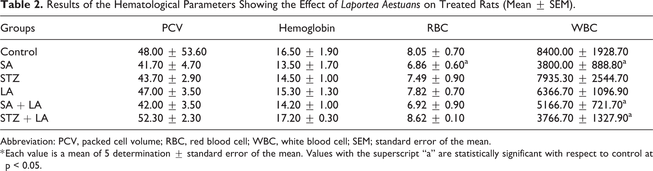
Abbreviation: PCV, packed cell volume; RBC, red blood cell; WBC, white blood cell; SEM; standard error of the mean.
* Each value is a mean of 5 determination ± standard error of the mean. Values with the superscript “a” are statistically significant with respect to control at p < 0.05.
Effect of MeLELAon Sodium Arsenite-and Streptozotocin-Induced on the Micronucleus
The micronuleated polychromatic erythrocytes (mPCE/1000) significantly increased in the SA treated group, but decreased significantly in the SA + LA group at p < 0.05. Also, the mPCE/1000 in the STZ + LA group decreased significantly at p < 0.05 (Figure 5).

Micronucleated polychromatic erythrocytes per 1000 polychromatic erythrocytes (mPCE/1000 PCE), with and without Laportea aestuans treatment in rats, before and after exposure to SA and STZ. Values were expressed as the mean ± SEM (n = 5). Statistical significance was assessed using one-way ANOVA test: ap < 0.05 vs. control; bp < 0.05 vs. SA and cp < 0.05 vs. STZ. Abbreviation: SA, sodium arsenite; STZ, streptozotocin; LA, Laportea aestuans; SEM, standard error of the mean.
Modulatory Role of MeLELA Based on Histological Assessments of Sections of Kidney, Liver and Pancreatic Tissues
Figures 6 to 8 show the tissue architectural changes induced by treatment of the experimental animals with SA/STZ along with the influence of MeLELA on sections of kidney, liver and pancreatic tissues, respectively.

Photomicrographs of kidney sections at X400 magnification after exposure to SA/STZ and methanol leaf extract of Laportea aestuans. (A). No visible lesion-control group. (B). Severe tubular necrosis with desquamation – SA group. (C). Moderate peritubular inflammation – STZ group. (D). No visible lesion seen – LA group. (E). Partial glomerulonecrosis– SA + LA group. (F). Mild tubular necrosis–STZ + LA group.
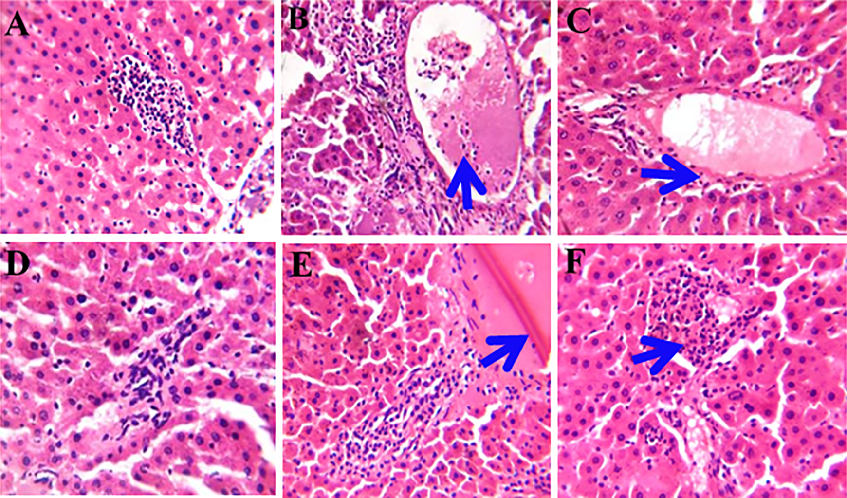
Photomicrographs of liver sections at X400 magnification after exposure to SA/STZ and methanol leaf extract of Laportea aestuans. (A). The architecture of the liver looks normal-control group. (B). Severe Periportal inflammation-SA group. (C). Slight periportal inflammation-STZ group. (D). No visible lesion seen (The architecture of the liver looks normal)–LA group. (E). Slight periportal inflammation with moderate congestion of hepatic vein SA + LA group. (F). Mild periportal inflammation – STZ + LA group).

Photomicrographs of pancreas sections at X400 magnification after exposure to SA/STZ and methanol leaf extract of Laportea aestuans (A). The islet of Langerhan looks normal – control group. (B). Decrease in number of islets of Langerhan with marked absence of centroacinar cells – SA group. (C). There is a decrease in the number of islet of Langerhans with infiltration of inflammatory cell – STZ group.(D). Adipocyte infiltration (accumulation of adipocyte in excessive amount) – LA group. (E). Pancreatic acini present in matured pancreas (It secretes digestive enzymes required for growth) – SA + LA group. (F). Pancreatic tissue showing pancreatic acini) – STZ + LA group.
Discussion
Phytochemicals present in plants with medicinal values have played remarkable roles in health care over the years, most especially in developing countries. Medical care based on the use of traditional plants is an avenue for the discovery of new drugs. 30,31 Scientific screening of medicinal plants has therefore become a worthwhile exercise. 32,33 The persistence of high mortality rate among cancer patients has become a pointer to the limited efficiency of the current cancer treatment modalities. 34 In exposed population, inorganic arsenic is associated with tumours of the lung, kidney, liver, bladder and skin. 35 The mechanism of inorganic arsenic-induced hepatotoxicity involves oxidative stress. 36 Laportea aestuans is an annual herb that is native to Africa and has been listed as one of the medicinal plants with active therapeutic agents and functions. 17 These properties are evident from the in vitro antioxidant and free radical scavenging activities of the methanol leaf extract of LA (MeLELA). 36,37 The MeLELA administered in this study has demonstrated strong reno-hepatic and antidiabetic properties. This is in conformity with the remarkable roles of medicinal plants used in the management of reno-hepatotoxicity 12,38 and diabetetes 9
Phytochemical screening of MeLELA reveals the presence of alkaloids, carotenoids, and oxalates in large quantities. Other constituents found in minute quantities include tannins, flavonoids, phytates, steroids, phlobatannins, terpenoids and anthroquinnone, while cardiac glycosides and cyanides were not detected. The phytochemicals present in the extract account for its medicinal properties and its applications in the treatment of several diseases. For instance, alkaloids exhibit antimicrobial, antihelminthic, antidiarrhoel activities, 17 while carotenoids exhibit anticancer activities. Furthermore, anthraquinones possess purgative, antitumor, antibacterial, and anti-inflammatory properties. Flavonoids also exhibit a wide range of biological activities such as anti-inflammatory, antimicrobial, anticancer and antiallergic properties, 17,37 while tannins are considered as primary antioxidants or free radical scavengers. 39
The assessment of the antioxidant activity of MeLELA based on 2, 2-diphenyl-1-picryhydrazyl radical (DPPH) free-radical-scavenging capacity reveals a reduction in absorption at 517 nm, indicating that the extract has a hydrogen-donating ability or can quench free radicals. As earlier reported, the presence of secondary metabolites in the extract is responsible for the antioxidant activity. 37 From our result, the potent antioxidant activity of the extract shows that it could be very useful in the treatment of ailments resulting from oxidative stress and, thus, mitigate cellular damages. 37
The extract of LA significantly reduced the nephrotoxicity induced by SA. This was demonstrated by reduction in the levels of urea and creatinine levels. 40 The hepatotoxicity of SA has been proven by various studies over the years and this was further validated in this study. 41,42 Activities of liver enzymes (AST, ALT, and ALP) were significantly high in the serum samples of the groups administered SA and STZ-treated groups, respectively. Methanol leaf extract of LA ameliorated the sodium arsenite-induced hepatotoxicity as seen in group IV. The significant reduction in hepatic damage biomarker enzymes’ activities by LA compares favorably with study by Abdallah et al. 12 The pathogenesis of reno-hepatotoxicityand diabetes can be attributed to ample production of free radicals. 43 Hence, compounds with anti-oxidant properties can ameliorate and inhibit the progression of reno-hepatotoxicity and diabetes. 43 LA is an ethno-medicinal plant possessing antioxidants, such as, alkaloids and flavonoids that exhibit reno-hepatoprotective effect against SA and antidiabetic effect against STZ. The medicinal properties of LA against diabetes have also been reported in traditional medicine. 17 Hence, based on the results of this study, the high fasting blood glucose level induced by STZ was significantly reduced by the administration of methanol leaf extract of LA.
Sodium arsenite significantly induced a higher frequency of micronucleated polychromatic erythrocytes when compared with control. However, methanol leaf extract of LA significantly reduced the frequency of micronucleated polychromatic erythrocytes induced by SA when co-administered. This is a demonstration of the anticlastogenic effect of the extract. There were also significant differences in the hematological parameters of treated rats.
The histological results of the kidney and liver further corroborate the toxic effects of SA (severe tubular necrosis with desquamation in the kidney and severe periportal inflammation of the liver), and the profound ameliorative effects of the methanol leaf extract of LA in which there was partial glomerulonecrosis and slight periportal inflammation with moderate congestion of hepatic vein. The histological results of the pancreas showed that STZ induces diabetes with direct effect on the beta cells of the pancreas (decrease in the number of islet of Langerhans with infiltration of inflammatory cell) which was ameliorated with co-administered methanol leaf extract of LA as seen in the pancreatic tissue showing pancreatic acini.
Conclusion
Overall, methanol leaf extract of LA (MeLELA) demonstrates protective potentials against sodium arsenite-induced toxicities and streptozotocin-induced diabetes in male albino Wistar rats. The protective effects of LA are due to the different bioactive components present in the extract and this warrants further studies to characterise the extract. The presence of these bioactive components may account for the folkloric use of the plant products.
Footnotes
Authors’ Note
OAA: Study design, performed the experiments, acquisition and analysis of data, and drafted the manuscript. JOO: Supervision of the study design and manuscript writing. AMA: Supervision the study design and manuscript writing. MAG: Study design, supervision of the study design and manuscript writing. OAO: Study design, supervision of the study design and manuscript writing. All the authors have accepted responsibility for the entire content of this submitted manuscript and approved submission.All animals used for the study were handled strictly in accordance with the University of Ibadan Ethics Committee guidelines for the use of experimental animals in research.
Acknowledgments
We acknowledge the technical supports from Cancer Research and Molecular Biology Laboratories of the Department of Biochemistry, University of Ibadan, Ibadan, Nigeria.
Declaration of Conflicting Interests
The authors declared no potential conflicts of interest with respect to the research, authorship, and/or publication of this article.
Funding
The authors received no financial support for the research, authorship, and/or publication of this article.