
Abstract
Herbal products from Paullinia pinnata Linn are widely used in African folk medicine to treat several infectious diseases. Although the extracts from this plant has been shown to possess antimicrobial potential, their activity in infectious diarrhea is less reported. Diarrhea was induced by oral administration of 1.2 × 10⁹ CFU/mL of Shigella flexneri to the rats. The infected rats were treated for 5 days with the doses of 111.42, 222.84, and 445.68 mg/kg of P pinnata. The level of biochemical parameters was assessed and histology of organs examined by 14 days subacute toxicity. S flexneri stool load was considerably reduced after 4 days of treatment with the dose of 445.68 mg/kg, 5 days at the dose of 222.84 mg/kg for the extract, and 2 days with ciprofloxacin. The dose of 111.42 mg/kg appeared efficient after 5 days of treatment. The creatinine level increased at the dose of 445.68 mg/kg in both male and female rats and decrease at the dose of 222.84 mg/mL in female rats while an increase was noted in the male rats. Liver and kidney histology were modified at the dose of 445.68 mg/kg while no change was observed at the doses of 111.42 and 222.84 mg/kg. P pinnata leaf extract is efficient against infectious diarrhea at 111.42 mg/kg without side effect.
Diarrhea is defined as having loose or watery stools at least 3 times per day, or more frequently than normal for an individual. 1 Diarrheal disease is a leading cause of mortality and morbidity, especially among children in developing countries due to significant dehydration resulting in death or other severe consequences. 2 Shigella is the predominant organism in bloody diarrhea in developing countries where it causes 10% of acute diarrhea in children younger than 5 years. 3 Shigella flexneri is endemic in most developing countries and causes more mortality than any other Shigella species. 4 Shigella is transmitted efficiently at low dose via the fecal-oral route in areas with poor hygienic conditions and limited access to clean and potable water. 5 S flexneri causes inflammatory diarrhea and the cellular response to various steps of the invasion process are the primary cause of inflammation of the colon with micro abscess and ulcerations, which cause mucous and bloody appearance of diarrheal stool. 6 Antibiotherapy has been the main way to treat diarrhea and the antibiotics commonly used are ampicillin, ceftriaxone, azithromycin, trimethoprim/sulfamethoxazole, nalidixic, fluoroquinolone, and ciprofloxacin. 7 The extensive use of current antibiotics, has increased the pathogenic bacteria resistance that constitute a serious health problem nowadays. The pathogens developed resistance to antibiotics, which opened the door to the exploration of herbs for antimicrobial activity. 8,9
Several African medicinal plants have been reported to be useful in the treatment, management, and/or control of infectious disease, including diarrhea. 10 Paullinia pinnata Linn leaf, root and bark extracts are widely used in African folk medicine to treat several diseases including rheumatism, weakness, impotence, 11 ulcers, haemorrhoids, wounds, 12 malaria, 13 fever, hypertension, contraceptive, 14 syphilis, 15 convulsion, 16 typhoid, 17 and diarrhea. 18 Previous works have reported the antianalgesic and anti-inflammatory activities, 19 anxiolytic, 20 antibacterial and antioxidant, 18,21 –24 antityphoidal, 17 and antidiarrheal properties of P pinnata. 25 The acute and subacute toxicity of the methanolic extract of the leaves of P pinnata was reported as well as the repeated toxicological study and cardiotoxicity of hydroalcoholic root extract. 26,27 Phytochemical studies of P pinnata have demonstrated the presence of phenols, alkaloids, flavonoid, sterols, tannins, and other classes of secondary metabolites. 18,28,29 Despite the fact that P pinnata has been intensively screened for pharmacological activities, few studies revealed its properties on infectious diarrhea. Therefore, the present study investigates the in vivo antidiarrheal activity and subacute toxicity of the ethanolic extract of P pinnata in Wistar rats.
Materials and Methods
Collection and Identification of Plant Materials
P pinnata leaves were collected at Nkoupa-Matapit in the Western region of Cameroon in December 2013. The plant identification was done at the Cameroon National Herbarium by comparison with specimen No. 20022/SRF Cam of P pinnata Linn.
Preparation of Ethanolic Extracts
Leaves of P pinnata were removed from the twigs, rinsed gently to remove dust and dirt, air-dried. The samples were then ground in a mortar and 200 g of dried powder of each sample was soaked for 48 hours in 600 mL of 95% ethanol. The mixture was filtered with Whatman No. 1 filter paper and concentrated using a rotary evaporator (Rotavapor R-300, Buchi) at 55°C.
Microorganism
S flexneri, associated with diarrheal infection was used for this study. This clinical isolate was obtained from the Centre Pasteur of Cameroon. The bacterial strain kept at +4°C was subcultured before any test.
Experimental Animals
Healthy male and female albino rats of Wistar breed (140-180 g) not genetically modified were purchased from the Laboratory of Food Science and Metabolism and bred at the animal house of the Laboratory of Pharmacology and Toxicology, Department of Biochemistry, University of Yaoundé I at room temperature with a 12-hour light/dark photoperiod cycle. The animals were acclimatized to laboratory conditions for 1 week prior to the experiments. They were kept in plastic bowls covered with metal gratings and received food and tap water ad libitum. The used litter was the sawdust, renewed twice per week to ensure the good hygienic condition of the animals. All procedures performed conformed to the rules and regulations of the European Union on Animal Care (EEC Council 86/609) 30 that were adopted by the Institutional Committee of the Ministry of Scientific Research and Innovation of Cameroon.
Antidiarrheal Activity
Thirty-six animals were divided into 6 groups containing 6 animals each. Animals in each group were numbered and for treatment, the control groups were treated first and thereafter the test groups from the lower to the higher doses.
The animals were treated as follows: Group 0 rats were not infected and not treated and served as neutral control. Group I rats were infected and not treated and served as negative control. Group II rats were infected and treated with ciprofloxacin at the dose of 2.50 mg/kg and served as positive control. Groups III, IV, and V rats were infected and treated with extract solution of P pinnata leaf at the doses of 111.42, 222.84, and 445.68 mg/kg, respectively.
Food and water were given to the animals before and during the treatment. Every morning, the stool was collected just before administration of plant extract or ciprofloxacin or nothing for the negative and neutral control.
The antidiarrheal activity was carried out on S flexneri–induced diarrhea rats. A S flexneri suspension was prepared at 4 McFarland standard. The bacterial suspension was prepared by transferring 3 colonies of an overnight S flexneri culture from nutrient agar with a loop into 5 mL of normal saline water in capped test tube and vortex. One milliliter of this solution containing about 1.2 × 10⁹ cells was orally administered to each animal. 31 Infected animals were selected appropriately based on increase in the fluidity and volume of stool, presence of blood or mucus in stool, fecal bristling coat, less aggressiveness, and reduced feed intake in animals. Treatment was done by administering the extracts orally, every morning for 5 days in the laboratory. The test solution of P pinnata was prepared in distilled water at the doses of 111.42, 222.84, and 445.68 mg/kg corresponding to 8, 16, and 32 minimal inhibitory concentration, respectively selected on a trial basis 18 and administered orally by gavage to the animals. The extent to which the animals responded to treatment was studied by recording the number of bacterial colonies in the stool samples. 32 Hence, a sample of dried powdered diarrheal feces (0.1 g) was completely dissolved in 5 mL of sterile distilled water. Fifty microliters of the resulting solution was spread on the surface of solidified MacConkey Petri dishes. Then the Petri dishes were incubated for 24 hours at 37°C. The number of colonies following growth of S flexneri in each Petri dish was counted and recorded. The result was converted into the number of colonies per gram of feces per animal.
Fourteen-Day Repeated Dose Toxicity Test
This test was carried out with the therapeutic doses used for antidiarrheal activity according to the Organisation for Economic Co-operation and Development (OECD) guidelines on subacute toxicity
33
with slight modifications. Forty male and female rats were divided into 4 groups of 10 animals each (5 rats of each sex). Animals in each group were numbered and for treatment the control groups were treated first and thereafter the test groups from the lower to the higher doses. The animals were treated as follows: Group 0 was considered as the control group and only received distilled water. Groups I, II, and III received orally the doses of 111.42, 222.84, and 445.68 mg/kg of crude extract of P pinnata.
The solution made of plant extract was orally administered to the rats for 14 days. During administration of crude extract of P pinnata, the decrease in body weight, death, and behavior (salivation, sleepy, and lethargy) were recorded.
Blood Collection and Organ Isolation
At day 14, animals were fasted overnight and sacrificed after sub-cutaneous injection of ketamine at the dose of 1 mg/kg and blood samples were collected in the dried tubes. The rats were immediately subjected to general autopsy and the liver and kidneys were isolated, blotted with clean tissue paper, weighed, and kept in the fridge as previously described. 32 Relative organ-body weight ratios were thereafter evaluated.
Assessment of Biochemical Parameters
The collected samples were thereafter centrifuged at 3000 rpm to obtain the serum that served for the assessment of the biochemical parameters using commercial kits, Fortress Diagnostics reviewed in October 2007. The amount of aspartate aminotransferase (AST), alanine aminotransferase (ALT), creatinine, glutathione, and total proteins were assessed.
Histopathological Examination
The liver and kidney fixed in formaldehyde 10% for 3 weeks were cut into small pieces of 5 to 10 mm and then dehydrated in cassettes by immersion in alcohol and acetone successively. The latter was eliminated by xylol before being flowed in molds containing paraffin melted by heating at 60°C. 34 Tissue sections were then prepared and stained with haematoxylin and eosin. The preparation was air-dried and the photomicrographs of the tissue sections were taken at ×400 using microscope. 32
Statistical Analysis
Data are expressed as mean ± standard error of the mean (SEM). The data were analyzed using SPSS version 20. Statistical significance was determined by 1-way analysis of variance (ANOVA) followed by post hoc Tukey’s multiple comparison test. The value of P < .05 was considered statistically significant.
Results
General Signs of Diarrhea
The volume of infected rat stools increased and appeared soft with mucus or watery or were molded and smooth. Sometimes, the presence of blood and mucus made the stool appear dark and shiny. Their coats were bristling and they were less aggressive and reduction in feed intake was observed. One rat of group III treated with the dose of 111.42 mg/kg of P pinnata and 1 rat of group I (not treated) died after 2 days of treatment. Signs of diarrhea appeared 24 hours after diarrheal induction. After treatment, stool appears normal depending on the dose and duration of treatment. Hence, rats treated with P pinnata extract presented normal stools 4 days after treatment with the dose 445 mg/kg and 5 days after treatment with the doses 222.84 and 111.42 mg/kg while normal stools were observed 2 days after treatment with ciprofloxacin.
Effects of the Crude Extract on the Fecal Load of S flexneri
The oral administration of the solution of P pinnata extract at the doses of 222.84 and 445.68 mg/kg improved the general condition of the animals. The S flexneri load in diarrheal stool dropped significantly in a dose-dependent manner during treatment (Figure 1). At the higher dose (445.68 mg/kg), the reduction in bacterial load was more pronounced compared to the lower dose (222.84 mg/kg). However, these reductions noted after treatment with extracts were less pronounced when compared with those obtained with ciprofloxacin. Animals stopped manifesting diarrhea after 4 and 5 days of treatment with the doses of 445.68 mg/kg and 222.84 mg/kg of ethanolic extract of P pinnata, respectively. The S flexneri density in stools of rats treated at the dose of 222.84 mg/kg significantly decreased after treatment but remained high. After day 5 of treatment with the dose of 111.42 mg/kg, condition of improvement was noted in rats even if the bacterial fecal load remained high. In the case of the negative control (untreated group), the S flexneri fecal load remained relatively high compared to those of the treated groups throughout the test period. However, it was observed that after day 1 of treatment, the number of colonies counted starts dropping gradually.

Variation of Shigella flexneri load in stools of treated and untreated rats during treatment. Rats were treated for 5 days with 111.42 mg/kg, 222.84 mg/kg, and 445.68 mg/kg of ethanolic extract of Paullinia pinnata or ciprofloxacin. Data are presented as mean ± SEM (n = 6). Significant difference: *P < .05 compared with negative control; a P < .05 compared with positive control; b P < .05 compared with initial point; day 1: diarrhea appearance and treatment start.
General Signs of Subacute Toxicity
In the subacute toxicity test, daily repeated oral treatment with the extract for 14 days at the doses of 111.42, 222.84, and 445.68 mg/kg had no clinical adverse effects of substance related toxicity. Neither death nor sign evoking the poisonous character of P pinnata extract such as convulsion, excessive grooming, salivation, tremor, change in step, coma, and the distress was observed throughout the study period. Hence, 10 animals (10/10) of each group were included in the analysis.
Effects of the Crude Extract on the Body Weight
The respective initial body weights of the treated rats and the control were compared with their final weights in order to ascertain a probable decrease in body weight, which is a sign of toxicity. A progressive weight gain was noted in all treated and control (noninfected and untreated) groups during this study with percentage weight gain varying from 9.32% to 15.36% for female and from 7.67% to 17.07% for male rats. However, male and female rats treated with the doses of 222.84 and 445.68 mg/kg of the ethanolic extract of P pinnata had lower weight gain (P < .05) when compared with that of the control groups (Table 1).
Variation of Body Weight Gain (g) for Treated and Untreated Rats During Treatment.a

a The results are mean ± standard error of the mean (SEM) (n = 5). Data in the same column in the same sex with asterisk (*) are significantly different (P < .05) when compared with the control.
Effects of the Crude Extract on Some Biochemical Parameters
Data obtained with regard to liver and kidney function parameters (ALT, AST, total protein, glutathione, and creatinine) evaluated in this study revealed that the ethanolic extract of P pinnata caused no significant difference (P < .05) in the levels of ALT, AST, total protein, and glutathione at different extract doses when compared with the control (Table 2). A significant increase (P < .05) of creatinine level (0.80 ± 0.03 g/L) was observed in female rats when compared with the control (0.41 ± 0.03 g/L) as well as in male rats with creatinine level of 1.03 ± 0.03 g/L when compared with the control (0.38 ± 0.00 g/L) at the dose of 445.68 mg/kg. At the dose of 222.84 mg/kg a decrease in creatinine level (0.34 ± 0.01 g/L) was observed in female rats when compared with the control (0.41 ± 0.03 g/L) while an increase (0.62 ± 0.03 g/L) was noted in males when compared with the control (0.38 ± 0.00 g/L).
Effects of the Ethanolic Extract of Paullinia pinnata on Biochemical Parameters.a

Abbreviations: ALT, alanine aminotransferase; AST, aspartate aminotransferase; TP, total proteins; GSH, glutathione; Crea, creatinine.
a The results are mean ± standard error of the mean (SEM) (n = 5). Data in the same column in the same sex with asterisk (*) are significantly different (P < .05) when compared with the control.
Effects of the Crude Extract on the Relative Weight (%) of Organs
In this study, the respective initial body weights of tests and control groups were compared with their final weight. No significant difference in the weight of liver and kidney of treated rats was observed at the dose of 222.84 and 445.68 mg/kg when compared with the control group irrespective of the sex of the animal (Table 3). The relative weight gain of the liver ranged between 3% ± 00% and 3.20% ± 0.3% compared with 3% ± 0.28% for control in male rats while varying between 3% ± 00% and 3.80% ± 0.33% compared with 3.24% ± 0.33% in female rats. Concerning the kidney, the relative weight gain ranged between 0.70% ± 0% and 0.76% ± 0.03% compared with 0.76% ± 0.12% in male rats while varying between 0.80% ± 0.03% and 0.90% ± 0.0% compared with 0.78% ± 0.33% in female rats.
Relative Weight (%) of Organs of Treated Rats and Control.a
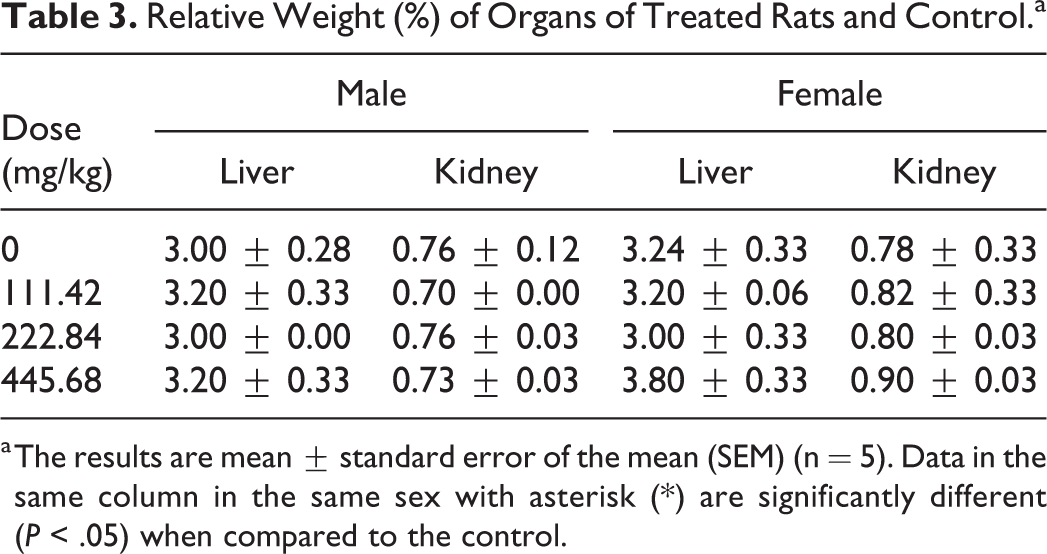
a The results are mean ± standard error of the mean (SEM) (n = 5). Data in the same column in the same sex with asterisk (*) are significantly different (P < .05) when compared to the control.
Histological Examination
The histological examination of the liver and kidney of the extract-treated animals revealed abnormalities in overall structural orientation and observable cellular injuries at the dose of 445.68 mg/kg while morphological features and tissue integrity of organs of the rats treated at the dose of 222.84 mg/kg presented slight abnormalities and comparable to the normal control (Figures 2 and 3).

Histological section of liver tissue (section stained with H&E, ×400). (a) Control. (b) Rats treated with dose of 222.84 mg/kg of Paullinia pinnata, without capillaries sinusoids dilatations. (c) Rats treated with dose of 445.68 mg/kg of P pinnata, with vascular congestion + slight capillaries sinusoids dilatations. (A) = male; (B) = female. CCLV. congestion of centrolobular vein; CLV, centrolobular vein; HP, hepatocites; CSD, capillaries sinusoids dilations; N, Necrosis; H&E, hematoxylin and eosin.

Histological section of kidney tissue (section stained with H&E, ×400) (a) Control. (b) Rats treated with dose of 222.84 mg/kg of Paullinia pinnata, without mesangial expansion. (c) Rats treated with dose of 445.68 mg /kg of P pinnata, with mesangial expansion. (A) = male; (B) = female. MEGl, mesangial expansion of glomerulus; UR, urinary room; Gl, glomerulus; H&E, hematoxylin and eosin.
The liver of male and female rats of control group and rats treated with the dose of 222.84 mg/kg appeared normal with preserved hepatic histology (Figure 2). However, in both female and male treated rats, slight degeneration and centrolobular necrosis were occasionally observed in the liver. These structural changes were more expressed in the group treated with the dose of 445.68 mg/kg. At this higher dose, the histological aspect of hepatic tissue is partially erased, sinusoids are dilated, and veins mark the vascular congestions of the liver. No structural change was observed at the dose of 111.42 mg/kg.
A mesangial expansion of the kidney regaining the urinary tract at the dose of 445.68 mg/kg was observed when compared with the control (Figure 3). No major damage was observed at the dose of 222.84 mg/kg.
Discussion
The manifestation of the diarrhea, associated to the febrility observed in infected rats, could be explained by the intestinal invasion of S flexneri, which have led to a generalized weakness of the animals. The infectious bacterium might have penetrated in the epithelial cells of the mucus membrane where it might have caused serious damage, and led to the bloody diarrhea, sometimes with mucus, which is accompanied by abdominal cramps and fever. 35 The decrease of S flexneri load in feces is an evidence of antidiarrheal activity, which is directly related to the antibacterial activity exhibited by the ethanolic extract of P pinnata on S flexneri. Animal’s response to treatment suggests that the doses of 222.84 and 445.68 mg/kg of ethanolic extract of P pinnata are most efficient for the treatment of diarrhea, and in accordance with the findings of Lunga et al 17 who demonstrated that methanolic leaf extract of P pinnata stopped Salmonella typhi–induced typhoid at the doses of 223 and 446.00 mg/kg after 5 and 4 days, respectively. However, their work revealed no significant difference (P ≥ .05) in bacterial load feces between the extract at the highest dose (446 mg/kg), and the reference antibiotics (ciprofloxacin). These findings are not in agreement with the present result, which shows that the reduction of bacterial load in stool by ciprofloxacin was more pronounced. This simply suggests that the activity of an extract depends on the microorganism species and nature of extracting solvent. In the case of the negative control (untreated group), it was observed that from the first day, the microbial load reduced gradually. This can be attributed to the immune response against the foreign microbe. 36 The antidiarrheal activity may be associated with the antimicrobial activity of this extract. 37 Tannins, alkaloids, saponins, steroids, and terpenoids found in this plant extract 18 are responsible for the antimicrobial activity and therefore justify its antidiarrheal activity. 38
In the subacute toxicity test, the less pronounced weight gain in the treated groups could be an indicator of undesirable side effects of this extract since weight loss is correlated to the physiological status of the animal. This could be associated with the reduction of the food consumption and the probable dose/absorption interactions. 39 Therefore, long-term exposure to higher doses of P pinnata could lead to weight loss.
ALT and AST are common markers that are associated to the hepatocellular damage and an increase in the levels of these enzymes is indicator of liver damage. 40 Since no significant difference (P < .05) was noted in the level of ALT and AST level it suggests that the ethanolic extract of P pinnata had no deleterious effect on liver function. In addition, it is known that an increase of glutathione level in rats’ hepatocytes indicates lipid peroxidation which results in oxidative stress. 41 Therefore, this study suggests that P pinnata ethanolic extract could not generate oxidative stress in rat. Creatinine is an indicator of renal function and an increase of the serum creatinine level is a sign of toxicity. The significant increase in creatinine level with 222.84 and 445.68 mg/kg indicates that the ethanolic extract of P pinnata could be toxic. This result is in accordance with a previous work that showed renal injuries by the increase in blood urea, creatinine, potassium, and chlorine. 27 However it is not in agreement with a previous finding that revealed that P pinnata aqueous ethanol roots showed no significant increase in biochemical parameters of the extract-treated animals at the doses of 375, 750, and 850 mg/kg. 13
Relative organ weight was determined to ascertain an eventual toxic effect of the extract on the liver and kidney. No significant difference in the organs weight of treated rats was observed but this finding is not sufficient to declare that this extract is nontoxic in rats.
Histopathological examinations were performed on the liver to assess whether organs or tissues had been damaged. The preserved hepatic histology and slight degeneration and centrolobular necrosis occasionally observed in the liver at the dose of 222.84 mg/kg suggest that the ethanolic extract of P pinnata is less toxic at that dose. These histological damages were more pronounced in rats treated with the dose of 445.68 mg/kg. Therefore, it is evident that the livers of both male and female rats treated with the dose of 445.68 mg/kg are modified. This result is not in accordance with that of Lunga et al 28 who found that only female rats presented liver damage at dose ≥223 mg/kg and therefore advise that this plant extract should thus be used with caution in male and should probably be eliminated in the treatment of female subjects at that dose.
A mesangial expansion of the kidney regaining the urinary tract at the dose of 445.68 mg/kg was observed when compared with the control. No major damage was observed at the dose of 222.84 mg/kg. However, the creatinine level increased at that dose. It has been shown that the observed hepato-renal toxicity of the high dose of plant extract is also supported by the increase in creatinine level which is an indicator of kidney function. 42 It suggests that at the dose of 222.84 mg/kg, the ethanolic extract of P pinnata affects kidney’s function, and not its structure. The subacute toxicity at repeated doses of 200, 400, and 800 mg/kg of hydroethanolic extract of P pinnata roots for 28 days showed significant increase in urea, creatinine, K+, and Cl− at the doses of 400 and 800 mg/kg and creatinine phosphokinase at the dose of 800 mg/kg in the serum. 43 However, no histological damage of the liver and kidneys was observed at these doses. Adeyemo and Makinde 26 showed in a subacute toxicity test of the methanolic extract of P pinnata leaves that oral administration of 200 mg/kg presents no evidence of toxicity by measurement of biochemical parameters and histopathological exam of organs. But at the doses higher than 400 mg/kg, a significant increase in biochemical parameters was noted as well as liver alterations and lymphocytic infiltrations of lungs of treated rats compared with the control group. The alteration of function and structure of the liver and kidney could be due to the presence of poisonous substance in the extract. It is evident from these observations that ethanolic crude extract of P pinnata is poisonous at the doses of 222.84 and 445.68 mg/kg.
Conclusion
Overall, it is evident from the present study that the ethanolic extract of P pinnata leaves is efficient against S flexneri–induced diarrhea at the dose of 111.42, 222.84, and 445.68 mg/kg by oral daily administration. Following its 14-day repeated daily oral dose administration in the animals, it may be concluded that it elicits treatment-related adverse effect on liver and kidney at the doses investigated. The findings of this study justify the use of P pinnata leaves to treat diarrhea in traditional medicine. The set of results gotten in this study suggests that the ethanolic extract of P pinnata could be used to treat infectious diarrhea at doses less or equal to 111.42 mg/kg without undesirable side effects.
Footnotes
Acknowledgments
The authors are thankful to the Cameroon National Herbarium (Yaounde) for plant identification and the University of Yaounde I for providing facilities.
Author Contributions
ADA and YNN conducted the practical work and prepared the first draft of the manuscript, SVD performed statistical analysis, MCF provided Shigella flexneri strain, MAN and EFX supervised the design of the research and implementation and edited the final draft of the manuscript. All authors read and approved the final manuscript.
Declaration of Conflicting Interests
The authors declared no potential conflicts of interest with respect to the research, authorship, and/or publication of this article.
Funding
The authors received no financial support for the research, authorship, and/or publication of this article.
Ethical Approval
All procedures performed conformed to the rules and regulations of the European Union on Animal Care (EEC Council 86/609) that were adopted by the Institutional Committee of the Ministry of Scientific Research and Innovation of Cameroon.