
Abstract
Medicinal plants over time have proven to have potential to manage a huge number of diseases and disorders and thus have become a great source of pharmaceutical drugs. One of such plants is Tapinanthus bangwensis (African mistletoe). It is a semiparasitic and epiphytic plant growing on citrus tree, obtaining its food photosynthetically while its nutrient and water is got from the host plant. The aim of this study was to determine the cytotoxicological and hepatocurative effect of aqueous fraction of T bangwensis in acetaminophen (paracetamol)-induced Wistar rats. The antioxidant potential of the plant was determined by 2,2-diphenyl-1-picrylhydrazine scavenging and ferric reducing power assays. The cytotoxic effect was determined using Allium cepa test while the liver biochemical indices were determined by standard protocols. Data obtained were analyzed by 2-way analysis of variance at 95% confidence level and reported as mean ± standard deviation. The concentrated aqueous fraction of T bangwensis was found to be 23.3 g (58.25%). Quantitative determination of some vital phytochemicals revealed the following: flavonoid (84.6 ± 0.41 mg/100 g), phenol (147.5 ± 1.07 mg/100 g), tannin (31 ± 0.85 mg/ 100 g), alkaloid (23.45 ± 0.09 mg/100 g), and saponin (0.146 ± 0.0 mg/100 g). Treatment of rats with the aqueous extract of T bangwensis significantly decreased paracetamol-induced elevation of activities of liver function indices, alkaline phosphatase, alanine aminotransferase, aspartate aminotransferase, triglyceride, total cholesterol level and increased the albumin, total protein, and high-density lipoprotein levels. The plant extract also attenuated the paracetamol elevated lipid peroxidation product, malondialdehyde. The research findings suggest that aqueous extract of T bangwensis is slightly cytotoxic, possesses appreciable antioxidant property and exhibited hepatocurative effect against paracetamol-induced hepatoxicity.
Phytomedicine sometimes called herbalism is the most ancient method of disease management, 1 and it has been known that plants are the first and only true medicines ever used by humans. 2 However, in Nigeria, until recently, the practice of the use of herbs has been kept in secrecy and shrouded in dreaded magical incantations, rituals, and sacrifices. It is now very clear that the potency of plants and its parts does not depend on such exhibition. The use of plants for medical purposes is an important part of the culture and tradition in Africa. 3 Plant extracts have continued to play significant roles in human health care particularly in developing countries as more than 80% of the world population had been shown to rely on phytomedicine as source of health care. 4 Decades of studies have shown promising returns from application of numerous plant parts either naturally or as extracts in the treatment of various ailments owing to the bioactive phytochemicals present. 5 –7 Phytochemicals are substances found in plants that exhibit a potential for modulating human metabolism in a manner beneficial for the prevention of chronic and degenerative diseases. 8 Tapinanthus bangwensis (also called African mistletoe), a well-known evergreen semiparasitic and photosynthetic plant belonging to the family Loranthanceae and possesses an excellent medicinal property. 9,10 The plant is commonly called bird lime, devil’s fuge, iscador, and locally as Afomo Onisana (Yoruba), Awurise (Igbo), and Kauchi (hausa). 11 Traditionally, extracts of T bangwensis have been used against a variety of diseases such as female reproductive disorders, oncological problems, rheumatoid arthritis, epithelial tumors, hypertension, asthma, nervousness, epilepsy liver disorders, and hypoglycemia. 1,12 –14 The host-plant relationship are of disease-curing specificity, for example, mistletoe grown on guava, kola nuts, and citrus are specific for curing diseases like cancer, hypertension, nervousness and insomnia, while those grown on cocoa is best against hyperglycemia. However, there is still dearth of information on the cytotoxic and hepatocurative effect of aqueous fraction of T bangwensis, which then necessitated this research investigation.
Materials and Methods
Plant Material
T bangwensis was obtained from a tradomedical practitioner in Lagos and was botanically identified and authenticated by Mr Adeleke in the Department of Pharmagnosy, College of Medicine, University of Lagos, Idi-Araba.
Preparation of Extract
The leaves of T bangwensis were removed from the twigs, washed, and air-dried for days. The dried leaves were blended into fine and loosely packed powdered form. A total of 2200 g of the powdered particles was macerated in 3500 cm3 of methanol and kept for 48 hours with intermittent shaking at intervals and then filtered with No. 1 Whatman filter paper. The pure filtrate was then concentrated to dryness in an oven at 40°C. The mass yield was 60.5 g. Forty grams of the dried extract was solvent partitioned in ethyl acetate–water mixture in 3:2 ratio. The aqueous portion was collected and concentrated and the yield was 23.3 g.
Experimental Animals
A total of 16 asymptomatic albino rats were used in this study. The rats were purchased from Biological Science Animal House, Bayero University, Kano. The animals were housed in clean and well-ventilated cages made of metal netting. The animals were allowed to acclimatize for 1 week prior to the experiment and had free access to food and clean water.
Experimental Design
Group A: Rats received water and feed only (normal control) Group B: Rats were induced with 600 mg/kg body weight paracetamol orally Group C: Rats were induced with 600 mg/kg body weight paracetamol and then administered with 200 mg/kg body weight of aqueous fraction of T bangwensis. Group D: Rats were induced with 600 mg/kg body weight paracetamol and then administered with 400 mg/kg body weight of aqueous fraction of T bangwensis.
Preparation of Animals
After induction, rats from groups C and D were administered with the extracts at concentrations 200 and 400 mg/kg body weight, respectively, for 9 days. On the ninth day, the rats were reinduced and treated for another 5 days before being anesthetically sacrificed using 10% chloroform solution. Their blood samples were collected and organs harvested for biochemical analysis.
Quantitative Phytochemical Compounds Determination
Estimation of Alkaloid
The method of Harbourne 15 was adopted. A given mass (0.2 g) of the sample was weighed into a conical flask and 100 cm3 of 10% acetic acid in ethanol was added and then allowed to stand for 4 hours. It was then filtered and filtrate concentrated on a water bath to one-quarter of the original volume. Concentrated ammonium hydroxide was added dropwise to the extract until the precipitate was completed. The whole solution was allowed to settle and the precipitated was collected and washed with dilute ammonium hydroxide and then filtered. The residue contained the alkaloid was dried and then weighed.
Estimation of Total Flavonoid Content
Total soluble flavonoid of the extract was determined with aluminum chloride using quercetin as standard. 16 A volume of 1.0 cm3 of sample solution (100 μg/cm3) was mixed with 3 cm3 of methanol, 0.2 cm3 of 10% aluminum chloride, 0.2 cm3 of 1 M potassium acetate, and 5.6 cm3 of distilled water. The resulting mixture was incubated at room temperature for 30 minutes and the absorbance of the reaction mixture was measured at 415 nm. The calibration curve was prepared by preparing quercetin solutions at various concentrations in methanol.
Estimation of Total Phenolic Compound
The amount of total phenolic content was determined by Folin-Ciocateu reagent method 17 using gallic acid as a standard following the method of Slinkard and Singleton. 18 A 0.5 cm3 sample of extract and 0.1 cm3 of Folin- Ciocalteu reagent (0.5 N) were mixed and incubated at room temperature for 15 minutes. After this, 2.5 cm3 sodium carbonate solution (7.5% w/v) was added and further incubated for 30 minutes at room temperature. The absorbance of the solution was measured at 760 nm. The concentration of total phenol was expressed as gallic acid equivalent (GAE) (mg/g of dry mass), which is a commonly used reference value.
Estimation of Tannin
This was based on the method described in Van Burden and Robinson. 19 A 0.5 g sample of the concentrate was added to 50 cm3 of distilled water and shaken for 1 hour. A 5 cm3 aliquot of the filtrate was mixed with 2 cm3 of 0.1 M FeCl3 in 0.1 N HCl and 0.008 M potassium ferrocyanide. The absorbance was measured at 720 nm within 10 minutes.
Determination of Saponin Content
Saponin content was estimated gravimetrically by the methods described by Obadoni and Ochuko. 20 The plant sample was weighed into a conical flask and ethanol added to it. The content was then vigorously shaken and kept for 5 hours in a rotary shaker maintained at 55°C. The solution was then filtered and the residue was resuspended with ethanol and the extracts were pooled. The pooled extracts were then concentrated in a water bath maintained at 90°C. The concentrate was mixed with diethyl ether in a separating funnel and shaken vigorously. The aqueous layer was collected while the diethyl ether layer was discarded. The process was repeated thrice and all the aqueous fractions pooled together and then butanol added. The total saponin content was expressed as mg/g sample.
In Vitro Antioxidant Property of Aqueous Extract of T bangwensis
DPPH Radical Scavenging Activity Assay
The free radical scavenging activity of the extracts based on the scavenging of the stable 2,2-diphenyl-1-picrylhydrazyl (DPPH) free radical was estimated according to the procedure described previously. 21 An aliquot of 0.5 cm3 of extract in ethanol (95%) at different concentrations (50, 100, 150, 200, and 250 μg/cm3) was mixed with 2.0 cm3 of reagent solution (0.004 g of DPPH in 100 cm3 methanol). The control contained only DPPH solution in place of the sample while methanol was used as the blank. The mixture was vigorously shaken and left to stand at room temperature. After 30 minutes the decrease in absorbance of test mixture (due to quenching of DPPH free radicals) was read at 517 nm. The scavenging effect was calculated using the expression:

where A 0 is the absorbance of the blank sample and A 1 is the absorbance of the extract.
Ferric Reducing Antioxidant Power Assay
The ferric reducing antioxidant power (FRAP) assay was done according to Benzie and Strain, 22 with some modifications. The stock solutions included 300 mM acetate buffer (3.1 g C2H3NaO2ċ3H2O) and 16 cm3 C2H4O2), pH 3.6, 10 mM TPTZ (2,4,6- tripyridyl-S-triazine) solution in 40 mM HCl, and 20 mM FeCl3ċ6H2O solution. The fresh working solution was prepared by mixing 25 cm3 acetate buffer, 2.5 cm3 TPTZ solution, and 2.5 cm3 FeCl3ċ6H2O solution and then warmed at 37°C before using. Plant extracts (150 cm3) was allowed to react with 2850 cm3 of the FRAP solution for 30 minutes in the dark condition. Readings of the colored product (ferrous tripyridyltriazine complex) were then taken at 593 nm.
Cytotoxicity Study Using Plant Model (Allium cepa)
The cytotoxic effect of the plant crude extract was verified using the protocol of Fiskesjo. 23 Different concentrations of the aqueous extracts of T bangwensis were prepared (20, 40, 60, 80, and 100 mg/100 cm3) alongside the control (distilled water). Onion bulbs were then placed on each of the solutions already prepared and left for 72 hours. At every 24 hours, the root lengths for each onion bulb were measured using a ruler. After 72 hours, the root tips were cut off with scissor and then fixed in a fixative solution (acetic acid–alcohol mixture in 3:1 ratio). The root tips were placed on a clean, plain glass slide and a drop of 1.0 N HCl was added to dehydrate and soften the tissue for easy maceration for 2 minutes. A dissection needle was then used to macerate the root tip after which a drop of acetic orcein stain (2.2 g of orcein powder in 45 cm3 of glacial acetic acid and making up to 100 cm3 of distilled water) was added for 15 to 20 minutes. The slide content was then covered with a cover slide to allow the stain to spread evenly. The slide with the specimen was then squashed and the edge sealed with white nail hardener. The slides were microscopically examined and the various mitotic stages and chromosomal aberrations recorded.
Analysis for Liver Indices
The following parameters were determined with their protocols: serum aspartate aminotransferase (AST) level, 24 serum alanine aminotransferase (ALT) level, 24 serum alkaline phosphatase (ALP) level, 25 serum albumin level, 26 serum total protein (STP) level, 27 serum high-density lipoprotein cholesterol (SHDL-c) level, 28 serum cholesterol (SCHOL) level, 29 serum triacylglycerol concentration (STRIG), 28 and malondialdehyde (MDA) level. 30
Results and Discussion
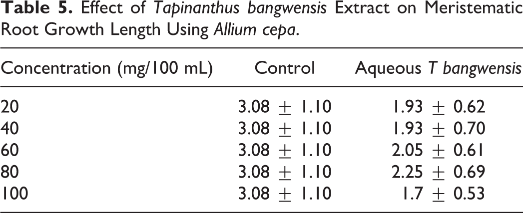
The mass yield of methanol extract of T bangwensis was 60.5 g (2.73%) while the aqueous fraction was 23.3 g (58.5%) (Table 1). Phytochemical screening reveals the presence of flavonoids (84.6 ± 0.41 mg/100 g), alkaloids (23.45 ± 0.09 mg/100 g), saponins (0.146 ± 0.01 mg/100 g), tannins (31 ± 0.85) mg/100 g), and phenols (147.5 ± 1.07 mg/100 g) (Table 2). These phytochemicals were reported to have medicinal properties and health promoting effects. 31,32 Phenol was found to be the most abundant and exhibits antioxidant and hepatocurative properties. 33 Flavonoid was also abundantly present and may ascribe to its powerful antioxidant properties. 34 The extract also has an appreciable concentration of tannin and alkaloid, which also exhibit antioxidant properties. 35 The extract also contains saponins, which are known to have hypocholesterolemic activities. 36 Hence, they may aid in lessening the metabolic burden that would have been placed on the liver during metabolism. 37 Total antioxidative capacity of any extract is contributed by the phytochemical components present in the extract and a higher phenolic content mostly makes the plant material to act as antioxidative agent. 38 The antioxidant activity of the aqueous extract of T bangwensis using DPPH and FRAP methods showed increased antioxidant activity as concentration increases. FRAP and DPPH results (Tables 3 and 4) showed appreciable antioxidant activity (IC50 = 2.908 ± 0.90 and 11.744 ± 0.88 µg/cm3) compared with ascorbic acid (IC50 = 2.746 ± 0.75 and 5.428 ± 0.90 µg/cm3). The cytotoxic effect of T bangwensis extract on meristematic growth length and mitotic index as presented in Tables 5 and 6 revealed marked decrease compared with the control. This suggests the extract exerts some inhibitory effect. Hence, the decrease in the mitotic index of A cepa meristematic cells could be interpreted as cellular death. 39 Figure 1 shows normal cellular mitotic division pattern (control) while Figure 2 (treated) represents some chromosomal abnormalities at different concentrations of the extracts. There were observable chromosomal aberrations, most frequently were stickiness and bridge chromosomes. Stickiness is one of the major indicators of cytotoxicity. This may result from improper folding of chromosome fibers or subchromatid bridges 40 or interpreted as a result of depolymerisation of DNA, partial dissolution of nucleoproteins, and the stripping of the protein covering of DNA in chromosomes. 41 Sticky chromosomes may indicate irreversible effect, probably leading to cell death. 42 Kabarity et al 43 reported that bridge chromosomes may be caused by stickiness of chromosomes, which made their separation and free movements incomplete thus they remained connected by bridges. Other chromosomal abnormalities observed were vagrant chromosomes, attached bridges, and C-mitosis. Their presence may be attributed to the failure of the spindle apparatus to organize and function optimally; or could be due to the blockage of DNA synthesis or inhibition of spindle formation. 39 Most of the aberrations are due to spindle failure, which indicates the interaction of the phytochemical constituents of the plant with the spindle apparatus. 44 Alkaloids have been reported to inhibit mitosis and also bind to tubulin, preventing spindle formation of the mitotic spindle. 45 The presence of alkaloid at an appreciable concentration in the plant extract may be responsible for its cytotoxicity.
The Yield of Aqueous Extract of Tapinanthus bangwensis Leaves.

Quantification of Phytochemical Compounds Present in Aqueous Extract of Tapinanthus bangwensis.a
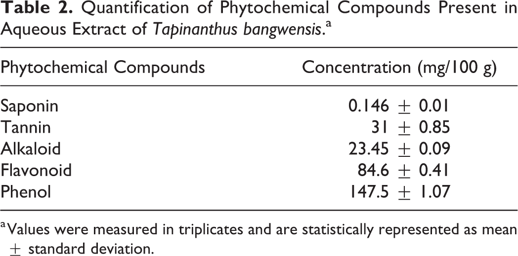
a Values were measured in triplicates and are statistically represented as mean ± standard deviation.
In Vitro Antioxidant Property of Tapinanthus bangwensis Using Ferric Reducing Antioxidant Power (FRAP).a
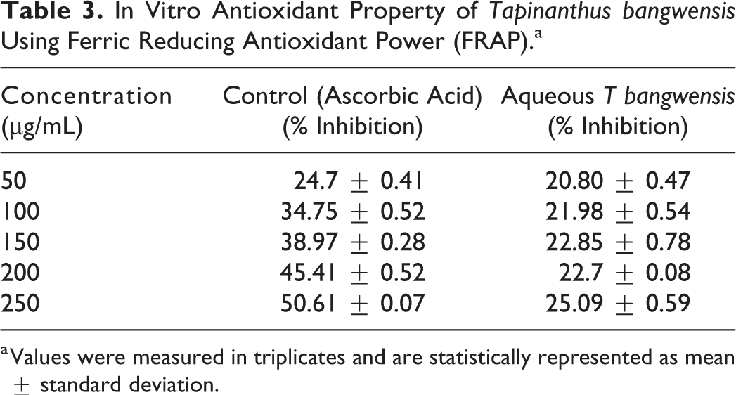
a Values were measured in triplicates and are statistically represented as mean ± standard deviation.
In Vitro Antioxidant Property of Aqueous Extract of Tapinanthus bangwensis Using 2,2-Diphenyl-1-Picrylhydrazine (DPPH).a

a Values were measured in triplicates and are statistically represented as mean ± standard deviation.
Effect of Tapinanthus bangwensis Extract on Meristematic Root Growth Length Using Allium cepa.
Cytotoxic Effect of Aqueous Extract of Tapinanthus bangwensis on the Chromosome Structure Using Allium cepa Test.


Photomicrographs of normal chromosomal structures.

Photomicrographs of the different chromosomal aberrations at different concentration of aqueous fraction of Tapinanthus bangwensis.
In living systems, liver is considered to be highly sensitive to toxic agents. The study of different enzyme activities and markers such as ALT, AST, albumin, and total protein have been found to be of great value in the assessment of clinical and experimental liver damage. 46 In this study, hepatotoxicity was induced by administering paracetamol (600 mg/kg body weight) and it was observed that the liver function indices of hepatic damage were elevated and some decreased as illustrated in Table 7. The results showed an increase in ALP, AST, ALT, HDL-c, MDA, and TRIG while the TC, TP, levels decreased in the rats in group 1 (paracetamol only). The hepatocurative activity of aqueous extract of T bangwensis at doses of 200 and 400 mg/kg were assessed by measuring the levels of various biochemical parameters. Animals treated with 200 mg/kg reduced the elevated levels of ALT, ALP, AST MDA, and HDL-c. While the aqueous extract at 400 mg/kg did not show a reduction in the level but rather, a high level of these serum markers was observed. The increase in the levels of MDA and HDL-c observed in the various target organs could be linked to the generation of free radicals, resulting in the peroxidation of membrane lipids. 47 The reduction in the elevated level of the enzymes due to administration of 200 mg/kg of the extract is in agreement with the commonly accepted view that serum levels of transaminase return to normal with healing of hepatic parenchyma and the regeneration of hepatocytes. 48 Similarly, treatment with the aqueous extract at 200 and 400 mg/kg dosage also elevated the levels of total protein and total cholesterol bringing the values close to normal. A fall in protein concentrations has been reported in severe parenchymal liver damage. Such a situation is found in severe acute insufficiency such as may occur in acute hepatic necrosis, poisoning from carbon tetrachloride and also in advanced stages of liver cirrhosis. 49 In severe acute hepatic necrosis, the total serum cholesterol is usually low. 49 Treatment with aqueous extract elevated the cholesterol and protein levels. It is thus possible that aqueous extract of T bangwensis elevated the reduced cholesterol and protein level caused by paracetamol.
Effect of Aqueous Tapinanthus bangwensis on Some Liver Functions.*
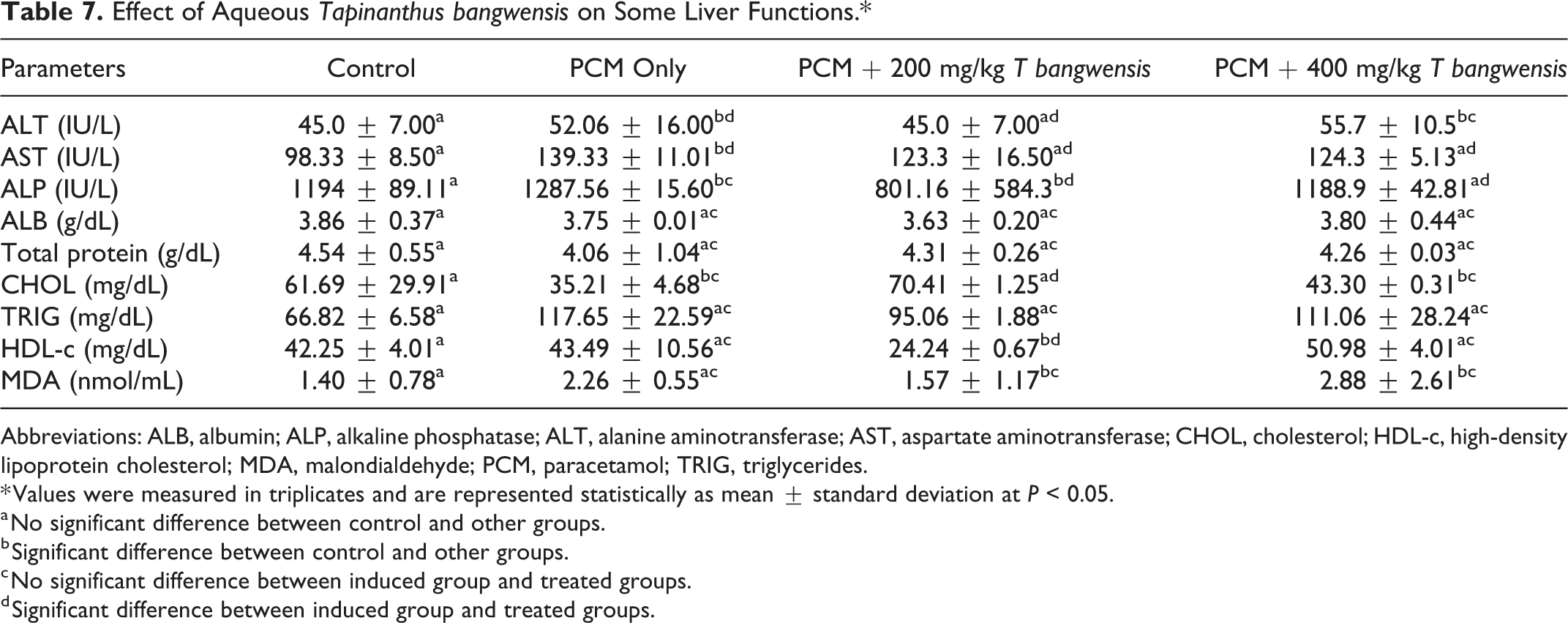
Abbreviations: ALB, albumin; ALP, alkaline phosphatase; ALT, alanine aminotransferase; AST, aspartate aminotransferase; CHOL, cholesterol; HDL-c, high-density lipoprotein cholesterol; MDA, malondialdehyde; PCM, paracetamol; TRIG, triglycerides.
* Values were measured in triplicates and are represented statistically as mean ± standard deviation at P < 0.05.
a No significant difference between control and other groups.
b Significant difference between control and other groups.
c No significant difference between induced group and treated groups.
d Significant difference between induced group and treated groups.
Conclusion
The research findings reveal that aqueous extract of T bangwensis leaves is slightly cytotoxic though exhibited hepatocurative effect against paracetamol-induced toxicity at both low and high doses although 200 mg/kg body weight was more potent.
Footnotes
Acknowledgments
The authors appreciate Mrs Lilian Ekeson, Cadet(ASP) Olua Moses and Cadet(ASP) Unah Paul of Department of Biochemistry & Forensic Science, Nigeria Police Academy, Wudil, for providing the required technical assistance and support for the success of this study.
Author Contributions
GOI and CJO conceived and supervised the work. DGO carried out the benchwork. EA carried out the statistical analysis and relevant interpretations. All authors contributed to drafting the manuscript.
Declaration of Conflicting Interests
The authors declared no potential conflicts of interest with respect to the research, authorship, and/or publication of this article.
Funding
The authors received no financial support for the research, authorship, and/or publication of this article.
Ethical Approval
The experimental protocols and procedures used in this study were approved by the Ethics Committee for the Care and Use of Laboratory Animals of Nigeria Police Academy, Wudil, Kano State, Nigeria.