
Abstract
Introduction
The cuneiforms facilitate gait by providing critical stabilization and support to the medial longitudinal arch. Damage to the cuneiforms may compromise midfoot stability. The lack of data regarding medial cuneiform fracture and treatment may be attributed to the rarity of such injuries because they make up less than 2% of all tarsal fractures. 2 Very few isolated medial cuneiform fractures have been reported in the literature, mainly involving high-energy trauma or Lisfranc fracture-dislocations. 5,10,11,13,14 Segmental or complete bone loss of the cuneiforms is even more challenging to treat. Currently available techniques to replace or regenerate structural bone include autograft or allograft implantation, use of growth factors, osteoconductive scaffolds, osteoprogenitor cells, and distraction osteogenesis. 4 Although these procedures have been thoroughly studied in the treatment of segmental loss in the long bones, there is still little information on techniques for treating loss of the tarsal bones. Thus, the treatment and healing process of the medial cuneiform fracture has not been fully researched or understood. Here, we present the case of medial cuneiform reformation that occurred in a 34-year-old man who had a gunshot wound to the foot resulting in complete loss of the medical cuneiform, which was treated with an antibiotic bone cement spacer that led to spontaneous regeneration of the bone with nearly anatomic configuration.
Case Report
A 34-year-old man with no significant past medical history accidentally inflicted a gunshot wound onto the dorsum of his left foot. The 12-gauge ballistic injury created a wound approximately 10 cm in length, 4 cm in width, and about 4 cm in depth (Figure 1A). Radiographs and operative debridement showed a disruption of the tibialis anterior tendon with maintenance of the extensor hallucis longus tendon and fracture of the navicular and extremely comminuted fracturing of the medial, middle, and lateral cuneiforms (Figure 2). He underwent open reduction with internal screw fixation of the navicular bone and application of a VAC dressing.

Left foot images showing injury progression: (A) Initial presentation; (B) 5 months, exposed cement is seen; (C) 7 months; (D) 15 months; (E) 20 months; (F) 24 months.

Left foot plain films (Anteroposterior, oblique, lateral) showing progression of medial cuneiform reformation: (A) 5 months postinjury, (B) 7 months, (C) 9 months, (D) 15 months.
Subsequent irrigation and debridement procedures were performed, and a sural flap was applied on the fifth day postinjury. During the flap surgery, bone cement with 1 g antibiotic vancomycin was applied for infection prophylaxis and to fill the area of the medial cuneiform bone. The bone defect was planned to be repaired with a bone allograft via Masquelet technique once the wound was stabilized by debridement of all dead tissue and subsequent development of healthy granulation. One month postinjury, he underwent another irrigation and debridement procedure that included a partial flap debridement because of venous congestion, which resulted in partial flap loss distally and resulted in exposure of underlying structures. Two months postinjury, the wound reopened, exposing the bone cement. After the cement was removed for debridement, the sural flap showed necrotic tissue at the cuneiform area, which was completely removed, creating a defect in the cuneiform area. The cement was then replaced. Debridement of the sural flap was done in addition to removal and replacement of antibiotic bone cement in the medial cuneiform area. Two and a half months postinjury, the bone cement was once again removed and new bone cement with vancomycin antibiotic applied during an irrigation and debridement procedure.
Around 5½ months postinjury, the cement had fallen out because of the partial sural flap loss and weightbearing radiographs (3 views) of his foot demonstrated a well-preserved arch with comminuted fracture of the medial cuneiform with ossification connecting the multiple fragments. About 8 months postinjury, his superficial wound was measured at approximately 3 cm × 2 cm with healthy-appearing granulation tissue with the exception of a small area of gray-colored epithelialization on the dorsum of the foot. Radiographs at this time demonstrated near complete reconstitution of the medial cuneiform with slight shortening.
One year postinjury, he underwent a left extensor hallucis longus tendon repair and tenolysis of tibialis anterior tendon, and removal of the navicular screw. It was found that he had re-formed bone in the area of the medial cuneiform, providing a stable bony architecture of his foot. The bone was left alone and proceeded to soft tissue reconstruction. Around 1 year 4 months postinjury, weightbearing radiographs (3 views) of his foot demonstrate neutral alignment of his arch with reconstitution of his medial cuneiform (Figures 3 and 4). One year 5 months postinjury, a free flap was applied to the left foot from the groin to close the wound. Subsequent tendon repair procedures followed. Most recent follow-up showed a well-healing injury with a structure that resembles the uninjured foot.
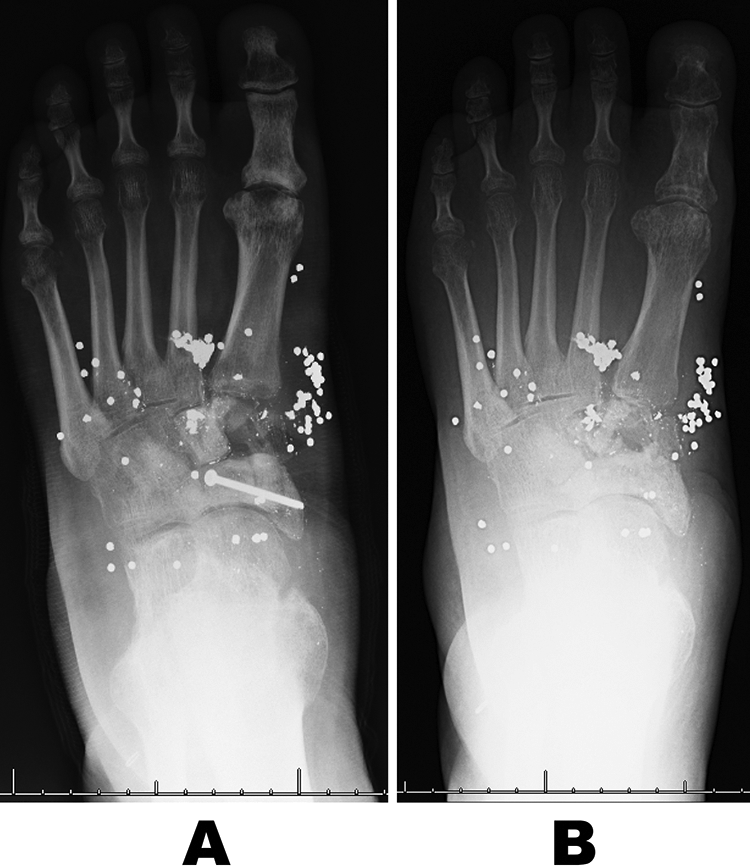
Anteroposterior radiographs at (A) 5 months and (B) 15 months postinjury.

Lateral weightbearing radiograph at (A) 5 months and (B) 15 months postinjury.
Discussion
We present a case where the damaged and partially debrided medial cuneiform area ossified enough to support the medial arch of the foot and allowed the patient to have stable walking. Medial cuneiform fracture is a rare occurrence that is mainly related to high-energy trauma, and its healing process has not been fully studied. Restoration of the medial cuneiform is important for gait and weightbearing as it is an essential structure of the first ray, the ray with the heaviest load, 7 and the medial longitudinal arch. 3,6 On literature review, reconstruction of the medial cuneiform has involved iliac allografts and plate fixation as a result of an aneurysmal bone cyst and a giant cell tumor. 1,8 However, there have been no reported cases of stable reformation of the medial cuneiform without grafting. Reformation likely resulted from natural bone healing, but it is important to evaluate the role of the bone cement in preparation for Masquelet technique procedure.
The induced membrane technique (Masquelet technique) is a 2-step operative procedure used to treat bone defects, nonunion of fractures, and infection eradication that involves debridement and placement of a cement spacer, which is replaced by cancellous bone in a second operative procedure. A systemic review of this technique has shown an 89.7% union rate. 9 In this case, the technique differed from the norm as the cancellous bone was not implanted after the cement spacer was removed because the cuneiform showed reformation. It is possible the spacer induced formation of a pseudo-synovial membrane that provided blood supply, growth factors like VEGF and TGF-beta1, and osteoinductive factors like BMP-2. 12 It is possible that in this case the supply of the previously mentioned factors were enough to induce the reformation of the damaged cuneiform. In a similar manner, it is important to consider that although bone cement has not been fully linked with osteoconduction, research on PMMA-based bone cement has shown osteoconduction properties when modified with γ-methacryloxypropyl-trimethoxysilane and calcium acetate. 15
Limitations of this report is that it did not have advanced imaging modalities. 3-D imaging could have provided a more detailed view of the bone regeneration. Magnetic resonance angiography was performed showing normal vasculature above the wound, but lead pellets in the wound bed created artifacts that did not allow full analysis of the blood supply to the medial cuneiform.
In conclusion, we report a case of a 34-year-old man who had an accidental self-inflicted gunshot whose medial cuneiform regenerated with only the use of a cement spacer. This case demonstrates that spontaneous bone regeneration in the foot is possible if the space is maintained during healing.
Supplemental Material
Supplemental Material, FAO876261-ICMJE - Spontaneous Regeneration of Medial Cuneiform Following Gunshot Wound
Supplemental Material, FAO876261-ICMJE for Spontaneous Regeneration of Medial Cuneiform Following Gunshot Wound by Ricardo Reyes, Tolga Türker and L. Daniel Latt in Foot & Ankle Orthopaedics
Footnotes
Declaration of Conflicting Interests
The author(s) declared no potential conflicts of interest with respect to the research, authorship, and/or publication of this article. ICMJE forms for all authors are available online.
Funding
The author(s) received no financial support for the research, authorship, and/or publication of this article.
References
Supplementary Material
Please find the following supplemental material available below.
For Open Access articles published under a Creative Commons License, all supplemental material carries the same license as the article it is associated with.
For non-Open Access articles published, all supplemental material carries a non-exclusive license, and permission requests for re-use of supplemental material or any part of supplemental material shall be sent directly to the copyright owner as specified in the copyright notice associated with the article.